
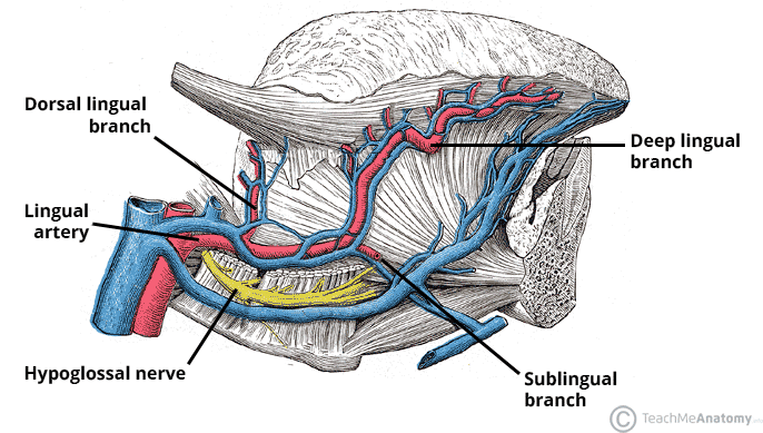
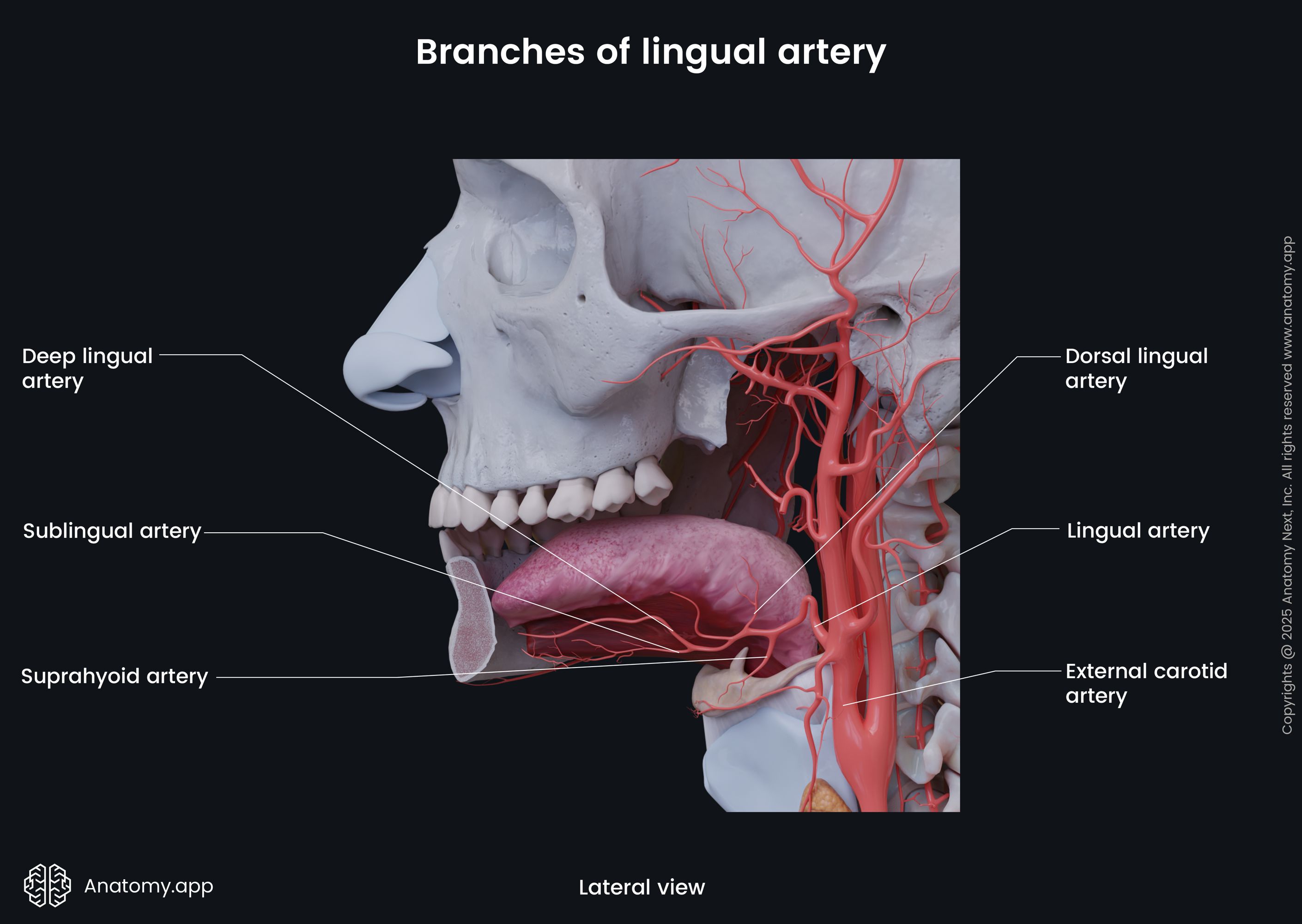
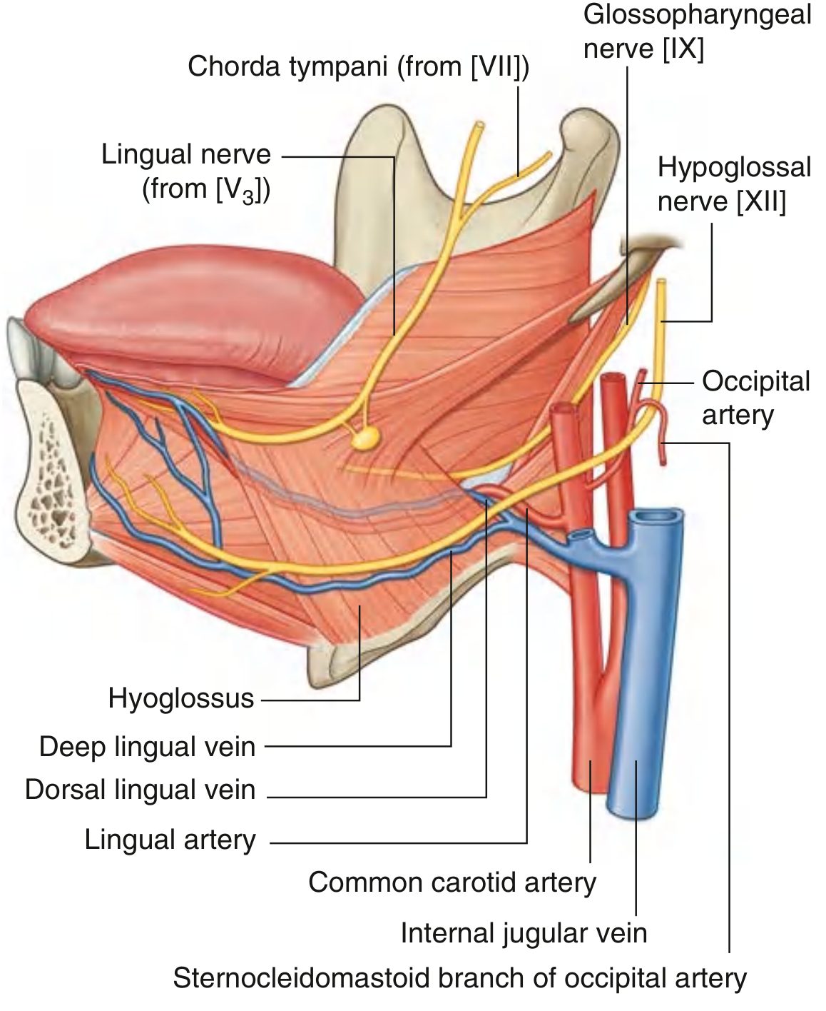
diagram of arterial supply of mouth for lingual artery.
lingual artery branches arterial supply mouth diagram anatomy
Arterial Supply of the Mouth - Lingual Artery
Diagrams
Origin and Course
- Forms an upward loop then passes deep to the hyoglossus muscle
- Passes through the oropharyngeal triangle (bounded by mylohyoid, superior constrictor, and middle constrictor muscles)
- Enters the floor of the oral cavity
- Runs forward between the hyoglossus and genioglossus muscles toward the tongue apex
Branches of the Lingual Artery
| Branch | Origin | Distribution |
|---|---|---|
| Suprahyoid artery | Near hyoid | Muscles above the hyoid bone |
| Dorsal lingual arteries (2-3) | Medial to hyoglossus | Posterior dorsum of tongue, palatoglossal arch, soft palate, tonsil, epiglottis |
| Sublingual artery | At anterior margin of hyoglossus | Sublingual gland, genioglossus, mylohyoid; sends a branch through the lingual foramen of the mandible |
| Deep lingual artery | Terminal branch | Inferior surface of tongue (near lingual frenulum) - runs forward to apex |
- Scott-Brown's Otorhinolaryngology Head & Neck Surgery, p. 683
Structures Supplied by the Lingual Artery
- Tongue (principal artery - all parts)
- Sublingual gland
- Gingiva (lingual gingival surface of mandibular teeth)
- Oral mucosa of the floor of the oral cavity
- Soft palate and palatine tonsil (via dorsal lingual branches)
- Epiglottis (inferior epiglottic branches)
Additional Contributions to the Mouth/Root of Tongue
-
Tonsillar and ascending palatine branches of the facial artery
-
Ascending pharyngeal artery branches (root of tongue)
-
Superior laryngeal artery epiglottic branches (anastomose with inferior dorsal lingual branches in the vallecula region)
-
Scott-Brown's Otorhinolaryngology Head & Neck Surgery, p. 683
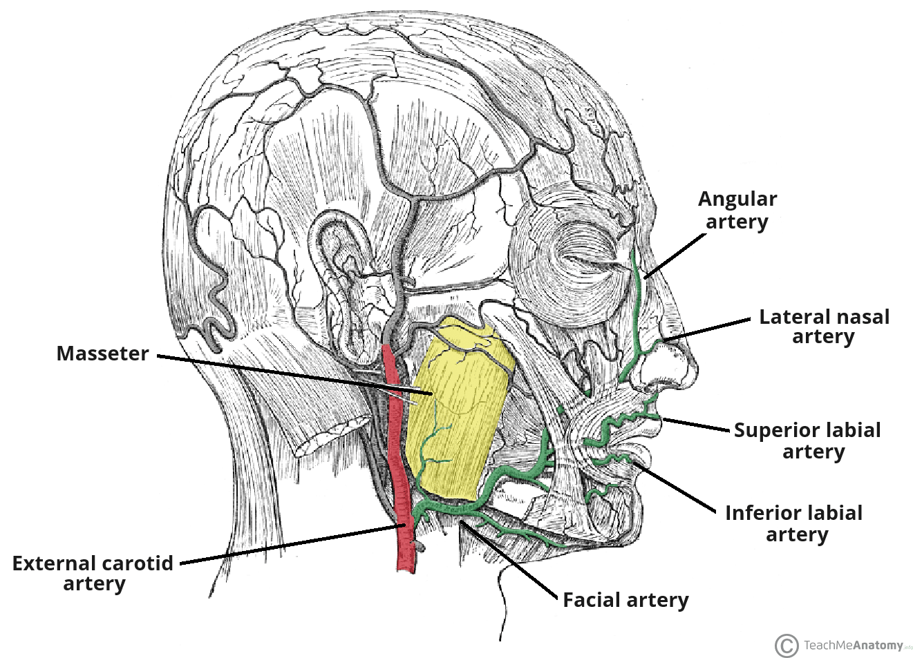
diagram of arterial supply of mouth for facial artery
facial artery branches arterial supply mouth diagram anatomy
Arterial Supply of the Mouth - Facial Artery
Diagrams
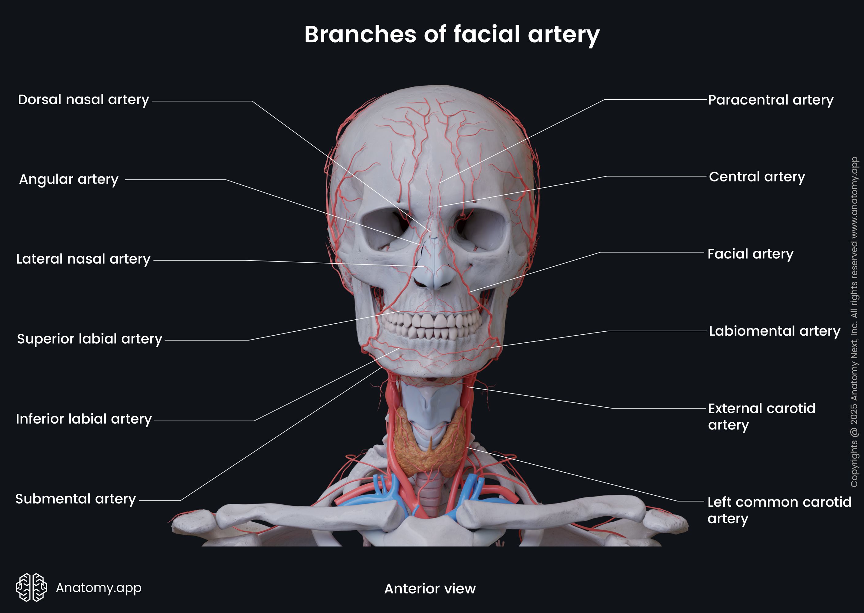
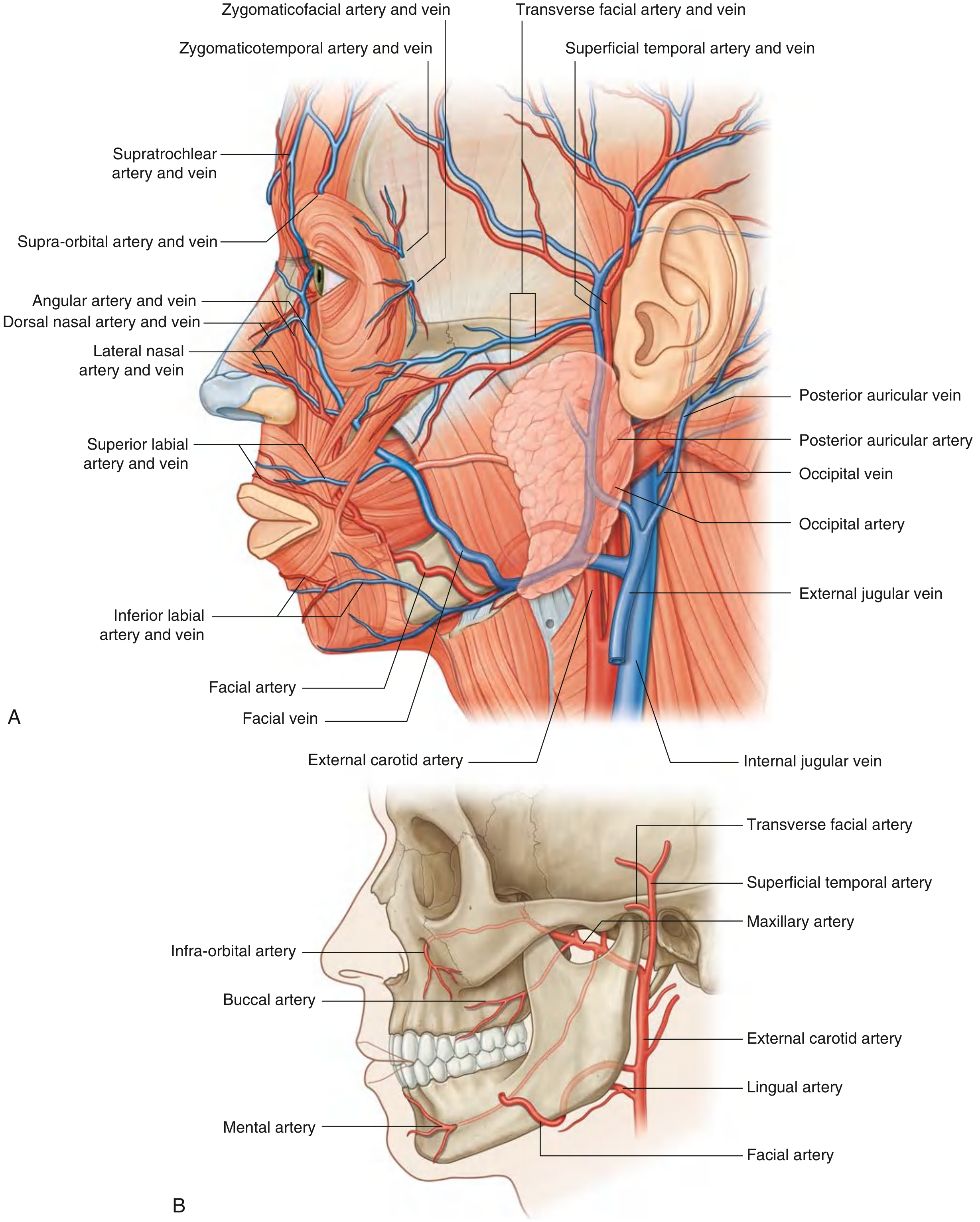
Origin and Course
- Arises from the external carotid artery, passes deep to the digastric and stylohyoid muscles
- Passes through (or deep to) the submandibular gland, giving off glandular branches
- Hooks over the inferior border of the mandible just anterior to the masseter (pulse palpable here)
- Enters the face, runs in a tortuous upward and medial course - deep to platysma, risorius, and zygomaticus; superficial to buccinator and levator anguli oris
- Passes lateral to the angle of the mouth giving off labial branches
- Ascends along the side of the nose
- Terminates as the angular artery at the medial canthus of the eye
- Gray's Anatomy for Students, p. 7257
Branches of the Facial Artery
Cervical Branches (given off in the neck)
| Branch | Distribution |
|---|---|
| Ascending palatine artery | Soft palate, palatine tonsil, auditory tube, superior constrictor |
| Tonsillar artery | Main arterial supply to the palatine tonsil; pierces superior constrictor |
| Submental artery | Mylohyoid, anterior belly of digastric, submental lymph nodes, chin skin |
| Glandular branches | Submandibular gland (3-4 branches) |
Facial Branches (given off on the face - relevant to the mouth)
| Branch | Distribution |
|---|---|
| Inferior labial artery | Arises near the angle of the mouth; supplies the lower lip, anastomoses with fellow of opposite side and mental artery |
| Superior labial artery | Supplies the upper lip and gives a nasal septal branch (supplies nasal septum and floor of nose); anastomoses with fellow of opposite side |
| Lateral nasal artery | Lateral surface and dorsum (ala) of the nose |
| Angular artery | Terminal branch; medial corner of eye; anastomoses with the dorsal nasal branch of the ophthalmic artery (linking external and internal carotid systems) |
- Gray's Anatomy for Students, p. 7264, 7275-7283; K.J. Lee's Essential Otolaryngology
Key Points for the Mouth Specifically
- Lips: The superior and inferior labial arteries are the principal supply. They form a labial arterial arcade around the lips by anastomosing with their counterparts from the opposite side. This rich anastomosis means lip lacerations bleed profusely but heal well.
- Labial gingiva (anterior): Supplied by the mental artery (branch of inferior alveolar), which also receives perforating branches from the incisive artery; labial branches of the infraorbital artery supply the upper labial gingiva.
- Tonsil and soft palate: Via the tonsillar and ascending palatine cervical branches.
- The labial branches anastomose across the midline, providing an important link between the facial arteries of both sides (and thus between both external carotid arteries). - Scott-Brown's Otorhinolaryngology Head & Neck Surgery, p. 681
Mnemonic for Branches

diagram of arterial supply of mouth for facial artery using this picture as a reference.
Medical anatomy diagram titled "FACIAL ARTERY:" in bold red text at the top center. Style: clean medical illustration on white background, matching this exact layout and style: LEFT SIDEBAR (blue table): Header "BRANCHES" in white on dark blue. Two sections with light blue background - "CERVICAL" (vertical label) listing: - ASCENDING PALATINE, - TONSILLAR TONSIL, - GLANDULAR, - SUBMENTAL. Then "FACIAL" (vertical label) listing: - INFERIOR LABIAL, - SUPERIOR LABIAL, - LATERAL NASAL, - ANGULAR. CENTER/RIGHT: Semi-transparent skull and cervical vertebrae illustration in light gray. A prominent dark red arterial trunk arises from the EXTERNAL CAROTID ARTERY (labeled bottom left in bold red) in the neck. The artery courses upward past the SUBMANDIBULAR SALIVARY GLAND (labeled, small orange-tan gland shown), hooks over the mandible anterior to the MASSETER muscle (large pink muscle, labeled), passing deep to STYLOHYOID (labeled, slender muscle) and DIGASTRIC (labeled) muscles. From the main trunk, branches labeled in bold dark red with arrows: - SUBMENTAL ARTERY: runs horizontally along the inferior border of the mandible above the MYLOHYOID muscle (labeled flat muscle) - INFERIOR LABIAL ARTERY: branches to lower lip region - SUPERIOR LABIAL ARTERY: branches to upper lip region - LATERAL NASAL ARTERY: ascending branch to side of nose - ANGULAR ARTERY: terminal branch at top right, reaching medial commissure of eye (small label in handwriting style: "medial commissure of the eye") TOP CENTER INSET: Small circular cutaway showing ASCENDING PALATINE artery supplying SOFT PALATE (labeled) and PALATINE TONSIL (labeled). Color scheme: arteries in dark red/maroon, muscles in pink/salmon, bones in light gray, labels in dark red for arteries and dark gray/black for structures. Style matches a professional Gemini AI generated medical illustration.
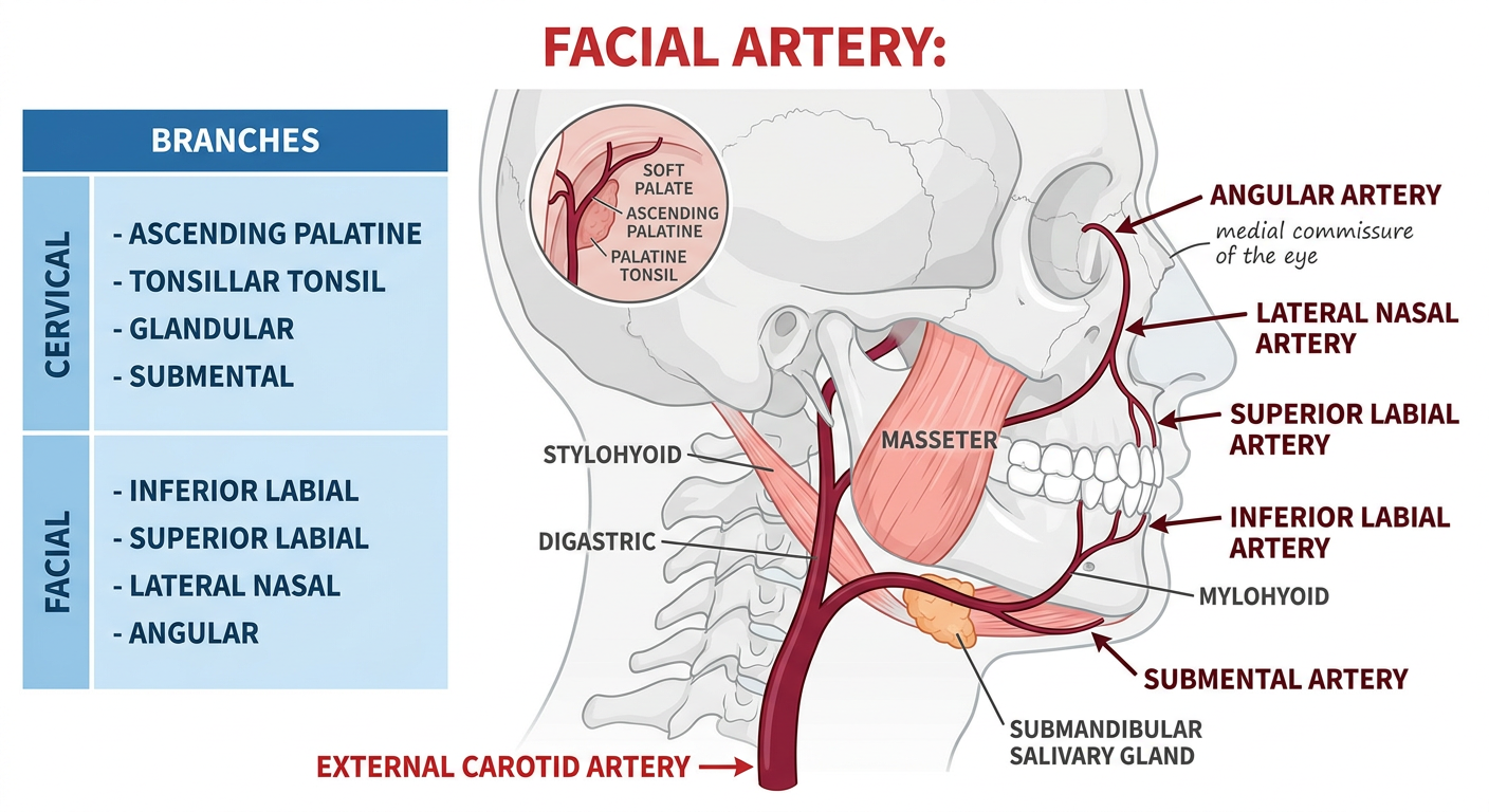
- Cervical: Ascending palatine, Tonsillar, Glandular, Submental
- Facial: Inferior labial, Superior labial, Lateral nasal, Angular
- Origin from the external carotid artery in the neck
- Course past the submandibular gland, deep to digastric and stylohyoid, hooking over the mandible anterior to the masseter
- Submental artery running along the underside of the jaw above mylohyoid
- Inferior and superior labial arteries at the angle of the mouth
- Lateral nasal artery up the side of the nose
- Angular artery as the terminal branch at the medial corner of the eye
make it handwritten
Handwritten medical anatomy sketch of the FACIAL ARTERY on white/cream paper background. Hand-drawn style like a medical student's notebook or lecture notes, using blue and red ink pens. Title "Facial Artery:" written in large handwritten red ink at the top. LEFT SIDE - handwritten blue ink table/box titled "BRANCHES" with two sections: CERVICAL (written vertically in blue): - ascending palatine, - tonsillar, - glandular, - submental FACIAL (written vertically in blue): - inferior labial, - superior labial, - lateral nasal, - angular CENTER/RIGHT - hand-drawn sketch of skull outline and cervical spine in light pencil/gray ink. Arterial tree drawn in red ink: - EXTERNAL CAROTID ARTERY labeled at bottom with handwritten arrow - Facial artery trunk curving up, hooking over mandible anterior to masseter - MASSETER muscle sketched as hatched rectangle, labeled in blue handwriting - STYLOHYOID and DIGASTRIC muscles roughly sketched with blue handwritten labels - SUBMANDIBULAR SALIVARY GLAND sketched as small oval, handwritten label - MYLOHYOID sketched as flat muscle, handwritten label Branches in red ink with handwritten arrow labels: - SUBMENTAL ARTERY (below jaw) - INFERIOR LABIAL ARTERY (to lower lip) - SUPERIOR LABIAL ARTERY (to upper lip) - LATERAL NASAL ARTERY (side of nose) - ANGULAR ARTERY (terminal, near medial corner of eye) with small note "= terminal branch" TOP INSET - small hand-drawn circle showing ascending palatine to soft palate and palatine tonsil, labeled in handwriting. Overall aesthetic: authentic medical school notebook sketch, slightly messy but clear, hand-lettered labels with ink pen, anatomical structures roughly but recognizably drawn, some labels with underlines or small arrows, cream/white paper texture.
400, message: Got more than 4194304 bytes when reading: b'data: {"candidates": [{"content": {"role": "model","parts": [{"inlineData": {"mimeType": "image/png"...'.
We encountered an error while processing your request. Please try again or reach out to us for help.
draw a hand drawn diagram
Hand-drawn medical notebook sketch of the facial artery and its branches. Drawn on cream/off-white paper with blue and red ballpoint pen, like a medical student's anatomy notes. Title "Facial Artery" written in large cursive red ink at top with an underline. Left side: a hand-drawn box in blue ink labeled "Branches" with two sections written in neat handwriting: Cervical: ascending palatine, tonsillar, glandular, submental Facial: inferior labial, superior labial, lateral nasal, angular Right side: rough pencil sketch of skull profile and jaw. Red ink artery drawn as a thick wavy line: - Starts at bottom labeled "External Carotid A." with arrow - Curves up behind submandibular gland (small oval sketched, labeled) - Hooks over mandible near masseter (hatched box labeled "Masseter") - Gives off branches drawn as thinner red lines with handwritten labels and arrows: * "Submental a." along underside of jaw * "Inferior labial a." to lower lip * "Superior labial a." to upper lip * "Lateral nasal a." up nose * "Angular a." at inner corner of eye, with note "terminal branch" - Small inset circle sketch showing ascending palatine to soft palate and tonsil Style: authentic hand-drawn medical notes, slightly imperfect lines, blue ink for labels and structure names, red ink for arteries, pencil for bone outlines, cream paper background with subtle paper texture.
400, message: Got more than 4194304 bytes when reading: b'data: {"candidates": [{"content": {"role": "model","parts": [{"inlineData": {"mimeType": "image/png"...'.
We encountered an error while processing your request. Please try again or reach out to us for help.
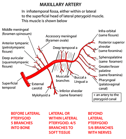
diagram of arterial supply of mouth for maxillary artery
maxillary artery branches arterial supply mouth diagram anatomy
Arterial Supply of the Mouth - Maxillary Artery
Diagrams
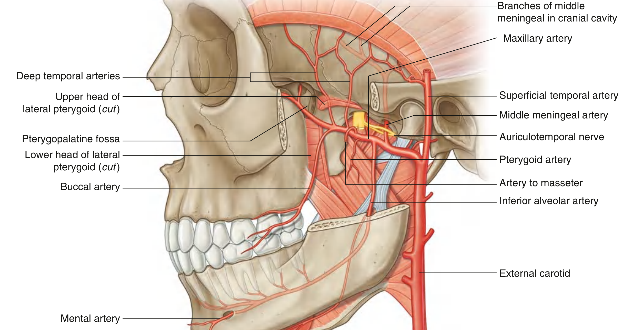
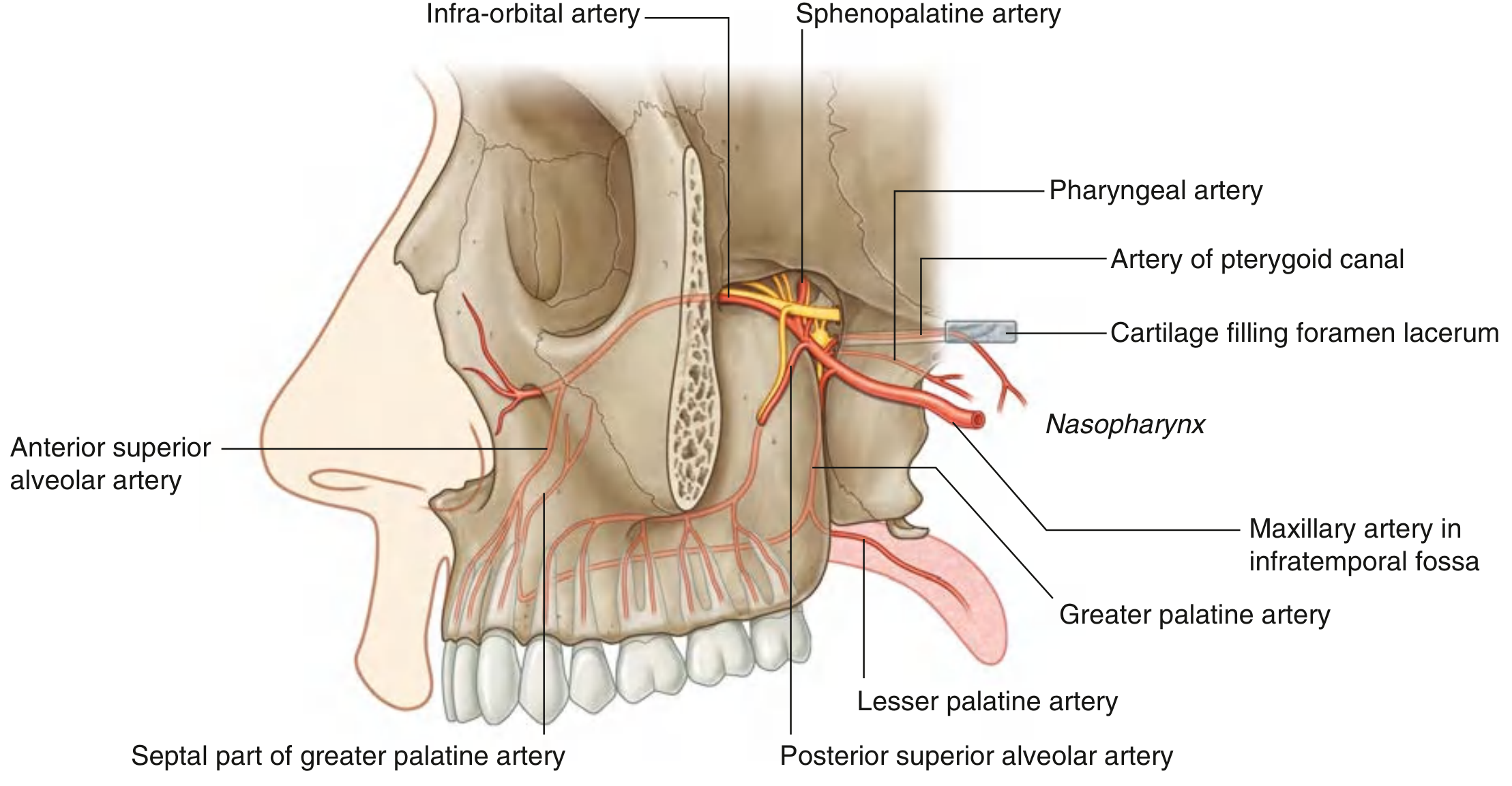
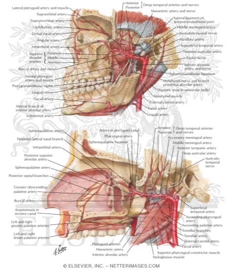
Origin and Course
| Part | Location | Key relation |
|---|---|---|
| 1st (Mandibular) | Neck of mandible to lateral pterygoid | Between ramus of mandible and sphenomandibular ligament |
| 2nd (Pterygoid) | Related to lateral pterygoid muscle | Lateral or medial to lower head of lateral pterygoid |
| 3rd (Pterygopalatine) | In pterygopalatine fossa | Anterior to pterygopalatine ganglion |
- Gray's Anatomy for Students, p. 2440-2442
Branches Relevant to the Mouth
From the 1st Part (Mandibular)
| Branch | Course | Supply to mouth |
|---|---|---|
| Inferior alveolar artery | Enters mandibular foramen with inferior alveolar nerve; runs in mandibular canal | All lower teeth (molars, premolars via dental branches); divides into: Incisive branch (lower incisors/canines) + Mental artery (exits mental foramen - supplies chin and labial gingiva) |
| Mylohyoid branch | Branches before entering mandibular foramen | Mylohyoid muscle and anterior belly of digastric |
From the 2nd Part (Pterygoid)
| Branch | Supply to mouth |
|---|---|
| Buccal artery | Buccinator muscle and mucosa; buccal gingiva of posterior lower teeth |
| Deep temporal arteries | Temporalis muscle (not directly mouth) |
| Masseteric artery | Masseter muscle |
From the 3rd Part (Pterygopalatine) - Most Important for Mouth
| Branch | Course | Supply to mouth |
|---|---|---|
| Posterior superior alveolar artery | Descends through alveolar foramina on infratemporal surface of maxilla | Upper molar and premolar teeth, adjacent gingiva, maxillary sinus |
| Infra-orbital artery | Runs in infra-orbital groove/canal; exits infra-orbital foramen | Gives off anterior superior alveolar arteries → upper incisor and canine teeth, maxillary sinus |
| Greater palatine artery | Descends through palatine canal | Hard palate - runs forward on palate to incisive fossa, anastomoses with nasopalatine artery |
| Lesser palatine artery | Branch of greater palatine; exits lesser palatine foramen | Soft palate and palatine tonsil |
| Sphenopalatine artery | Enters nasal cavity through sphenopalatine foramen | Nasal cavity; septal branch (nasopalatine) descends to supply anterior hard palate via incisive canal |
| Pharyngeal artery | Passes through palatovaginal canal | Roof of nasopharynx, auditory tube, sphenoidal sinus |
- Gray's Anatomy for Students, p. 2771-2781; Scott-Brown's Otorhinolaryngology, p. 681-683
Summary: Maxillary Artery Supply to Each Part of the Mouth
| Structure | Artery |
|---|---|
| Lower teeth | Inferior alveolar a. (from 1st part) |
| Lower labial gingiva (anterior) | Mental a. + incisive a. (terminal branches of inferior alveolar) |
| Lower buccal gingiva (posterior) | Buccal a. (2nd part) + perforating branches of inferior alveolar |
| Upper molars/premolars | Posterior superior alveolar a. (3rd part) |
| Upper incisors/canines | Anterior superior alveolar a. (from infra-orbital, 3rd part) |
| Upper premolars | Middle superior alveolar a. (when present, from 3rd part) |
| Upper palatal gingiva | Greater palatine a. (3rd part) |
| Hard palate | Greater palatine a. + nasopalatine/sphenopalatine a. (anastomose at incisive fossa) |
| Soft palate | Lesser palatine a. (3rd part) |
| Cheek mucosa | Buccal a. (2nd part) |
- Scott-Brown's Otorhinolaryngology Head & Neck Surgery, p. 681-683; Gray's Anatomy for Students, p. 2766
pterygoid fossa of maxillary artery and its supplies
pterygopalatine fossa maxillary artery third part branches diagram anatomy
Pterygopalatine Fossa - 3rd Part of the Maxillary Artery
Diagrams
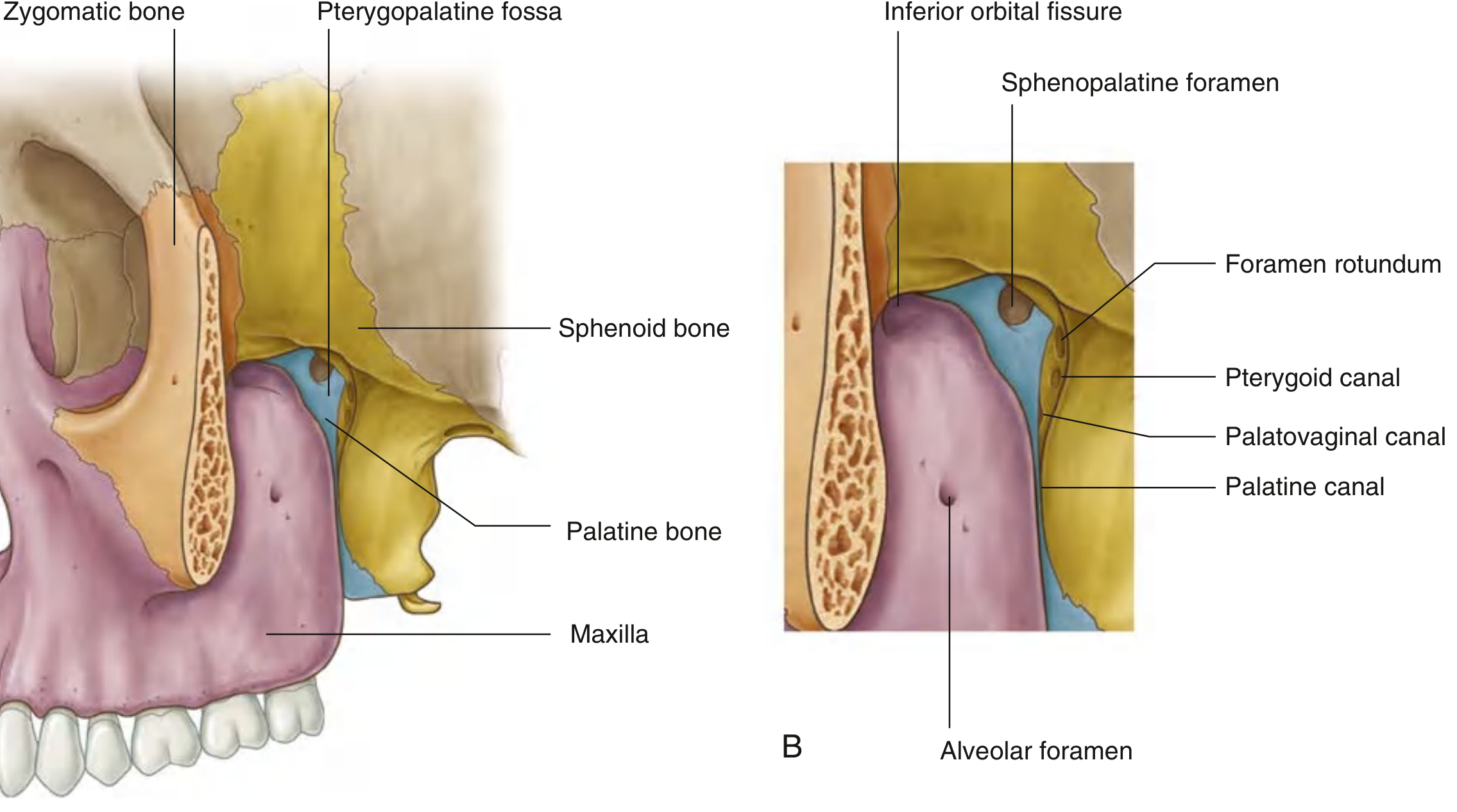
What is the Pterygopalatine Fossa?
| Wall | Bone |
|---|---|
| Anterior | Posterior surface of maxilla |
| Medial | Lateral surface of palatine bone |
| Posterior/roof | Sphenoid bone (pterygoid process) |
- Middle cranial fossa - via foramen rotundum (maxillary nerve V2) and pterygoid canal
- Infratemporal fossa - via pterygomaxillary fissure (entry of maxillary artery)
- Floor of orbit - via inferior orbital fissure (infra-orbital artery/nerve)
- Lateral nasal cavity - via sphenopalatine foramen (sphenopalatine artery)
- Hard palate/oral cavity - via greater/lesser palatine canals
- Nasopharynx - via palatovaginal canal (pharyngeal branch)
3rd Part of the Maxillary Artery - The 6 Branches
1. Posterior Superior Alveolar Artery (PSA)
- Origin: Given off as the maxillary artery passes through the pterygomaxillary fissure (just before entering the fossa)
- Course: Descends on the posterolateral surface of the maxilla; enters through alveolar foramina
- Supply: Upper molar and premolar teeth, adjacent gingiva, maxillary sinus
2. Infra-orbital Artery
- Course: Exits fossa through inferior orbital fissure → runs in infra-orbital groove and canal → exits at infra-orbital foramen
- Supply:
- Anterior superior alveolar arteries (branch within infra-orbital canal) → upper incisor and canine teeth + maxillary sinus
- Inferior rectus and inferior oblique muscles, lacrimal sac (orbital floor structures)
- Face below orbit (skin of cheek, lower eyelid)
- Note: A middle superior alveolar artery (when present) also arises from this vessel to supply upper premolars
3. Greater Palatine Artery
- Course: Descends with palatine nerves through palatine canal; exits at greater palatine foramen; runs forward on inferior surface of hard palate to incisive fossa; ascends through incisive canal
- Supply:
- Hard palate (mucosa and glands)
- Palatal gingiva of all upper teeth
- Anterior nasal septum (via incisive canal - anastomoses with nasopalatine/sphenopalatine artery = Kiesselbach's plexus/Little's area)
4. Lesser Palatine Artery
- Origin: Branch of the greater palatine artery within the palatine canal
- Course: Exits through lesser palatine foramen
- Supply: Soft palate, palatine tonsil
5. Pharyngeal Artery
- Course: Passes posteriorly through palatovaginal canal (with pharyngeal nerve of V2)
- Supply: Posterior roof of nasal cavity, sphenoidal sinus, pharyngotympanic (Eustachian) tube, nasopharynx
6. Sphenopalatine Artery (Terminal Branch)
- Course: Exits medially through sphenopalatine foramen into nasal cavity
- Supply:
- Posterior lateral nasal arteries → lateral wall of nasal cavity, paranasal sinuses
- Posterior septal branches → nasal septum; largest branch descends septum anteriorly to anastomose with greater palatine artery at incisive fossa
7. Artery of Pterygoid Canal (Vidian Artery)
- Course: Passes posteriorly through pterygoid canal (with nerve of pterygoid canal/Vidian nerve); terminates in nasopharyngeal mucosa via cartilage of foramen lacerum
- Supply: Pharyngotympanic tube, upper nasopharynx, middle ear
Summary Table
| Branch | Exit from Fossa | Main Supply |
|---|---|---|
| Posterior superior alveolar | Alveolar foramina (maxilla) | Upper molars/premolars, gingiva, maxillary sinus |
| Infra-orbital | Inferior orbital fissure → infra-orbital foramen | Upper incisors/canines (via ASA), orbit floor, face |
| Greater palatine | Greater palatine foramen | Hard palate, palatal gingiva |
| Lesser palatine | Lesser palatine foramen | Soft palate, tonsil |
| Pharyngeal | Palatovaginal canal | Sphenoidal sinus, Eustachian tube, nasopharynx |
| Sphenopalatine | Sphenopalatine foramen | Nasal cavity walls, nasal septum |
| Artery of pterygoid canal | Pterygoid canal → foramen lacerum | Upper nasopharynx, Eustachian tube |
- Gray's Anatomy for Students, p. 2755-2793; Scott-Brown's Otorhinolaryngology, p. 681-683
Clinical Note
1st part of maxillary artery
first part maxillary artery mandibular part branches diagram anatomy
1st Part of the Maxillary Artery (Mandibular Part)
Diagrams
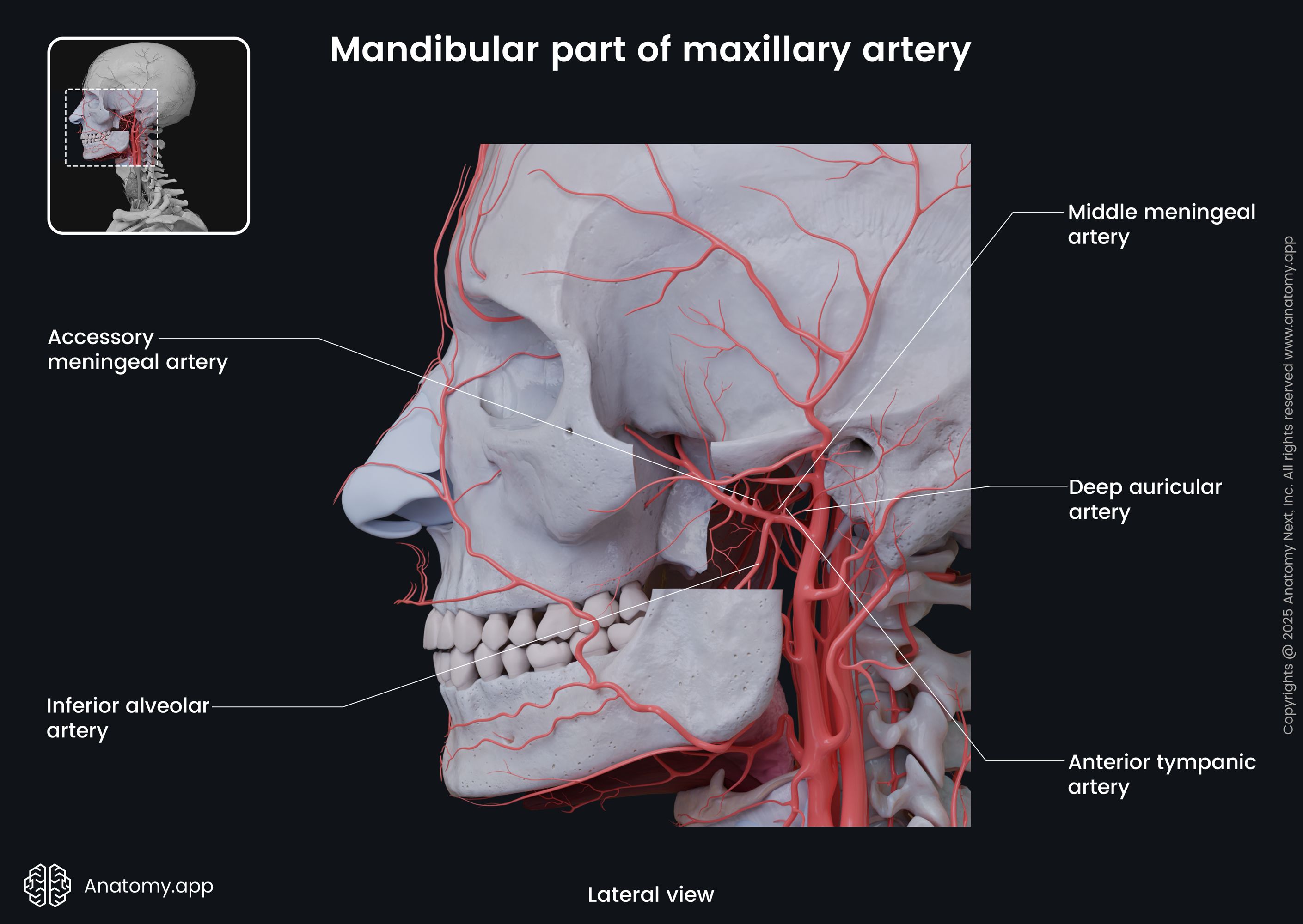
Location and Relations
- Laterally: Neck/ramus of the mandible
- Medially: Sphenomandibular ligament
5 Branches of the 1st Part
1. Deep Auricular Artery
- Course: Ascends superiorly toward the temporomandibular joint (TMJ)
- Supply: External acoustic meatus (skin lining), outer surface of the tympanic membrane
- Small branch; passes through or near the parotid tissue
2. Anterior Tympanic Artery
- Course: Passes through the petrotympanic fissure
- Supply: Inner (deep) surface of the tympanic membrane; mucosa of the middle ear (tympanic cavity)
- Anastomoses with the stylomastoid branch of the posterior auricular artery
3. Middle Meningeal Artery (Largest and Most Important)
- Course: Ascends vertically → passes between the two roots of the auriculotemporal nerve → enters cranial cavity through foramen spinosum; divides into anterior and posterior branches within the dura
- Supply: Largest meningeal artery; supplies dura mater, cranial bone and bone marrow of the lateral cranial wall
- Clinical: Torn by a blow to the pterion (thinnest part of skull) → extradural (epidural) hematoma. Anterior branch most commonly injured. CT shows biconvex/lenticular hyperdense collection.
4. Accessory Meningeal Artery
- Course: Passes through foramen ovale (alongside V3/mandibular nerve)
- Supply: Cranial dura mater (accessory supply), trigeminal ganglion (Gasserian ganglion), walls of the cavernous sinus
- Also gives small branches to the infratemporal fossa structures (pterygoid muscles, tensor veli palatini)
5. Inferior Alveolar Artery (Major Branch)
- Course: Descends with the inferior alveolar nerve (V3) → enters mandibular foramen → traverses the mandibular canal → gives off:
- Dental branches to all lower molar/premolar teeth
- Incisive branch (terminal) → lower incisors and canines; continues to midline
- Mental artery → exits mental foramen → supplies chin, lower lip, labial gingiva
- Before entering mandible: gives off mylohyoid branch → supplies mylohyoid muscle and anterior belly of digastric
- Supply: All lower teeth, mandibular bone, lower labial gingiva, chin, mental region
Summary Table
| Branch | Foramen/Canal | Supply |
|---|---|---|
| Deep auricular | External acoustic meatus | External ear canal, outer tympanic membrane |
| Anterior tympanic | Petrotympanic fissure | Inner tympanic membrane, middle ear |
| Middle meningeal | Foramen spinosum | Dura mater, cranial walls (anterior + posterior branches) |
| Accessory meningeal | Foramen ovale | Dura mater, trigeminal ganglion |
| Inferior alveolar | Mandibular foramen → mandibular canal | All lower teeth; mental br. → chin/lower lip; mylohyoid br. → mylohyoid muscle |
- Gray's Anatomy for Students, p. 2449-2476
Key Clinical Points
| Vessel | Clinical Importance |
|---|---|
| Middle meningeal a. | Rupture from lateral skull trauma (pterion) → extradural hematoma ("lucid interval" then rapid deterioration) |
| Inferior alveolar a. | Must identify in mandibular surgery, implants, and IAN blocks; risk during third molar extraction |
| Mental artery | Landmark in chin/lower lip reconstruction; exits mental foramen below premolars |
| Accessory meningeal a. | Embolised in some interventional radiology procedures targeting the cavernous sinus or trigeminal neuralgia |
2nd part
second part maxillary artery pterygoid part branches diagram anatomy
2nd Part of the Maxillary Artery (Pterygoid / Muscular Part)
Diagrams

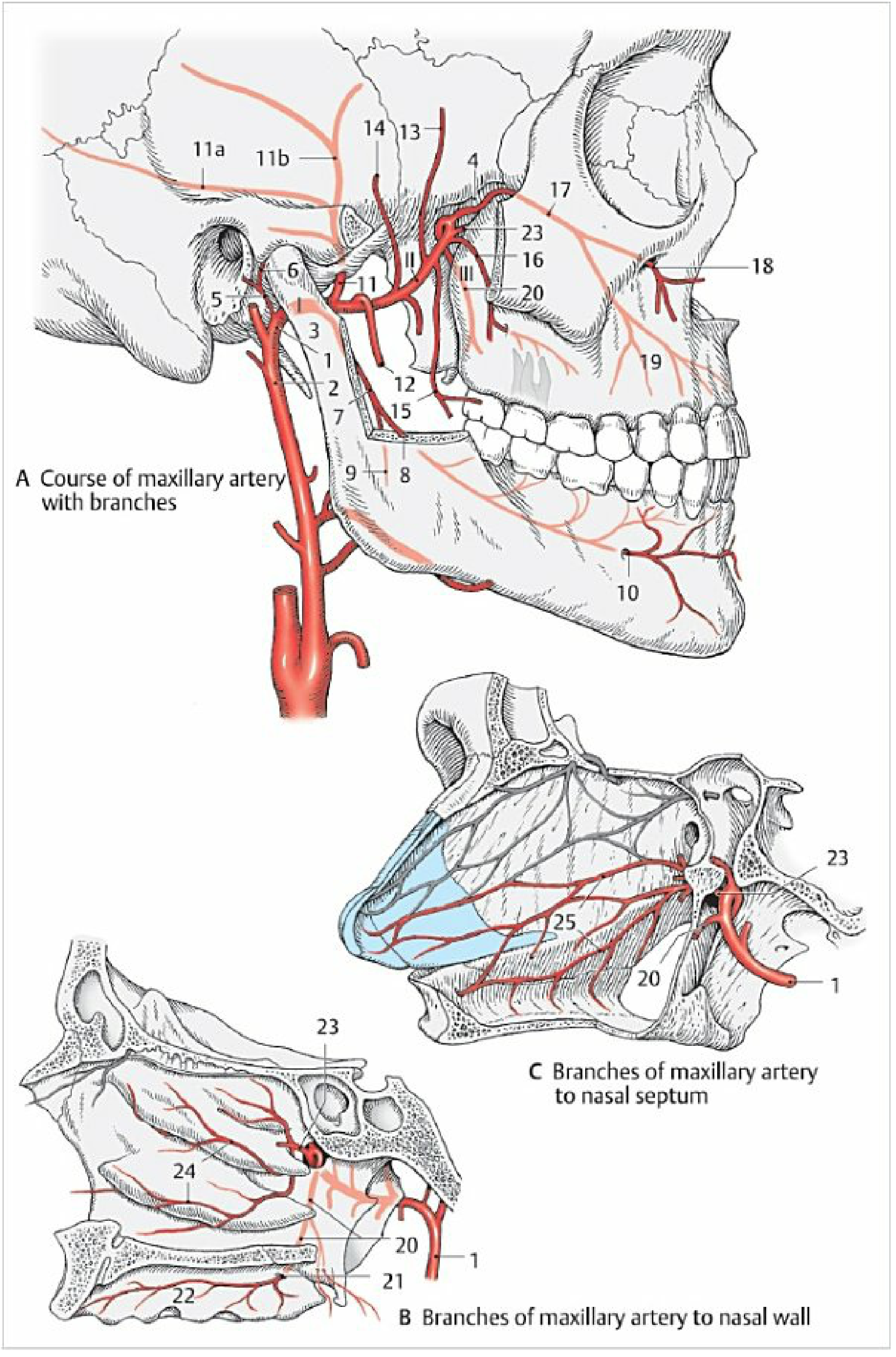
Location and Relations
- Branches of the mandibular nerve [V3] (lingual, inferior alveolar, buccal, deep temporal nerves)
- The pterygoid venous plexus
- Medial and lateral pterygoid muscles
4-5 Branches of the 2nd Part
1. Anterior Deep Temporal Artery
- Course: Ascends between the temporalis muscle and the pericranium (periosteum of temporal fossa)
- Accompanies: Anterior deep temporal nerve (V3)
- Supply: Temporalis muscle (anterior part)
- Also sends a small branch through the zygomatico-orbital foramen to the orbit
2. Posterior Deep Temporal Artery
- Course: Also ascends between temporalis and pericranium (posterior to the anterior branch)
- Accompanies: Posterior deep temporal nerve (V3)
- Supply: Temporalis muscle (posterior part)
- Together, both deep temporal arteries are the principal blood supply of the temporalis muscle
3. Pterygoid Branches (multiple)
- Course: Multiple small branches to both medial and lateral pterygoid muscles
- Supply: Medial pterygoid and lateral pterygoid muscles (muscles of mastication)
- These do not have named nerve companions but follow pterygoid muscle planes
4. Masseteric Artery
- Course: Passes laterally through the mandibular notch (sigmoid notch) between the coronoid process and neck of condyle
- Accompanies: Masseteric nerve (V3)
- Supply: Masseter muscle (deep surface); also supplies the TMJ joint
5. Buccal Artery
- Course: Passes forward and downward between the two heads of the lateral pterygoid → runs with the buccal nerve (V3) to reach the cheek
- Supply: Buccinator muscle, buccal mucosa and skin of the cheek, buccal gingiva (posterior lower teeth)
- Anastomoses with the facial artery and infraorbital artery - important collateral connection
Summary Table
| Branch | Course | Accompanies (nerve) | Supply |
|---|---|---|---|
| Anterior deep temporal | Ascends to temporal fossa | Anterior deep temporal n. | Temporalis (anterior) |
| Posterior deep temporal | Ascends to temporal fossa | Posterior deep temporal n. | Temporalis (posterior) |
| Pterygoid branches | Direct to pterygoid muscles | - | Medial and lateral pterygoid |
| Masseteric | Through mandibular notch | Masseteric n. | Masseter + TMJ |
| Buccal | Forward with buccal nerve to cheek | Buccal n. | Buccinator, buccal mucosa, buccal gingiva |
- Gray's Anatomy for Students, p. 2479-2485; Color Atlas of Human Anatomy Vol. 2, p. 93
Key Points
- The 2nd part is the "muscular part" - all branches supply muscles of mastication or adjacent soft tissue
- No branch passes through a bony foramen (a key distinguishing feature from parts 1 and 3)
- The buccal artery is the only branch with relevance to oral mucosa
- The deep temporal arteries are important in temporal arteritis and in temporal muscle flap surgery
- The pterygoid venous plexus surrounding this part has communications with the cavernous sinus via emissary veins through the foramen ovale - a route for spreading dental infections intracranially - Gray's Anatomy for Students, p. 2501