
Investigation of ankylosing spondyloarthritis
I'll read the medical library skill first, then research ankylosing spondylitis investigations thoroughly.
Reading File
Now I'll run parallel searches across headings and paragraphs for ankylosing spondylitis investigations, along with a PubMed search for recent guidelines.
Finding Sources
Finding Sources
Searching PubMed
"ankylosing spondylitis" AND "diagnosis"
Excellent - I have rich sources. Now let me read the most relevant sections in parallel - the radiology textbook (Grainger & Allison), the rheumatology textbooks, Goldman-Cecil, and Henry's Clinical Lab textbook for investigations.
Reading File
Reading File
Reading File
Reading File
The radiology textbook is rich. Let me read more of that radiology section plus a dedicated AS investigations/diagnosis section from the rheumatology and Goldman-Cecil textbooks.
Reading File
Finding Sources
Finding Sources
Let me now read the Goldman-Cecil chapter on AS and the Rosen's emergency medicine section, plus search for the diagnostic criteria section in the rheumatology textbook.
Reading File
Reading File
Finding Sources
Finding Sources
Excellent! Found the Modified New York Criteria section in Firestein & Kelley. Let me read that and the ASAS criteria section, plus the radiology sacroiliitis grading section.
Reading File
Reading File
Reading File
Now let me read the Modified New York criteria continuation and the MRI section for complete details.
Reading File
Reading File
Loading Image
I now have comprehensive data from multiple authoritative textbooks. Let me also fetch a couple of the key radiology images and then compile the full answer.
Loading Image
Loading Image
Loading Image
I now have comprehensive material from multiple authoritative textbooks with imaging. Here is the full, structured answer:
Investigation of Ankylosing Spondylitis (AS)
AS is a multisystem inflammatory spondyloarthropathy primarily affecting the axial skeleton. HLA-B27 positive males under 40 are most commonly affected, presenting with chronic inflammatory back pain. The hallmark is sacroiliitis and enthesitis of the axial skeleton, with peripheral joint involvement in up to 30% of patients.
1. Classification / Diagnostic Criteria
Modified New York Criteria (1984) - Still the reference standard for definite AS
| # | Criterion |
|---|---|
| 1 | Low back pain of ≥3 months' duration, improved by exercise, NOT relieved by rest |
| 2 | Limitation of lumbar spine in both sagittal and frontal planes |
| 3 | Chest expansion decreased relative to normal for age and sex |
| 4 | Bilateral sacroiliitis grade 2-4 (radiographic) |
| 5 | Unilateral sacroiliitis grade 3 or 4 (radiographic) |
Definite AS = Criterion 4 or 5 (radiographic sacroiliitis) + any 1 clinical criterion
Note: Criteria 2 and 3 reflect disease duration and are typically absent in early disease - making modified New York criteria poorly suited for early diagnosis. - Firestein & Kelley's Textbook of Rheumatology
ASAS (Assessment of SpondyloArthritis International Society) Criteria
Developed to capture the full spectrum including non-radiographic axial SpA (nr-axSpA). For patients with chronic back pain and age of onset <45 years:
- Imaging arm: Sacroiliitis on imaging (radiograph or MRI) + ≥1 SpA feature
- Clinical arm: HLA-B27 positive + ≥2 additional SpA features
Sensitivity 83%, specificity 84%. Note: these are classification criteria, not diagnostic criteria - applying them diagnostically risks ~4 false positives per 1 true positive in a chronic back pain population. - Firestein & Kelley's Textbook of Rheumatology
2. Laboratory Investigations
HLA-B27 Testing
- ~90% of Caucasian AS patients are HLA-B27 positive (vs. 5-10% of healthy controls)
- Performed by flow cytometry (quick, cost-efficient) - but lower sensitivity/specificity than molecular typing due to inability to recognise all B27 subgroups
- Molecular typing is more sensitive/specific but complicated by high polymorphism (>200 alleles described)
- Key subtypes associated with AS: HLA-B*27:05 and *27:02 (Caucasians), *27:04 (Asians)
- NOT diagnostic alone: only ~2% of HLA-B27 positive individuals ever develop AS - best used as a rule-out test (high negative predictive value)
- Henry's Clinical Diagnosis and Management by Laboratory Methods
Inflammatory Markers
- ESR and CRP - elevated in active disease; CRP levels improve with TNF inhibitor therapy and are used to monitor treatment response
- May be normal in up to 40% of AS patients despite active disease
- Firestein & Kelley's Textbook of Rheumatology
Other Bloods
- FBC: normocytic anaemia of chronic disease in active AS
- Rheumatoid factor (RF): negative - AS is a seronegative spondyloarthropathy
- ANA: typically negative
- Alkaline phosphatase: may be mildly elevated in active disease
- Serum IgA: often elevated (reflects mucosal immune activation)
3. Imaging Investigations
A. Plain Radiography (X-Ray) - First-line
Sacroiliac Joints - AP pelvis or Ferguson view (angled AP)
Graded according to the Modified New York Criteria:
| Grade | Findings |
|---|---|
| 0 | Normal |
| 1 | Suspicious - slight blurring of cortical margins |
| 2 | Minimal - small areas of erosion or sclerosis; joint space normal |
| 3 | Moderate - definite erosion/sclerosis, pseudowidening or partial ankylosis |
| 4 | Ankylosis - complete fusion |
Disease is bilateral and symmetrical in AS (distinguishes from psoriatic/reactive arthritis which are asymmetric).
Spinal Changes on X-Ray (lateral views):
- Shiny corners (Romanus lesions): earliest spinal change - sclerosis at corners of vertebral bodies at Sharpey fibre insertion
- Squaring of vertebral bodies: loss of normal concave anterior surface due to erosion + anterior longitudinal ligament ossification
- Syndesmophytes: thin, vertically oriented bony outgrowths at disc margins - distinguishable from osteophytes (horizontal) and psoriatic coarse syndesmophytes
- Bamboo spine: complete fusion of vertebral bodies + facet joints in advanced disease
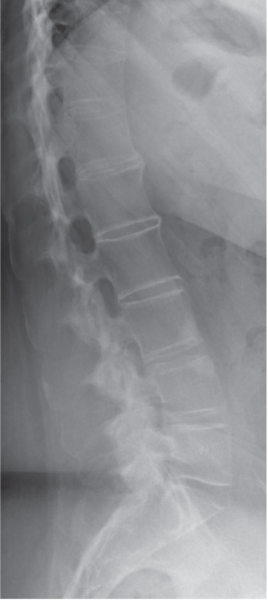
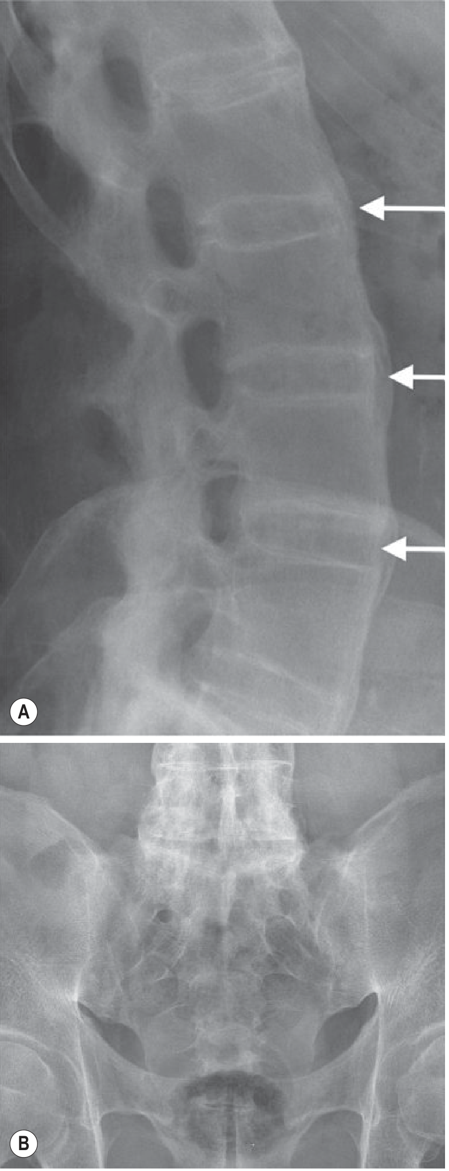
Scoring systems for radiographic spinal progression (primarily used in trials):
- Bath Ankylosing Spondylitis Radiology Index (BASRI)
- Stoke Ankylosing Spondylitis Spine Score (SASSS)
- Modified SASSS (most sensitive to change)
B. MRI - Investigation of Choice for Early Disease
MRI is the most sensitive modality for early axial SpA, detecting inflammatory changes before any radiographic sacroiliitis becomes apparent. - Grainger & Allison's Diagnostic Radiology; Firestein & Kelley's Textbook
Key sequences:
| Sequence | Purpose |
|---|---|
| STIR / T2 fat-suppressed (water-sensitive) | Detects active inflammation - bone marrow oedema (BME) |
| T1-weighted | Detects structural damage - erosions, fat metaplasia, sclerosis, ankylosis |
| Gadolinium contrast | Does NOT enhance diagnostic accuracy over conventional MRI sequences |
Imaging planes: Semicoronal/oblique coronal (parallel to posterior S2), supplemented by semiaxial views for SI joints; sagittal views for spine.
SI Joint MRI Findings:
- Active (inflammatory): Juxta-articular bone marrow oedema (STIR) - most indicative of active sacroiliitis; capsulitis; enthesitis
- Structural (damage): Erosions, sclerosis, periarticular fat metaplasia (T1 bright "fat lesions"), ankylosis
Spinal MRI Findings (axial SpA):
- Bone oedema at vertebral body corners = MRI equivalent of Romanus lesion (detected before sclerosis on X-ray)
- Spondylodiscitis (inflammation along vertebral end plate)
- Costovertebral joint oedema
- Spinous process and facet joint oedema
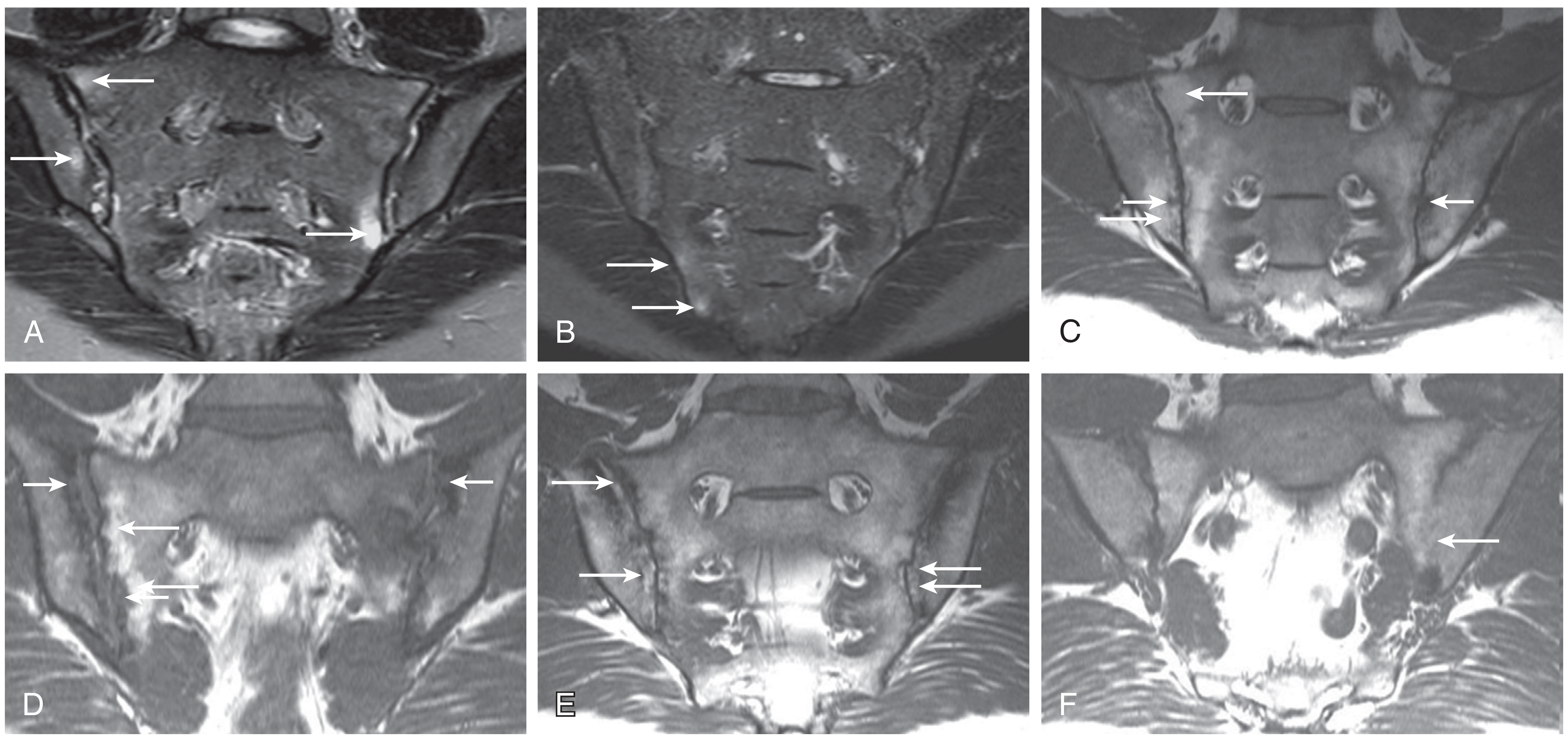
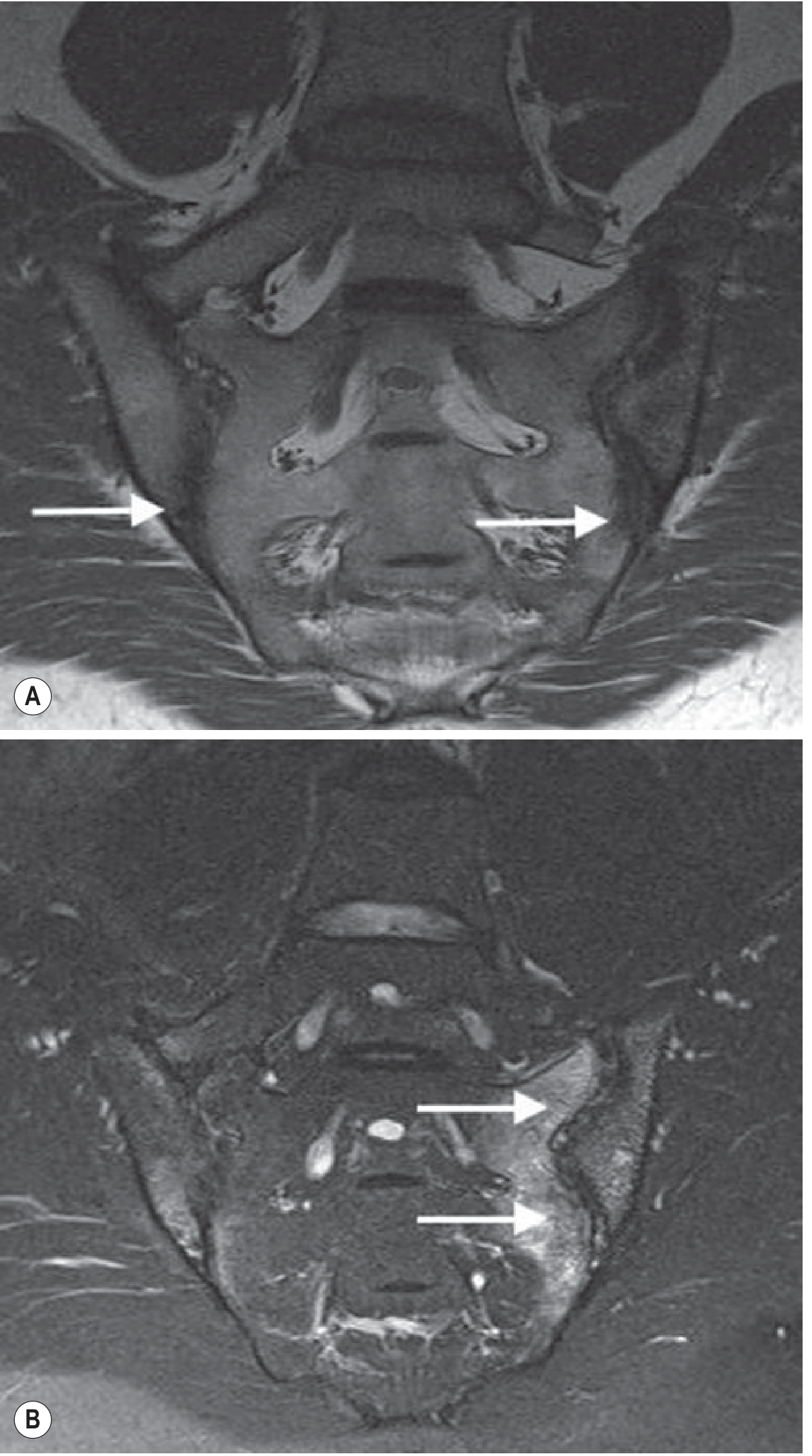
C. CT Scan
- Gold standard for detecting structural damage (erosions, sclerosis) - more detail than plain X-ray
- Detects bony changes sooner and with greater resolution than conventional radiography
- Limited by: radiation dose; inability to assess soft tissue or bone marrow (so cannot detect early inflammatory changes)
- Low-dose CT techniques now available at radiation doses similar to plain X-ray - increasingly used as an alternative to CR for SI joint and spinal structural assessment
- Firestein & Kelley's Textbook of Rheumatology
D. Bone Scintigraphy (Skeletal Scintigraphy)
- Historically used for early detection of sacroiliitis
- Now generally superseded - MRI has greater sensitivity and specificity than scintigraphy, conventional radiography, and CT for early sacroiliitis detection
- Grainger & Allison's Diagnostic Radiology
4. Functional Assessments
These are validated indices used in clinical practice and trials:
| Assessment | Measures |
|---|---|
| BASDAI (Bath AS Disease Activity Index) | Disease activity (patient-reported) |
| BASFI (Bath AS Functional Index) | Physical function |
| BASMI (Bath AS Metrology Index) | Spinal mobility (includes lumbar flexion, cervical rotation, tragus-to-wall, lumbar lateral flexion, intermalleolar distance) |
| ASDAS (AS Disease Activity Score) | Composite score using CRP/ESR + patient-reported items |
Specific physical examination tests:
- Schober's test - lumbar flexion (mark 10 cm above + 5 cm below L5; normal expansion >5 cm)
- Occiput-to-wall distance - forward stooped posture in advanced disease
- Chest expansion - should be >5 cm at the 4th intercostal space (reduced in thoracic disease)
- FABER test (Flexion, ABduction, External Rotation) and Gaenslen's test - provoke sacroiliac joint pain
5. Investigations for Extra-Articular Manifestations
AS is a multisystem disease; investigations should be targeted:
| Extra-Articular Feature | Investigation |
|---|---|
| Anterior uveitis (most common; 25-40%) | Slit-lamp examination; HLA-B27 |
| Aortic regurgitation / cardiac conduction defects | ECG; echocardiogram |
| Apical pulmonary fibrosis (fibrobullous disease) | CXR; HRCT chest; PFTs (restrictive pattern due to chest wall rigidity) |
| Osteoporosis | DEXA scan (significant complication due to chronic inflammation + mechanical loading) |
| IBD-related arthritis | Colonoscopy, faecal calprotectin |
| Cauda equina syndrome (late) | MRI spine; lumbar puncture |
The most common pulmonary manifestation is upper lobe bilateral reticulonodular infiltrates with cyst formation due to parenchymal destruction (fibrobullous disease) - there is no effective therapy for this. - Goldman-Cecil Medicine
6. Summary Algorithm
Chronic inflammatory back pain <45 years + morning stiffness >30 min
                         ↓
        X-ray SI joints + HLA-B27 + CRP/ESR
                         ↓
    ┌─────────────────────────────────────┐
    │ X-ray sacroiliitis grade 2-4 bilateral  │  → Modified New York Criteria = Definite AS
    │ or grade 3-4 unilateral              │
    └─────────────────────────────────────┘
                         ↓ (if X-ray negative/equivocal)
              MRI SI joints (STIR + T1)
                         ↓
    ┌──────────────────────────────────────────────────────┐
    │ Active BME on STIR + SpA feature = Axial SpA (nr-axSpA) │
    └──────────────────────────────────────────────────────┘
                         ↓ (if MRI equivocal)
         HLA-B27 + ≥2 SpA features = Clinical arm AxSpA
Sources:
- Grainger & Allison's Diagnostic Radiology (9780323760751), Chapter 44
- Firestein & Kelley's Textbook of Rheumatology (9780323935364), Chapters 75-76
- Henry's Clinical Diagnosis and Management by Laboratory Methods (9780323673204), Chapter on HLA-B27
- Goldman-Cecil Medicine International Edition (9780323930345)
- ROSEN's Emergency Medicine (9780323757898)
- Rheumatology 2-Volume Set (Elsevier 2022) (9780702081330)
Investigations of seronegative spondyloarthropathy
Finding Sources
Finding Sources
Finding Sources
Good. Now let me read the key sections in parallel - the orthopaedics textbook on seronegative SpA, the ASAS criteria, Goldman-Cecil criteria, and search for each subtype's investigations.
Reading File
Reading File
Reading File
Finding Sources
Now let me search for each subtype's specific investigations in parallel.
Finding Sources
Finding Sources
Finding Sources
Finding Sources
Reading File
Reading File
Excellent - the Goldman-Cecil section has a very rich differential diagnosis table and clinical details. Let me continue reading it for reactive arthritis and enteropathic arthritis investigations, and also fetch the psoriatic arthritis radiology images.
Reading File
Reading File
Loading Image
Now let me fetch the remaining key images and read the reactive arthritis investigation section more fully.
Reading File
Loading Image
Loading Image
Loading Image
I have comprehensive material from multiple textbooks with excellent imaging. Now I'll compile the complete, well-structured answer.
Investigations of Seronegative Spondyloarthropathy (SpA)
Seronegative spondyloarthropathies are a group of inflammatory arthritides that share:
- Absence of rheumatoid factor (seronegative)
- Involvement of entheses (the bone-ligament/tendon interface)
- Association with HLA-B27
- Tendency to affect the axial skeleton and sacroiliac joints
- Destruction of fibrocartilage and collagen more than synovial cartilage
The group includes:
- Ankylosing Spondylitis (AS) - covered separately; see previous answer
- Psoriatic Arthritis (PsA)
- Reactive Arthritis (formerly Reiter syndrome)
- Enteropathic Arthritis (associated with IBD)
- Undifferentiated Spondyloarthritis
Comparative Differential Table
(Goldman-Cecil Medicine)
| Feature | RA | AS | Enteropathic | Psoriatic | Reactive |
|---|---|---|---|---|---|
| Sex ratio (M:F) | 1:3 | 3:1 | 1:1 | 1:1 | 10:1 |
| HLA association | DR4 | B27 | B27 (axial) | B27 (axial) | B27 |
| Joint pattern | Symmetrical, peripheral | Axial | Axial + peripheral | Axial + asymmetric peripheral | Axial + asymmetric peripheral |
| Sacroiliac | Absent | Symmetrical | Symmetrical | Asymmetrical | Asymmetrical |
| Syndesmophyte | Absent | Smooth, marginal (vertical) | Smooth, marginal | Coarse, non-marginal | Coarse, non-marginal |
| Eye | Scleritis | Iritis | +/- | +/- | Iritis + conjunctivitis |
| Skin | Vasculitis | Rare | Erythema nodosum | Psoriasis | Keratoderma blennorrhagica |
| Rheumatoid factor | >80% | Rare | Rare | Rare | Rare |
I. COMMON INVESTIGATIONS FOR ALL SERONEGATIVE SpA
A. Laboratory
| Test | Relevance |
|---|---|
| Rheumatoid factor (RF) | Negative - this is the defining feature of the group |
| Anti-CCP antibodies | Negative (distinguishes from RA) |
| HLA-B27 | Positive in ~90% AS, ~70-80% reactive arthritis, ~50-70% PsA with axial disease, ~50% enteropathic axial disease |
| ESR / CRP | Elevated in active disease; monitor response to treatment |
| FBC | Anaemia of chronic disease; thrombocytosis in active disease |
| ANA | Negative |
| Uric acid | To exclude gout (important differential) |
| LFTs / Renal function | Baseline before NSAID/DMARD therapy |
B. Imaging - General Principles
| Modality | Role |
|---|---|
| Plain X-ray | First-line; sacroiliac joints + spine + affected peripheral joints |
| MRI | Gold standard for early/active inflammation - bone marrow oedema before X-ray changes |
| CT | Best for bony structural detail (erosions, sclerosis) but limited soft-tissue assessment |
| Ultrasound | Detects enthesitis, synovitis, bursitis, tendinopathy - particularly in peripheral disease |
C. ASAS Classification Criteria (for Axial SpA)
For chronic back pain ≥3 months, age of onset <45 years:
Imaging arm: Sacroiliitis on imaging (X-ray or MRI) + ≥1 SpA feature
OR
Clinical arm: HLA-B27 positive + ≥2 SpA features
SpA features: inflammatory back pain, peripheral arthritis, enthesitis (heel), uveitis, dactylitis, psoriasis, Crohn's/UC, good NSAID response, family history of SpA, HLA-B27, elevated CRP.
II. PSORIATIC ARTHRITIS (PsA)
Clinical Subtypes (Moll & Wright)
- Asymmetric oligoarthritis (most common, ~70%)
- Symmetric polyarthritis (RA-like but seronegative)
- Distal interphalangeal (DIP) arthropathy
- Spondylitis with or without sacroiliitis
- Arthritis mutilans (severe destructive form)
Laboratory Investigations
- RF: Negative (distinguishes from RA)
- Anti-CCP: Usually negative
- HLA-B27: Positive in ~50% with axial/spinal disease
- ESR, CRP: Elevated in active disease
- Skin biopsy: If psoriasis diagnosis is uncertain
Imaging Investigations
X-ray (hands, feet, pelvis, spine):
- No periarticular osteopenia (unlike RA - useful distinguishing feature)
- Marginal entheseal erosions with new bone formation at the erosion site - "fluffy, whiskered" appearance
- "Pencil-in-cup" deformity: Erosions on distal side of joint merge together; proximal phalanx has pointed "pencil" shape sitting in the cup of the distal phalanx - considered pathognomonic
- Acro-osteolysis: Distal tuft resorption giving peg-like appearance to terminal phalanges
- Dactylitis ("sausage digit"): Global soft-tissue swelling of a digit on plain film
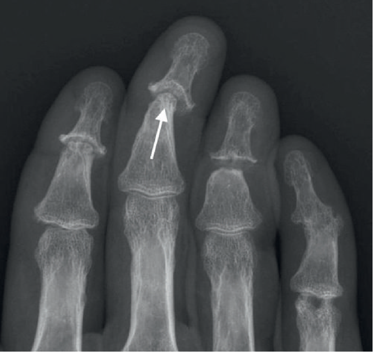
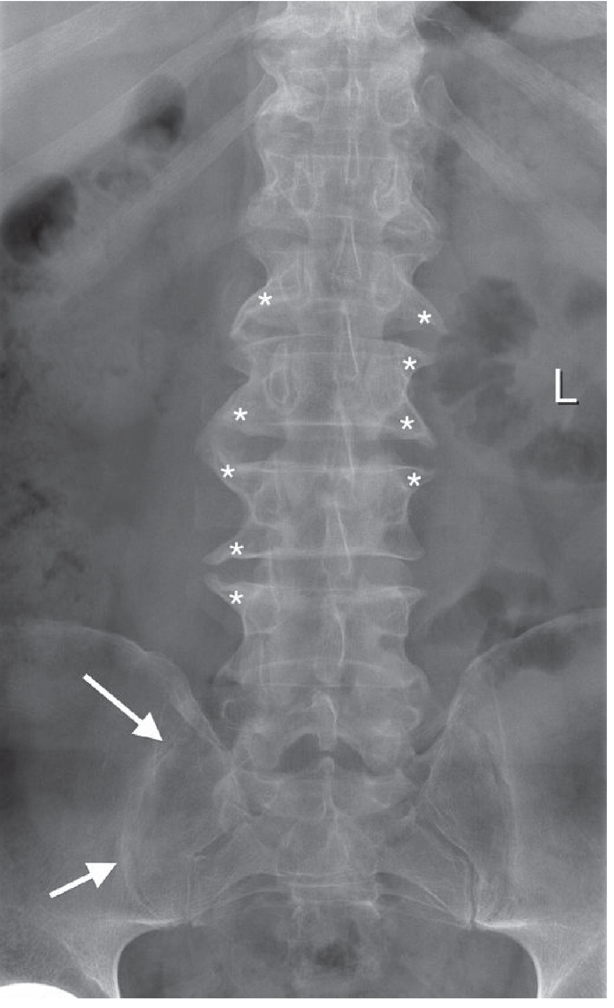
Spinal/SIJ X-ray findings (psoriatic spondylitis):
- Asymmetric sacroiliitis (unlike the bilateral symmetrical sacroiliitis of AS)
- Coarse, non-marginal (bulky) syndesmophytes in a horizontal orientation - contrast with the fine, vertical syndesmophytes of AS
- Asymmetric distribution about the spine
- Costovertebral disease is rare in PsA (common in AS)
MRI: Enthesitis, synovitis, bone marrow oedema - useful when X-ray is negative or equivocal
Ultrasound: Detection of subclinical enthesitis and synovitis at multiple sites
Validated Assessment Tools
- CASPAR criteria (ClASsification criteria for Psoriatic ARthritis) - used for classification
- DAPSA (Disease Activity in PSoriatic Arthritis)
- MDA (Minimal Disease Activity)
III. REACTIVE ARTHRITIS
Background
Sterile inflammatory arthritis triggered by extra-articular infection (typically genitourinary or gastrointestinal), occurring 2-4 weeks after infection. Classic triad (Reiter): urethritis + arthritis + conjunctivitis/uveitis.
Common triggers: Chlamydia trachomatis (GU), Salmonella, Shigella, Yersinia, Campylobacter (GI)
Laboratory Investigations
- RF: Negative
- HLA-B27: Positive in ~70-75%
- ESR, CRP: Elevated
- FBC: Neutrophilia during acute phase
- Urethral/cervical swabs: Chlamydia culture, NAAT (nucleic acid amplification test)
- Stool cultures / serology: For Salmonella, Shigella, Yersinia, Campylobacter
- Throat swab: If streptococcal infection suspected
- HIV serology: Important - HIV alters the course, leading to more aggressive and refractory joint disease
Synovial Fluid Analysis
- Sterile inflammatory pattern (WBC 10,000-50,000, predominantly neutrophils)
- Cultures negative (reactive, not septic arthritis)
- Chlamydia antigen / PCR: May be positive in synovial fluid even when peripheral cultures are negative
Imaging Investigations
- X-ray feet: New bone formation at calcaneal entheses (plantar and posterior insertions) - prominent feature; also seen at other entheseal sites
- Soft-tissue swelling, joint space narrowing, proliferative marginal erosions in small joints of the lower limbs
- Sesamoid enlargement due to periostitis
- Sacroiliitis: Bilateral asymmetric (unlike AS which is symmetric) - erosions, pseudowidening, ileal sclerosis
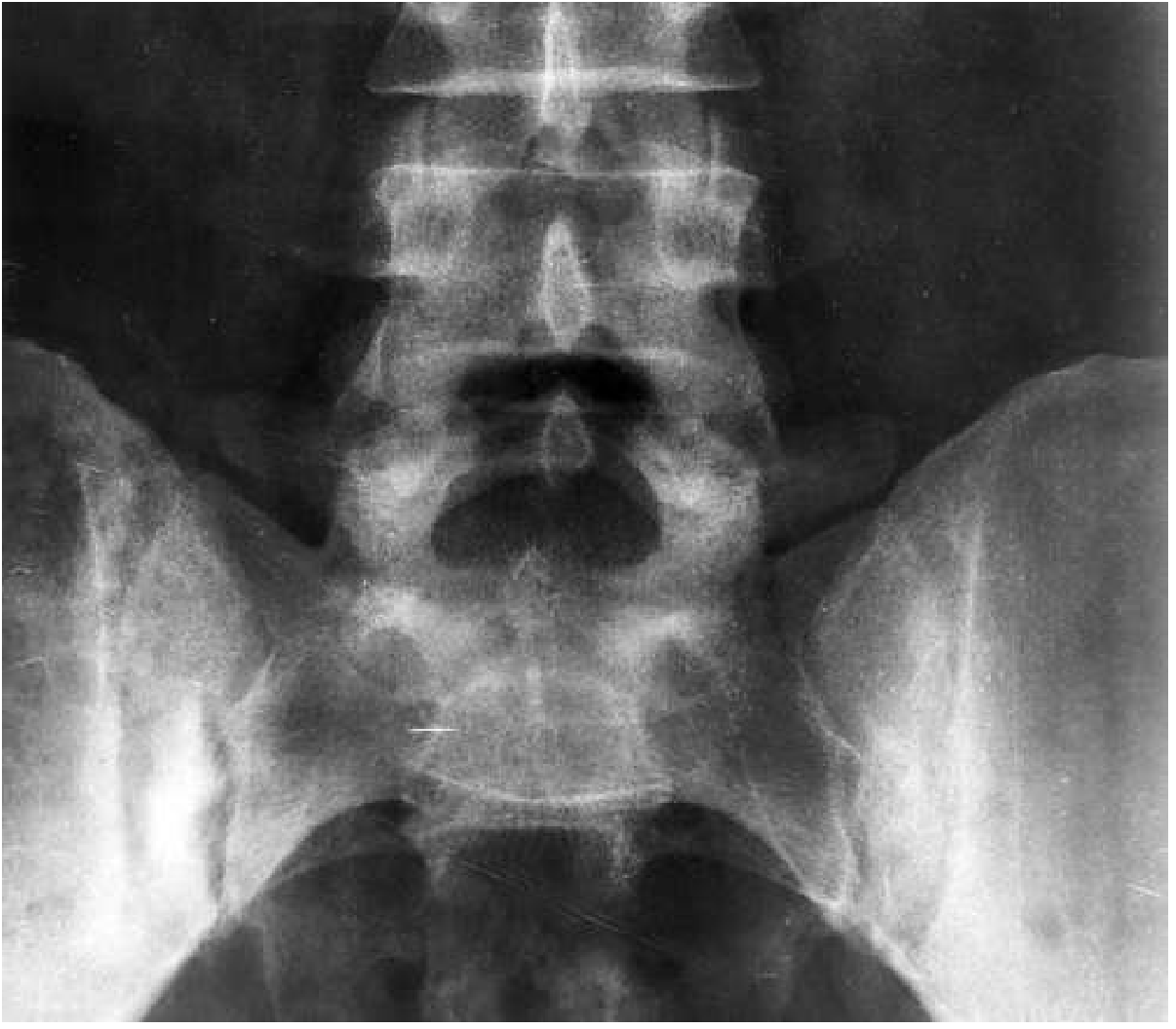
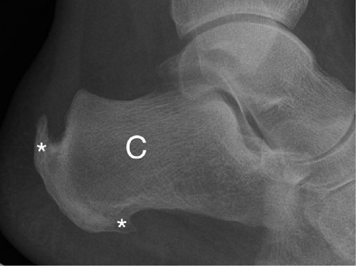
IV. ENTEROPATHIC ARTHRITIS (IBD-associated)
Associated with Crohn's disease and ulcerative colitis. Two distinct patterns:
| Pattern | Peripheral arthritis | Axial/Sacroiliac arthritis |
|---|---|---|
| Frequency (Crohn's) | 10-20% | 2-7% |
| Frequency (UC) | 5-10% | 2-7% |
| HLA-B27 | Not associated | Associated |
| Course | Parallel to bowel disease activity | Independent of bowel disease |
| Pattern | Transient, migratory | Chronic |
(Goldman-Cecil Medicine)
Laboratory Investigations
- RF: Negative
- HLA-B27: Positive in axial disease; negative in peripheral disease
- ESR, CRP, faecal calprotectin: Monitor IBD activity
- FBC: Anaemia (multifactorial - chronic disease, B12/folate malabsorption in Crohn's)
- Colonoscopy / biopsy: Confirms IBD diagnosis - axial disease may precede GI symptoms
Imaging Investigations
- Pelvic X-ray: Sacroiliitis - relatively asymmetric, with bone oedema progressing to erosion, joint space loss, and fusion
- Peripheral arthritis is characteristically non-destructive - wrists, knees, ankles, elbows
- MRI SI joints: Active sacroiliitis; spinal involvement similar to AS
- Small bowel radiology / MRI enterography: For Crohn's disease assessment
V. SUMMARY: KEY DIFFERENTIATING RADIOLOGICAL FEATURES
| Feature | AS | PsA | Reactive | Enteropathic |
|---|---|---|---|---|
| Sacroiliitis | Bilateral symmetric | Asymmetric | Asymmetric | Relatively asymmetric |
| Syndesmophytes | Fine, vertical, marginal | Coarse, horizontal, non-marginal | Coarse, non-marginal | Fine, marginal (AS-like) |
| Peripheral erosions | Uncommon | Pencil-in-cup, entheseal | Proliferative, entheseal | Non-destructive |
| Dactylitis on X-ray | Rare | Typical | Rare | Absent |
| Acro-osteolysis | Absent | Present | Absent | Absent |
| Calcaneal enthesophytes | Present | Present | Prominent | Rare |
| Periarticular osteopenia | Present | Absent | Present | Present |
VI. INVESTIGATION ALGORITHM
Seronegative inflammatory arthritis
(RF negative, asymmetric, lower limb, enthesitis)
               ↓
    ┌──────────────────────────────────┐
    │         FIRST LINE               │
    │  FBC, ESR, CRP, RF (neg),        │
    │  anti-CCP (neg), uric acid       │
    │  HLA-B27                         │
    │  X-ray: SI joints + affected     │
    │         peripheral joints        │
    └──────────────────────────────────┘
               ↓
    ┌──────────────────────────────────────────────────────┐
    │ Are there SpA features?                               │
    │  Axial pain + HLA-B27: MRI SI joints + spine         │
    │  Preceding infection: Chlamydia/stool cultures,       │
    │                       synovial fluid culture + PCR    │
    │  Psoriasis/nail changes: skin biopsy if needed        │
    │  GI symptoms: faecal calprotectin, colonoscopy        │
    │  Uveitis: slit-lamp examination                       │
    └──────────────────────────────────────────────────────┘
               ↓
    ASAS criteria: imaging arm or clinical arm for axial SpA
    CASPAR criteria: for psoriatic arthritis
    Modified New York criteria: for definite AS
Sources:
- Goldman-Cecil Medicine International Edition (9780323930345), Chapter 244
- Grainger & Allison's Diagnostic Radiology (9780323760751), Chapter 44
- Rheumatology 2-Volume Set, Elsevier 2022 (9780702081330)
- Miller's Review of Orthopaedics 9th Edition (9780443112140)
- Firestein & Kelley's Textbook of Rheumatology (9780323935364)
- ROSEN's Emergency Medicine (9780323757898)
This is a shared conversation. Sign in to Orris to start your own chat.