
Write a note on buccal e drg delivery system.
buccal drug delivery system pharmaceutical overview
https://pmc.ncbi.nlm.nih.gov/articles/PMC3968279
buccal drug delivery system oral mucosa diagram
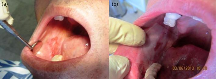
Two-panel clinical photograph demonstrating intraoral imaging and the application of a drug delivery system. Panel (a) shows the oral cavity being examined with a metallic circular dental mirror. The mirror is retracting the buccal mucosa to reveal an erythematous, mottled lesion with raised textures and irregular borders on the internal cheek surface, consistent with oral inflammatory or ulcerative conditions. Panel (b) illustrates the clinical application of a translucent, rectangular mucoadhesive patch (containing 2% Mycophenolate Mofetil) placed directly over the affected mucosal tissue. A gloved hand provides cheek retraction for clear visualization. The patch appears thin and well-adhered to the moist oral surface, positioned near the maxillary teeth. These images demonstrate a therapeutic protocol for localized treatment of oral mucosal lesions, highlighting the preparation phase (mouth breathing to dry the site) and the subsequent one-minute post-application adherence of the polymeric drug-delivery film.
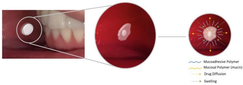
This educational graphic illustrates the pharmacokinetics and physical behavior of a mucoadhesive buccal tablet within the oral cavity. The image is divided into three sequential panels. The first panel shows a clinical photograph of a white, disc-shaped tablet placed against the buccal mucosa near the lower teeth. The second panel provides a magnified view of the tablet beginning to hydrate and swell, losing its distinct circular edges as it adheres to the mucosal tissue. The third panel is a schematic diagram detailing the molecular mechanism of adhesion and drug release. Key components identified in the legend include mucoadhesive polymers (blue wavy lines) and mucosal polymers/mucin (yellow wavy lines) interpenetrating at the interface. Green arrows indicate the physical swelling of the polymer matrix, while yellow radial arrows represent drug diffusion from the tablet core through the swollen layer and into the systemic circulation or local tissue. The illustration highlights the importance of polymer hydration and chain entanglement in transmucosal drug delivery.
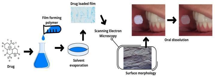
This educational diagram illustrates the pharmaceutical manufacturing and clinical application of a mucoadhesive oral drug-loaded film. The flowchart begins with the synthesis process, where a chemical drug molecule is combined with a 'film forming polymer' in a liquid solution. This is followed by 'solvent evaporation' in a casting dish, resulting in a 'drug loaded film' shown in a light-blue microscopic view with needle-like drug structures. Characterization is demonstrated through 'Scanning Electron Microscopy' (SEM), providing a high-magnification view of the film's surface morphology. The clinical sequence features two intraoral clinical photographs showing a circular white film applied to the buccal mucosa adjacent to the lower molars. The 'oral dissolution' phase depicts the gradual degradation and size reduction of the film over time in the oral cavity. This schematic is designed to teach the solvent casting technique and the mechanism of transmucosal drug delivery for oral healthcare applications.
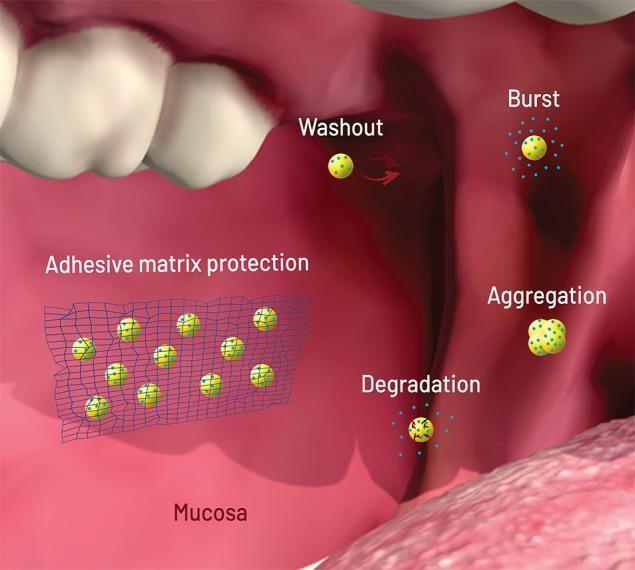
This medical illustration depicts the pharmacokinetics and failure modes of drug-loaded nanoparticles (NPs) within the oral cavity environment. The background features anatomical structures of the mouth, including teeth, tongue, and the pink mucosal surface. Nanoparticles are represented as yellow spheres containing blue dots, which signify the loaded drug. The diagram illustrates four primary failure modes: 'Washout' (removal of NPs from the site by saliva flow), 'Burst' (uncontrolled rapid drug release upon contact with saliva), 'Aggregation' (clumping of individual NPs into larger, less effective complexes due to pH or protein binding), and 'Degradation' (physical breakdown of the carrier by enzymes or acidic pH). On the left, 'Adhesive matrix protection' is demonstrated, showing a blue net-like polymer matrix adhered to the mucosa. This matrix serves as a protective delivery system, physically shielding the nanoparticles from external stressors to eliminate or reduce the aforementioned failure modes. This content is relevant for pharmacology and dental students focusing on advanced drug delivery systems and buccal drug administration strategies.
Buccal Drug Delivery System
1. Introduction
- Mucosal (local) effect - site-specific drug release on the mucosa (e.g., treatment of aphthous ulcers, oral candidiasis, gingivitis)
- Transmucosal (systemic) effect - drug absorption through the mucosal barrier into systemic circulation, bypassing hepatic first-pass metabolism
2. Anatomy of the Buccal Mucosa
- Non-keratinized surface - more permeable than the keratinized gingiva and palate, making it a preferred site for drug absorption
- Rich blood supply - venous drainage via the superior vena cava, bypassing the portal circulation and hepatic first-pass effect
- Surface area - approximately 50 cm², providing a reasonable area for drug contact
- Salivary environment - the oral cavity is kept moist by saliva (pH 5.5-7.5, volume ~0.5-1 L/day), which aids dissolution but can also cause premature drug washing away
3. Mechanism of Drug Absorption
a) Transcellular (Intracellular) Route
b) Paracellular (Intercellular) Route
4. Advantages of the Buccal Route
| Advantage | Explanation |
|---|---|
| Bypass of first-pass metabolism | Venous drainage from buccal mucosa goes to the superior vena cava, not the portal vein |
| Avoidance of GI degradation | No exposure to gastric acid, proteolytic enzymes, or gut-wall metabolism |
| Rapid onset (for immediate-release) | Highly vascularized mucosa allows fast drug entry into systemic circulation |
| Sustained release possible | Mucoadhesive formulations can provide prolonged drug contact (4-6 hours) |
| Patient-friendly | Non-invasive, painless; suitable for patients who cannot swallow (geriatric, pediatric, dysphagia patients) |
| Easy drug withdrawal | Formulation can be removed if adverse effects occur |
| Low enzymatic activity | Compared to the GI tract, the buccal mucosa has relatively low enzyme activity |
| Reduced dose and toxicity | Lower drug content needed compared to oral tablets |
5. Disadvantages and Limitations
- Limited surface area (~50 cm²) restricts the amount of drug that can be absorbed
- Saliva washout - continuous salivary flow dilutes and washes away drug; patient swallowing limits residence time
- Barrier function - even non-keratinized buccal mucosa is less permeable than intestinal epithelium
- Taste and irritation - some drugs have unpleasant taste or irritate the mucosa
- Patient discomfort - prolonged retention of a dosage form in the mouth may feel uncomfortable
- Eating, drinking, and talking can dislodge the formulation
- Only suitable for potent drugs - the limited absorption area makes it unsuitable for high-dose drugs
- Mucus turnover - approximately every 4-6 hours, limiting residence time of mucoadhesive devices
6. Dosage Forms (Formulation Types)
a) Buccal Tablets / Mucoadhesive Tablets
- Most common solid dosage form
- Compressed tablets containing mucoadhesive polymers (carbopol, HPMC, sodium CMC, polycarbophil)
- Placed between cheek and gum; hydrate and swell, adhering to mucosa
- Can be monolayer (drug released on all sides) or bilayer (unidirectional drug release)
- Example: Striant® (testosterone), Buccastem® (prochlorperazine), Nitrogard® (nitroglycerin)
b) Buccal Films / Patches
- Thin, flexible polymeric films applied to the buccal mucosa
- Better patient compliance than tablets due to small size and flexibility
- Can be made as monolayer or bilayer (backing layer + drug layer to prevent drug loss to saliva)
- Polymers used: HPMC, HPC, PVA, PVP, carbopol
- Example: Suboxone Film® (buprenorphine + naloxone) using PharmFilm technology (FDA-approved 2010)
c) Gels and Ointments
- Semisolid formulations easily dispersed over the oral mucosa
- Mucoadhesive polymers (sodium CMC, carbopol, hyaluronic acid, xanthan gum) undergo sol-gel transition on contact with mucosa, enhancing viscosity
- Useful for local therapy (e.g., oral ulcers, stomatitis)
- Less accurate dosing compared to solid forms
d) Oral Sprays
- Drug dissolved in liquid vehicle and delivered as a fine mist onto buccal/sublingual mucosa
- Multidose pump or aerosol systems
- Rapid absorption due to large surface area coverage
- Example: NitroMist® (nitroglycerin spray for angina), ZolpiMist® (zolpidem for insomnia), both FDA-approved
e) Chewing Gums
- Drug incorporated into a gum base; chewing releases the drug, which contacts buccal mucosa
- Example: Nicorette® (nicotine chewing gum) for smoking cessation
f) Lozenges / Troches
- Dissolve slowly in the mouth, releasing drug for local or systemic absorption
- Example: Loramyc®/Lauriad® (miconazole) for oropharyngeal candidiasis
g) Wafers
- Porous or non-porous lyophilized structures; absorb moisture rapidly
- Useful for fast-dissolving applications or wound-based local delivery
7. Mucoadhesive Polymers Used
| Polymer | Type | Properties |
|---|---|---|
| Carbopol (Carbomer) | Anionic, crosslinked | High mucoadhesive strength; pH sensitive |
| Polycarbophil | Anionic | Similar to carbopol; resists enzyme degradation |
| HPMC | Non-ionic | Good film-forming, water-soluble |
| Sodium CMC | Anionic | Good mucoadhesion, water-swellable |
| Chitosan | Cationic | Bioadhesive, biodegradable; also acts as penetration enhancer |
| HPC | Non-ionic | Flexible films |
| Hyaluronic acid | Anionic | Biocompatible; good for gels |
| Xanthan gum | Anionic | Viscosity modifier |
8. Penetration Enhancers
- Bile salts (sodium deoxycholate, sodium glycocholate) - disrupt lipid bilayers
- Fatty acids (oleic acid, caprylic acid) - increase membrane fluidity
- Surfactants (sodium lauryl sulfate, polysorbates) - solubilize membrane lipids
- Cyclodextrins - improve drug solubility and membrane partitioning
- Chitosan - opens tight junctions (paracellular enhancement)
- EDTA - chelates calcium, loosening tight junctions
9. Ideal Characteristics of a Drug for Buccal Delivery
- Have a low molecular weight (ideally <1000 Da)
- Be potent at small doses (due to limited absorption area)
- Have adequate lipophilicity (log P 1-3) for membrane partitioning
- Have a neutral or un-ionized form at oral pH (5.5-7.5)
- Undergo significant first-pass metabolism if given orally (greatest benefit from bypassing it)
- Have a short half-life requiring sustained release
- Be stable in salivary environment
10. Commercially Available Buccal Products
| Drug | Brand | Indication |
|---|---|---|
| Nitroglycerin | Nitrogard® | Angina pectoris |
| Testosterone | Striant® SR | Hypogonadism |
| Prochlorperazine | Buccastem® | Nausea/vomiting, migraine |
| Miconazole | Loramyc®/Lauriad® | Oropharyngeal candidiasis |
| Buprenorphine + Naloxone | Suboxone Film® | Opioid dependence |
| Nicotine | Nicorette® gum, Nicotinell® lozenge | Smoking cessation |
| Nitroglycerine spray | NitroMist® | Angina |
| Zolpidem spray | ZolpiMist® | Insomnia |
11. Evaluation of Buccal Drug Delivery Systems
In Vitro Tests:
- Mucoadhesive strength - tensile or shear force measurements
- Drug release - using Franz diffusion cell or dissolution apparatus
- Swelling index / erosion
- Surface pH
- Folding endurance (for films/patches)
- Thickness and weight uniformity
Ex Vivo/In Vivo Tests:
- Permeation studies - using porcine or bovine buccal mucosa (closest to human in structure)
- Residence time - measured in animals or human volunteers
- Bioavailability studies - pharmacokinetic studies in animals/humans
12. Recent Advances
- Nanoparticle-based buccal systems - liposomes, nanostructured lipid carriers (NLCs), polymeric nanoparticles for improved drug loading and permeation. Adhesive matrix protection is used to shield nanoparticles from saliva washout, burst release, aggregation, and enzymatic degradation.
- Bilaminated/Bilayer films - one mucoadhesive layer facing the mucosa, one backing layer preventing drug loss to saliva, ensuring unidirectional drug flux
- Peptide and protein delivery - chitosan-based films for insulin, myoglobin, and other macromolecules
- 3D-printed buccal devices - precise control of drug loading and geometry
- In situ gelling systems - liquid preparations that gel on contact with buccal mucosa
13. Summary
- Madhav NVS et al. A clinical perspective on mucoadhesive buccal drug delivery systems. J Adv Pharm Technol Res, 2009.
- Reddy PC et al. A review on bioadhesive buccal drug delivery systems. J Pharm Bioallied Sci, 2011.
- Research and developments in buccal and sublingual drug delivery. The Pharmaceutical Journal, 2011.
I need a note on a nasal drug dg delivery system.
nasal drug delivery system pharmaceutical overview advantages formulations
https://eprints.hud.ac.uk/id/eprint/26635/3/Nasal%20review%2…
nasal drug delivery system anatomy olfactory mucosa diagram
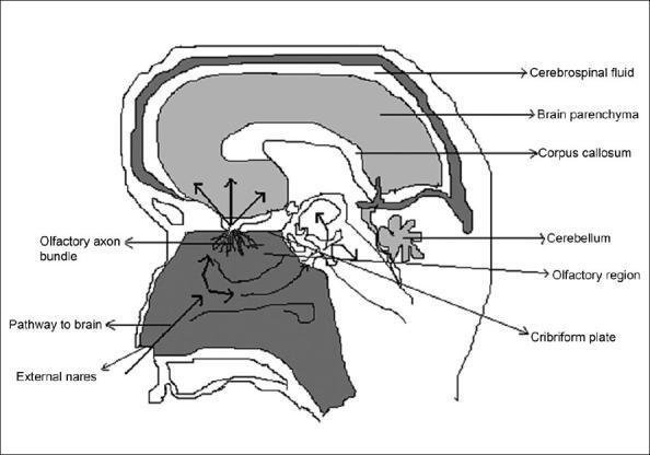
A sagittal schematic diagram illustrating the intranasal olfactory pathway for drug delivery to the central nervous system (CNS). The diagram displays a cross-section of the human head, highlighting the relationship between the nasal cavity and the brain. Key labeled anatomical structures include the external nares, the olfactory region of the nasal mucosa, the cribriform plate of the ethmoid bone, and the olfactory axon bundle. The brain is shown with labels for the brain parenchyma, corpus callosum, cerebellum, and the surrounding cerebrospinal fluid (CSF) space. Functional arrows trace a 'pathway to brain,' demonstrating the movement of substances from the external nares, through the nasal cavity, and across the cribriform plate via the olfactory nerves. The illustration emphasizes both intraneuronal (axonal transport) and extraneuronal (bulk flow through perineural channels) routes, bypassing the blood-brain barrier to reach various cerebral regions. This content is designed for pharmacy and medical students studying neuropharmacology and non-invasive therapeutic delivery systems.
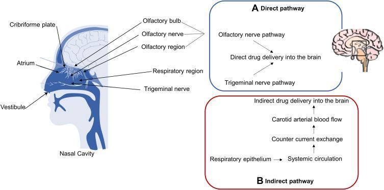
An anatomical and pathophysiological diagram illustrating the nose-to-brain drug delivery pathways. The left side features a sagittal cross-section of the human head, specifically detailing the nasal cavity anatomy. Labeled landmarks include the vestibule, atrium, and respiratory region, as well as the superior olfactory region adjacent to the cribriform plate and olfactory bulb. The diagram highlights two distinct mechanisms of drug transport to the central nervous system (CNS). (A) The 'Direct pathway' (blue box) shows intranasal drugs bypassing the blood-brain barrier (BBB) via the olfactory and trigeminal nerve pathways. (B) The 'Indirect pathway' (red box) describes systemic absorption through the respiratory epithelium into the circulation, involving countercurrent exchange and carotid arterial blood flow to reach the brain. This schematic is designed for pharmacology and neurology education, focusing on therapeutic delivery strategies for neurodegenerative conditions like Alzheimer's disease.
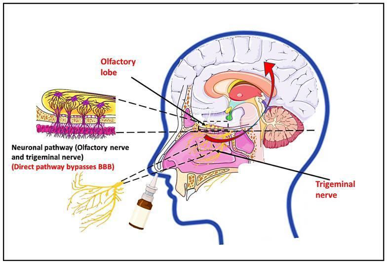
This medical illustration depicts the nose-to-brain drug delivery route, an anatomical pathway used to bypass the blood-brain barrier (BBB). The primary diagram shows a sagittal section of the human head, highlighting a nasal spray bottle administering a drug into the nasal cavity. Red arrows indicate the transport of therapeutics through two main neuronal pathways: the olfactory nerve pathway leading to the olfactory lobe and the trigeminal nerve pathway. An anatomical inset provides a microscopic view of the olfactory neuronal pathway, detailing the cellular arrangement of the olfactory epithelium. It shows columnar epithelial cells, olfactory sensory neurons with dendrites extending into the nasal mucosa, and axons projecting superiorly through the cribriform plate into the olfactory bulb. The diagram emphasizes the direct connection between the nasal mucosa and central nervous system structures, including the brainstem and olfactory lobe. This educational visual is used in pharmacology and neurobiology to explain non-invasive drug administration techniques for treating neurological disorders like Alzheimer's or Parkinson's disease.
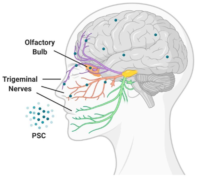
This medical anatomical diagram illustrates the nose-to-brain drug delivery route for Polysaccharide-based Carriers (PSC). The diagram shows a sagittal profile of a human head with the brain and neurological pathways highlighted. The drug, represented as teal dots (PSC), is shown entering through the nasal cavity. Two primary pathways are depicted for direct central nervous system access, bypassing the blood-brain barrier: the olfactory pathway (indicated in purple), originating from the olfactory bulb at the base of the frontal lobe, and the trigeminal nerve pathway (indicated in orange/green), showing the ophthalmic and maxillary branches providing a conduit from the nasal mucosa to the brainstem and higher brain regions. Small teal dots distributed throughout the cerebral cortex and cerebellum represent the subsequent distribution of the therapeutic agent within the brain tissue. This illustration is used to explain non-invasive administration strategies for treating neurological diseases and the pharmacological advantage of utilizing cranial nerve pathways for targeted brain delivery.
Nasal Drug Delivery System (NDDS)
1. Introduction
2. Anatomy and Physiology of the Nasal Cavity
Regions of the Nasal Cavity:
| Region | Area | Characteristics |
|---|---|---|
| Vestibule | ~0.6 cm² | Skin-lined, keratinized, poor absorption |
| Atrium | Transition zone | Connects vestibule to respiratory region |
| Respiratory region | ~130 cm² | Pseudostratified columnar ciliated epithelium; highly vascularized; main absorption site |
| Olfactory region | ~10 cm² | Located near cribriform plate; gateway to CNS via olfactory nerve |
Key Physiological Features:
- Pseudostratified ciliated columnar epithelium with goblet cells lining the respiratory region
- Mucus layer (10-15 µm thick) produced by goblet cells and seromucinous glands; consists of outer gel layer and inner sol layer
- Rich submucosal vasculature - capillaries, sinusoids, and venous plexus providing rapid drug uptake
- Mucociliary clearance (MCC): cilia beat at 700-1000 strokes/min, moving mucus (and entrapped drug) at 3-25 mm/min toward the nasopharynx - a major barrier to prolonged drug contact
3. Nose-to-Brain Pathway
- Direct (olfactory) pathway: Drug enters olfactory neurons in the olfactory epithelium → axonal transport → olfactory bulb → brain parenchyma; also via bulk flow through perineural channels in the olfactory nerve bundle
- Trigeminal nerve pathway: Drug from nasal mucosa travels along ophthalmic and maxillary branches of the trigeminal nerve to the brainstem and higher brain regions
- Indirect pathway: Drug absorbed into systemic circulation → crosses BBB via countercurrent exchange in carotid arterial blood → brain
4. Mechanism of Drug Absorption
a) Transcellular (Intracellular) Route
- Drug molecules pass directly through the epithelial cell membranes
- Favored by lipophilic, low molecular weight drugs
- Driven by passive diffusion down a concentration gradient
- Rate depends on lipophilicity (log P), degree of ionization, molecular size
b) Paracellular (Intercellular) Route
- Drug passes through aqueous channels between cells, via tight junctions
- Favored by hydrophilic and polar drugs
- Limited by tight junction integrity; absorption enhancers work mainly here
c) Transcytosis
- Vesicle-mediated transport across epithelial cells
- Important for macromolecules, peptides, and nanoparticles
d) Olfactory Nerve Transport (for N2B delivery)
- Drug taken up by olfactory sensory neurons → anterograde axonal transport → olfactory bulb
5. Advantages of NDDS
- Non-invasive and patient-friendly - avoids needles; suitable for self-administration
- Avoidance of hepatic first-pass metabolism - drug absorbed from nasal mucosa drains into systemic circulation without portal passage
- Rapid absorption and quick onset - highly vascularized mucosa gives rapid attainment of therapeutic blood levels; faster than oral route, sometimes comparable to IV
- Large surface area (~150-180 cm²) for adequate absorption
- Avoidance of GI degradation - no exposure to gastric acid, bile salts, or gut-wall enzymes
- Direct nose-to-brain delivery - bypass of the BBB for CNS-targeted therapy
- Suitable for vaccines - nasal-associated lymphoid tissue (NALT) provides direct contact with immune cells
- Good permeability for lipophilic and low molecular weight drugs
- Reduced dose and side effects compared to systemic routes
- No complex formulation requirement for some drugs
- Lower risk of overdose compared to IV administration
6. Disadvantages and Limitations
| Limitation | Details |
|---|---|
| Mucociliary clearance | Constantly moves drug to nasopharynx; limits contact time to ~15-20 minutes |
| Small delivery volume | Restricted to 25-200 µL per nostril; unsuitable for large doses |
| Molecular weight cutoff | Drugs >1 kDa (peptides, proteins) show poor permeation |
| Enzymatic degradation | Nasal mucosa contains proteases, peptidases, CYP450 enzymes |
| Nasal irritation | Some drugs (budesonide, azelastine) and excipients irritate the mucosa |
| Pathological conditions | Rhinitis, colds, nasal congestion, atrophic rhinitis alter absorption unpredictably |
| Drug loss | Drug may drip out anteriorly or drain posteriorly into the throat |
| Interspecies variability | Large difference in nasal anatomy between animals and humans; complicates preclinical models |
| Cilia toxicity | Some absorption enhancers may impair ciliary function |
| Limited for high-dose drugs | Small surface area and volume restrict applicability |
7. Ideal Drug Characteristics for Nasal Delivery
- Has a low molecular weight (<1 kDa ideal; up to ~6 kDa possible with enhancers)
- Is lipophilic (log P 1-4) for transcellular absorption
- Has a low dose requirement (high potency)
- Is unstable in the GI tract or subject to extensive first-pass metabolism
- Has a neutral or un-ionized form at nasal pH (5.5-6.5)
- Is non-irritating to the nasal mucosa
- Is stable in aqueous solution (for liquid formulations)
8. Dosage Forms and Formulations
a) Nasal Drops
- Simplest liquid formulation; delivered as drops directly into the nasal cavity
- Used for systemic (e.g., hormone therapy, desmopressin) and local effects
- Disadvantage: poor reproducibility of dose; risk of posterior drainage
b) Nasal Sprays
- Most commonly used formulation; delivered as a fine mist ensuring even distribution across nasal mucosa
- Metered-dose pump dispensers provide accurate, reproducible dosing
- Used for decongestants, corticosteroids, antihistamines, and systemic drugs
- Examples: Fluticasone nasal spray (allergic rhinitis), Sumatriptan nasal spray (migraine), Calcitonin nasal spray (osteoporosis), Desmopressin spray (diabetes insipidus)
c) Nasal Gels
- Semisolid formulations with prolonged contact time compared to liquids
- Mucoadhesive polymers (carbopol, HPMC, chitosan) increase viscosity and retention
- Reduces dripping and posterior drainage
- In situ gelling systems (thermosensitive or pH-sensitive) are liquid on instillation but gel on contact with nasal mucosa
d) Nasal Powders
- Solid dosage forms; improved stability compared to aqueous formulations
- Longer residence time; less drainage
- Can contain mucoadhesive excipients (cyclodextrins, starch, chitosan)
- Suitable for thermolabile or hygroscopic drugs
e) Nasal Ointments and Creams
- For local/topical treatment (e.g., nasal furunculosis treatment with mupirocin ointment)
f) Microspheres
- Microparticulate systems (5-500 µm) made from biopolymers (starch, albumin, PLGA, chitosan)
- Advantages: prolonged drug residence, protection from enzymatic degradation, controlled release
- Absorbed by NALT for vaccine delivery
g) Liposomes
- Phospholipid vesicles encapsulating drug in aqueous core or lipid bilayer
- Improve drug stability, reduce mucosal irritation, enhance permeation
- Bypass the BBB; suitable for CNS drug delivery
h) Nanoparticles and Lipid Nanoparticles
- Polymeric nanoparticles (PLGA, chitosan), solid lipid nanoparticles (SLNs), nanostructured lipid carriers (NLCs)
- High bioavailability; ability to cross BBB and reach brain parenchyma
- FDA-approved lipid nanoparticles are the most common nano-drug carrier class
- Liposomes can bypass the BBB; lipid solubility of encapsulated drug improves significantly
- Hyaluronic acid-coated micelles enhance permeability via receptor-mediated uptake
i) Microemulsions
- Thermodynamically stable isotropic systems; o/w type suitable for poorly water-soluble drugs
- High solubilization capacity; enhanced absorption potential via CSF
- Thermosensitive microemulsions gel in situ after nasal instillation
j) Hydrogels
- Porous, hydrated polymer networks; excellent for controlling release of macromolecules
- Mimic natural tissue microenvironment
- Can be combined with nanoparticles for enhanced efficiency
9. Mucoadhesive Polymers Used in NDDS
| Polymer | Type | Role |
|---|---|---|
| Carbopol (Carbomer) | Anionic | High mucoadhesive strength; forms gel with mucin |
| Chitosan | Cationic | Mucoadhesive + penetration enhancer (opens tight junctions) |
| HPMC | Non-ionic | Film-forming; moderate mucoadhesion |
| Sodium CMC | Anionic | Viscosity modifier, mucoadhesive |
| Hydroxypropyl cellulose (HPC) | Non-ionic | Flexible films |
| Hyaluronic acid | Anionic | Biocompatible; receptor-mediated mucosal interaction |
| Starch | Non-ionic | Used in microspheres |
10. Penetration Enhancers
- Bile salts (sodium deoxycholate, sodium glycocholate) - disrupt epithelial lipid bilayers
- Fatty acids (oleic acid, lauric acid) - increase membrane fluidity
- Surfactants (Brij, polysorbate, sodium lauryl sulfate) - solubilize membrane components
- Cyclodextrins - form inclusion complexes; improve drug solubility and membrane partitioning
- Chitosan - positive charge interacts with negative epithelial surface; opens paracellular tight junctions
- EDTA - chelates calcium; loosens tight junctions
- Saponins - cholesterol extraction from membranes
- Note: Enhancers must be evaluated for cilia toxicity before clinical use
11. Nasal Drug Delivery Devices
| Device | Description | Example Use |
|---|---|---|
| Metered-dose nasal pump | Most common; delivers fixed volume (100-150 µL/spray) | Fluticasone, Sumatriptan |
| Nasal drops (dropper bottle) | Simple; variable dose; used for local/systemic effects | Desmopressin |
| Pressurized MDI (metered-dose inhaler adapted) | For aerosol delivery to nasal cavity | Corticosteroids |
| Dry powder inhaler (DPI) for nasal use | Delivers powder formulations; breath-actuated | Vaccine delivery, insulin |
| Mucosal atomizer device (MAD) | Atomizes liquid to 10-50 µm particles for optimal mucosal deposition | Emergency: intranasal midazolam, naloxone, fentanyl |
| Unit-dose devices | Pre-filled single-use containers for sterile drugs | Migraine treatments |
| Bidirectional delivery devices | Uses positive pressure in one nostril to distribute drug to contralateral side and paranasal sinuses | Sinus disease treatment |
12. Marketed Nasal Products
| Drug | Brand Name | Indication |
|---|---|---|
| Desmopressin | DDAVP Nasal Spray | Diabetes insipidus, nocturnal enuresis |
| Sumatriptan | Imitrex Nasal | Migraine |
| Calcitonin | Miacalcin Nasal | Osteoporosis |
| Budesonide | Rhinocort | Allergic rhinitis |
| Fluticasone | Flonase | Allergic rhinitis |
| Zolmitriptan | Zomig Nasal | Migraine |
| Nicotine | Nicotrol NS | Smoking cessation |
| Ketamine | Spravato (esketamine) | Treatment-resistant depression |
| Naloxone | Narcan Nasal Spray | Opioid overdose reversal |
| Midazolam | Nayzilam | Acute seizures |
| Oxytocin | Syntocinon | Labor induction (adjunct) |
| Live attenuated influenza vaccine | FluMist | Influenza prophylaxis |
13. Special Applications
Nasal Insulin
- Insulin has been explored intranasally to overcome injections
- Low nasal bioavailability (~10%) remains a challenge
- Absorption enhancers (bile salts, chitosan) are being investigated to improve bioavailability
Nasal Vaccines
- Nasal-associated lymphoid tissue (NALT) acts as an inductive site for mucosal immunity
- Intranasal vaccination can generate both systemic IgG and local secretory IgA (sIgA)
- FluMist® (live attenuated influenza vaccine) is a successful commercial example
- Microspheres and nanoparticles enhance antigen delivery to NALT
CNS Drug Delivery (N2B)
- Intranasal delivery of drugs for Alzheimer's (insulin, NGF), Parkinson's (dopamine agonists), epilepsy (midazolam, diazepam), and depression (esketamine/Spravato®)
- Growing area due to failure of BBB-penetrant drug development
- Nanoparticles, liposomes, and cyclodextrin-complexed drugs are the most promising carriers
14. Evaluation of NDDS
In Vitro:
- Drug release studies (Franz diffusion cell, modified dissolution)
- Viscosity and gel strength measurements
- Mucoadhesive force (tensile testing)
- Particle size distribution (for sprays)
- pH, osmolarity, and isotonicity testing
Ex Vivo / In Vivo:
- Permeation studies using excised sheep, bovine, or porcine nasal mucosa
- Ciliotoxicity studies (frog palate model, cell culture models)
- Mucociliary clearance studies (gamma scintigraphy)
- Bioavailability and pharmacokinetic studies in animal models (rat, rabbit)
- Human pharmacokinetic studies for marketed products
15. Summary
- Ghori MU et al. Nasal Drug Delivery Systems: An Overview. Current Drug Delivery, 2015.
- Patel Z et al. A Review on Intranasal Drug Delivery System. JAPER, 2013.
- Eedara BB et al. Different Methods and Formulations of Drugs and Vaccines for Nasal Administration. Pharmaceutics, 2022.
- Roberts and Hedges' Clinical Procedures in Emergency Medicine, 9780323354783 (intranasal device use in emergency settings).
- Miller's Anesthesia, 10e (nasal mucosal opioid delivery).