
Comparison between three Neimann pick disease types
Niemann-Pick disease foam cells bone marrow histology
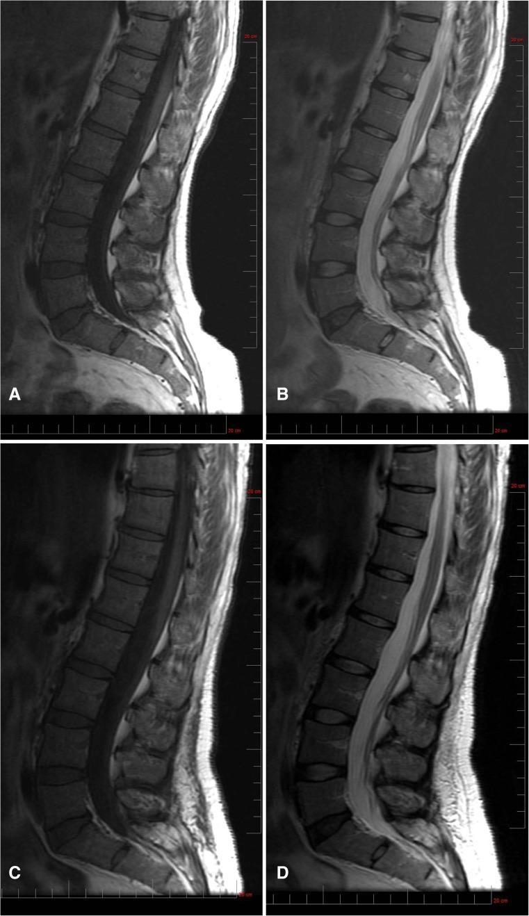
This diagnostic comparison uses Magnetic Resonance Imaging (MRI) to assess Bone Marrow Burden (BMB) in a patient with Niemann-Pick disease type B, specifically focusing on the lumbar spine. The panel presents sagittal views at baseline (A and B) and after 30 months of olipudase alfa treatment (C and D). Figures A and C are T1-weighted images, while B and D are T2-weighted. At baseline, the T1-weighted sequence (A) demonstrates diffuse infiltrative disease characterized by bone marrow isointensity relative to the intervertebral discs. The baseline T2-weighted image (B) shows hyperintense signal within the presacral fat. Following 30 months of enzyme replacement therapy, the T1-weighted image (C) indicates that marrow infiltration remains largely unchanged. However, the T2-weighted sequence (D) reveals improvement in the presacral fat signal, which has transitioned to a slightly hyperintense state. This longitudinal comparison illustrates the variable response of different anatomical compartments to treatment, emphasizing the use of signal intensity shifts as a metric for monitoring marrow infiltration and clinical progression in metabolic storage disorders.
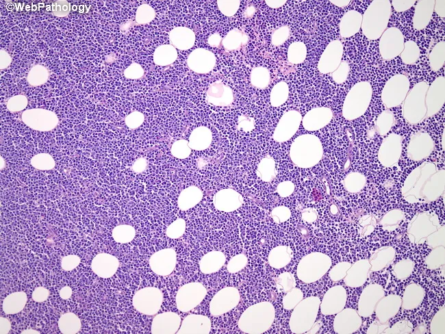
Bone marrow histology on this hematoxylin and eosin–stained section shows diffuse infiltration by mature B-lymphoid cells with prolymphocytic features, partially replacing normal adipocytic marrow. Infiltrating cells are medium to large with round to irregular nuclei, coarse chromatin, conspicuous nucleoli, and scant to moderate cytoplasm. Scattered prolymphocytes are present and background fat cells persist in residual islands; the overall architecture is variably effaced. This pattern is characteristic of bone marrow involvement by B-cell prolymphocytic leukemia (B-PLL). Although immunophenotype cannot be determined on this slide, typical profiles include bright surface immunoglobulin expression with CD19 and CD20 positivity, coexpression of CD5 and FMC7, and often absence of CD23. Clinically, B-PLL affects the elderly and presents with leukocytosis (often >100,000/μL), anemia, thrombocytopenia, and splenomegaly; B symptoms may be present. Relapsed disease can show central nervous system involvement, refractory hypercalcemia, or extramedullary masses. The histopathologic finding confirms marrow involvement and aids staging and management, while recognizing differential diagnoses such as chronic lymphocytic leukemia/small lymphocytic lymphoma, mantle cell lymphoma, hairy cell leukemia, and acute lymphoblastic leukemia. Integrate complete blood count, flow cytometry immunophenotyping, cytogenetics, and molecular studies for definitive classification and prognosis. This image depicts a bone marrow biopsy/aspirate specimen illustrating B-PLL–related marrow infiltration.
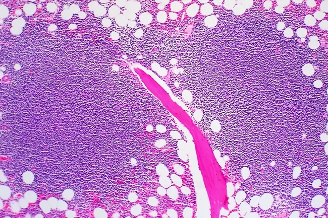
Bone marrow histology from a suspected CLL case illustrating marrow involvement by small mature lymphocytes. Modality: light microscopy; specimen type bone marrow core biopsy with aspirate smear; technique hematoxylin and eosin staining. Infiltration patterns are shown: focal, non-paratrabecular nodules; interstitial admixture of lymphocytes with normal hematopoietic elements; and a rare diffuse, solid pattern appearing dark on low magnification. Lymphocytes comprise more than 30% of marrow cellularity in aspirates, and their morphology mirrors circulating leukemic cells with scant cytoplasm and clumped chromatin. Proliferation foci and true germinal centers are extremely rare. Immunophenotypic profile typically shows clonal B cells expressing CD5 and CD23, with potential ZAP-70 association when diffuse infiltration or high disease burden is present. The presence of extensive solid infiltration correlates with cytopenias, higher clinical stage, and adverse prognosis. The described patterns reflect disease burden and marrow hematopoiesis compromise, with possible implications for therapy and monitoring. Differential considerations include other small B‑cell lymphomas such as mantle cell lymphoma, as well as reactive lymphoid aggregates. Clinical correlation is essential, as marrow involvement informs staging and helps evaluate response to therapy. This image underscores marrow histology’s role in CLL assessment and prognostication. These features aid pathologists, hematologists, and trainees in diagnosis and grading.
Niemann-Pick Disease: Comparison of Types A, B, and C
Overview Table
| Feature | Type A | Type B | Type C |
|---|---|---|---|
| Also called | Classic infantile NPD | Chronic visceral NPD | Neurovisceral lipid storage |
| Gene defect | SMPD1 (chr 11p15.4) | SMPD1 (chr 11p15.4) | NPC1 (chr 18q11.2) or NPC2 |
| Enzyme/protein | Acid sphingomyelinase (ASM) | Acid sphingomyelinase (ASM) | NPC1/NPC2 - cholesterol trafficking proteins |
| Storage substance | Sphingomyelin + cholesterol | Sphingomyelin | Free cholesterol + selected sphingolipids in lysosomes |
| Inheritance | Autosomal recessive | Autosomal recessive | Autosomal recessive |
| Ethnic predilection | Ashkenazi Jewish (~2/3 of cases) | No ethnic predilection | No ethnic predilection |
| Prevalence | Rare | Rare | 1-2% of all autosomal recessive cerebellar ataxias |
Age of Onset
| Type A | Type B | Type C |
|---|---|---|
| Onset 3-9 months (infancy) | Childhood (variable, later onset) | Variable - neonatal to adult |
Pathophysiology
-
Types A & B: Deficiency of acid sphingomyelinase (ASM) encoded by SMPD1 causes accumulation of sphingomyelin in cell membranes and myelin sheaths. Foam cells (Niemann-Pick cells) fill viscera. ASM activity is reduced to 1-10% of normal.
-
Type C: Mutations in NPC1 or NPC2 impair free cholesterol and sphingolipid transport out of the lysosome. There is only modest sphingomyelin accumulation - the mechanism is fundamentally different. Late-stage disease preferentially destroys cerebellar Purkinje cells.
Clinical Features
Type A (Severe Neurovisceral)
- Massive hepatosplenomegaly and lymphadenopathy
- Rapidly progressive CNS deterioration within first year
- Hypotonia, axial weakness, bilateral corticospinal signs
- Loss of spontaneous movements, lack of environmental interest
- Cherry-red macular spot (~25-50% of patients)
- Blindness, amaurotic nystagmus
- Pulmonary infiltration (lung involvement)
- Failure to thrive
- Seizures (late manifestation)
- Death usually by end of 2nd year from intercurrent infection
Type B (Chronic Visceral)
- Hepatosplenomegaly - slow, progressive (no or minimal CNS disease)
- Progressive pulmonary disease: dyspnea, hypoxemia, reticular infiltrative pattern on CXR
- Foam cells in alveoli, lymphatics, and pulmonary arteries
- Eventual cirrhosis from hepatic parenchymal and Kupffer cell replacement
- Spared or minimal neurological involvement (key distinguishing feature from A)
- An A/B variant exists with slowly progressive CNS involvement
- Many patients survive into adulthood; death can occur in adolescence/early adulthood from hepatic or pulmonary complications
Type C (Progressive CNS)
- Neonatal jaundice (may be presenting feature)
- Vertical supranuclear palsy - pathognomonic sign
- Progressive cerebellar ataxia (predominant across all age groups)
- Cognitive impairment / dementia
- Extrapyramidal dysfunction: dystonia, choreoathetosis
- Hypotonia and developmental delay (infantile)
- Psychiatric manifestations (adult onset)
- Only moderate visceromegaly - less visceral involvement than A/B
- Foam cells may be present in bone marrow
- Progressive CNS deterioration over 1-2 decades
Key Distinguishing Features
| Distinguishing Sign | Type A | Type B | Type C |
|---|---|---|---|
| Cherry-red spot | Present (~25-50%) | Absent | Absent |
| Neurodegeneration | Severe, rapid | Absent/minimal | Progressive over decades |
| Pulmonary disease | Present | Prominent | Absent/minimal |
| Vertical supranuclear palsy | Absent | Absent | Present (pathognomonic) |
| Ataxia | Absent | Absent | Prominent |
| Visceromegaly | Massive | Moderate-severe | Mild-moderate |
| Survival | <2 years | Into adulthood | Variable (childhood to mid-adult) |
Laboratory Diagnosis
| Test | Type A | Type B | Type C |
|---|---|---|---|
| ASM activity (leukocytes/fibroblasts/DBS) | Markedly decreased (1-10% of normal) | Markedly decreased (1-10% of normal) | Normal or near-normal |
| Cholesterol esterification in fibroblasts | Normal | Normal | Reduced (after LDL loading - traditional diagnostic method) |
| Bone marrow | Vacuolated foam cells (histiocytes) | Foam cells | Foam cells may be present |
| Blood | Vacuolated lymphocytes | Vacuolated lymphocytes | Variable |
| Genetic testing | SMPD1 mutation | SMPD1 mutation | NPC1 or NPC2 mutation |
Treatment
| Treatment | Type A | Type B | Type C |
|---|---|---|---|
| Enzyme replacement therapy (ERT) | Limited benefit (CNS involvement limits efficacy) | Olipudase alfa (IV ERT) - improves pulmonary and visceral disease | Not applicable (different mechanism) |
| Substrate reduction therapy | Not established | Not established | Miglustat - delays CNS progression |
| Bone marrow/stem cell transplant (HSCT) | Efficacy not established; complications reported | Efficacy not established | Not standard |
| Cyclodextrin | Not applicable | Not applicable | In clinical trials for NPC1 disease |
| Supportive | Supportive | Management of pulmonary/liver disease | Management of neuropsychiatric symptoms |
Pathology (Histology)
Summary Mnemonic
- Type A: Acute, Ashkenazi, Awful prognosis - death by age 2
- Type B: Better prognosis, Big liver/spleen, Big lungs involved, no Brain (or minimal)
- Type C: Cholesterol trafficking defect, Cerebellar ataxia, Characteristic vertical gaze palsy
- Harrison's Principles of Internal Medicine, 22E - Niemann-Pick Diseases section
- Tietz Textbook of Laboratory Medicine, 7th Ed. - Niemann-Pick Diseases
- Adams & Victor's Principles of Neurology, 12th Ed. - Infantile Niemann-Pick Disease
- Bradley and Daroff's Neurology in Clinical Practice - Niemann-Pick C