
PT management of fracture at shft of humerus
humeral shaft fracture physiotherapy rehabilitation exercises
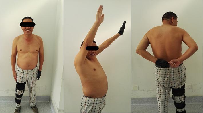
This set of clinical photographs demonstrates functional outcomes and range of motion (ROM) 6 months postoperatively following treatment for a humeral shaft fracture and associated deltoid palsy. The image displays three views of a male patient: 1) Anterior view showing a neutral standing posture with a leg brace on the left lower extremity. 2) Lateral view demonstrating active forward flexion and abduction of the shoulders, with the right arm showing near-full vertical elevation. 3) Posterior view illustrating internal rotation and adduction as the patient reaches behind the back to touch hands at the lumbar level. The series illustrates the clinical recovery of shoulder mobility and the resolution of postoperative nerve injury (deltoid palsy). A visible scar is noted on the lateral aspect of the right proximal arm, consistent with surgical access for internal fixation. These images serve as an educational resource for assessing orthopedic recovery and functional musculoskeletal rehabilitation.
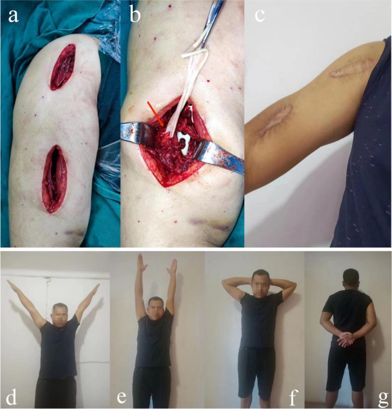
A multi-panel clinical photograph illustrating the surgical approach and functional outcomes of Minimally Invasive Plate Osteosynthesis (MIPO) for a humeral shaft fracture. Panel (a) shows two small, linear surgical incisions on the upper arm, characteristic of a minimally invasive approach. Panel (b) provides an intraoperative view of the distal anterior incision, where the radial nerve is identified and protected (indicated by a red arrow) using surgical retractors and vessel loops to ensure safety during plate insertion. Panel (c) displays the 12-month postoperative appearance of the arm, showing two well-healed, slightly hypopigmented linear scars with smooth texture and minimal inflammation. Panels (d) through (g) demonstrate successful functional recovery of the shoulder and elbow joints, showing the patient performing abduction, flexion, and rotation maneuvers with a full range of motion. This image set serves to educate on the benefits of MIPO in preserving soft tissue integrity, protecting critical neurovascular structures like the radial nerve, and facilitating rapid functional rehabilitation in orthopedic trauma surgery.
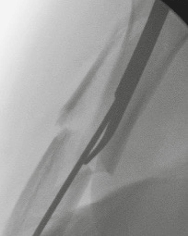
This diagnostic fluoroscopic image illustrates an intraoperative view of a humeral shaft fracture during an orthopedic surgical procedure. The fracture is localized to the mid-diaphysis and appears comminuted, with at least two primary bone fragments and visible smaller intermediary fragments. Significant displacement and lateral angulation are evident at the fracture site. A radiopaque, elongated intramedullary (IM) reduction rod is visualized within the medullary canal. The rod is positioned to span the fracture gap, serving as a joystick-like tool to assist in the anatomical alignment of the proximal and distal fragments. A thin, radiopaque guide wire is seen running parallel to and guided by the reduction tool, which is a critical step for subsequent reaming and the permanent placement of an intramedullary nail. This image demonstrates the clinical application of percutaneous reduction techniques in managing complex humeral fractures to minimize soft tissue disruption while achieving mechanical stability.
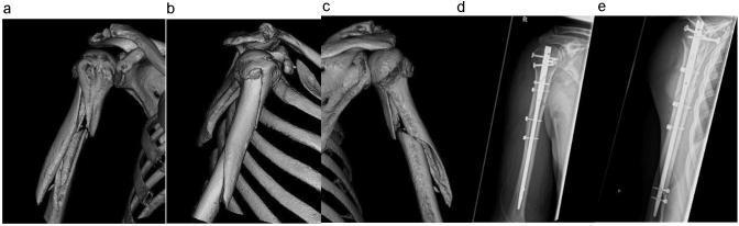
This composite medical image displays pre-operative and post-operative imaging of a humeral shaft fracture, classified as AO 12-B2. Panels a, b, and c show pre-operative 3D computed tomography (CT) reconstructions from three different rotational perspectives. These images reveal a displaced, wedge-type (comminuted) fracture located in the proximal to mid-diaphyseal region of the humerus. Significant angulation and separation of cortical fragments are visible. Panels d and e are post-operative X-ray radiographs in anteroposterior (AP) and lateral views. They demonstrate surgical stabilization using antegrade intramedullary nail osteosynthesis. The nail is centrally positioned within the medullary canal, spanning the fracture site to restore axial alignment. Proximal and distal stability is achieved via multiple locking screws. Additionally, radiopaque cerclage wiring is visible, providing supplementary fixation of the wedge fragment to the main humeral shaft. The combination of techniques illustrates a modern orthopedic approach to complex long-bone fractures, focusing on both intramedullary and extramedullary stabilization for optimal bony consolidation.
PT Management of Fracture of the Shaft of the Humerus (Humeral Diaphysis)
Overview
Conservative (Non-operative) Management
Phase 1: Immobilization Phase (Days 0-10)
- A coaptation splint or hanging arm cast is applied for the first 7-10 days to allow pain and swelling to subside.
- The use of a sling alone is discouraged as it promotes varus and internal rotation deformity.
- After 7-10 days, converted to a Sarmiento functional brace (prefabricated plastic clamshell with Velcro straps).
- Patient education on posture and positioning
- Hand and wrist active ROM exercises (finger fisting, wrist circles)
- Ice/cryotherapy for pain and swelling control
- Pendulum (Codman) exercises - started early to prevent shoulder stiffness. Gravity-assisted, patient leans forward and lets arm swing in small circles. The shoulder must not be immobile during this phase.
"Gravity-assisted pendulum exercises are instituted early on to prevent shoulder stiffness." - Bailey and Love's Short Practice of Surgery, 28th Ed.
Phase 2: Functional Bracing Phase (Weeks 1-6)
- The functional brace uses gravity and muscle compression to maintain alignment (gravity traction).
- Active shoulder abduction is strictly avoided during this phase to prevent varus deformity at the fracture site.
- Daily skin hygiene with brace reapplication is stressed to avoid maceration.
| Exercise | Purpose |
|---|---|
| Pendulum/Codman exercises | Prevent shoulder contracture |
| Active elbow flexion/extension | Maintain elbow ROM (gravity helps maintain fracture alignment) |
| Active forearm pronation/supination | Prevent radioulnar stiffness |
| Wrist and hand ROM and grip strengthening | Prevent distal stiffness, reduce swelling |
| Scapular retraction and depression | Postural correction, upper trapezius relaxation |
Phase 3: Progressive Mobilization Phase (Weeks 6-12)
- Active-assisted shoulder ROM - flexion, abduction, external/internal rotation
- Active shoulder ROM - full range
- Begin rotator cuff strengthening with isometrics, then isotonics (theraband/resistance exercises):
- Shoulder external rotation
- Shoulder internal rotation
- Deltoid (resisted abduction, avoiding extremes initially)
- Elbow strengthening - biceps curls, triceps extension
- Postural retraining and scapular stabilizer strengthening (serratus anterior, lower trapezius)
- Begin functional activities - ADL retraining (grooming, dressing, reaching)
- Low-intensity pulsed ultrasound (LIPUS)
- Vitamin D supplementation These support bone healing and PT can continue alongside.
Phase 4: Strengthening and Return to Function (Weeks 12+)
- Brace worn until patient is pain-free AND radiographic union is evident
- Progressive resistance training for entire upper extremity
- Sport-specific or work-specific retraining
- Proprioceptive/neuromuscular control training
- Acceptable healing criteria: union typically achieved in >80% within 12 weeks with non-operative treatment
Post-operative PT Management
Indications for Surgery (and thus post-op PT):
- Open fractures, vascular injury, progressive radial nerve palsy
- Polytrauma / floating elbow
- Failure to maintain adequate reduction (shortening >3 cm, angulation >20°, rotation >30°)
- Pathologic fracture
Post-op PT Protocol
- Arm sling for protection
- Immediate pendulum exercises
- Active ROM of hand, wrist, elbow
- Ice/elevation for swelling
- Active-assisted shoulder ROM
- Begin isometric deltoid and rotator cuff exercises
- Elbow and forearm active ROM
- Wound and scar management once healed
- Active shoulder ROM in all planes
- Progressive rotator cuff and deltoid strengthening
- Biceps and triceps strengthening
- Functional activities and ADL retraining
Surgery allows earlier initiation of physiotherapy and avoidance of joint stiffness, especially when ipsilateral joint injuries are present. - Rockwood and Green's Fractures in Adults, 10th Ed., 2025
- Full strengthening program
- Sport/work specific training
- Outcomes assessed using ASES score, DASH score, or Mayo Elbow Performance Index (MEPI)
Special Considerations in PT
Radial Nerve Palsy (Most common nerve complication - ~18%)
- Wrist drop is a hallmark finding
- Wrist splint in functional position during PT to prevent wrist contracture
- Active ROM exercises for all unaffected joints
- Nerve gliding / neural mobilization exercises
- Strengthening of proximal muscles that are unaffected
- Most cases recover spontaneously - PT monitors return of nerve function (wrist extensors first to recover)
- Neuromuscular electrical stimulation (NMES) may be used to maintain muscle bulk
Shoulder Stiffness Prevention
- A key risk with conservative management is posttraumatic shoulder stiffness
- Consistent pendulum exercises from day 1 are the main prevention strategy
- If stiffness develops despite PT, manipulation under anesthesia may be considered (contraindicated if fracture healing is uncertain or severe osteoporosis is present)
Distal Third Fractures
- More difficult to control with functional bracing; tendency toward varus deformity
- PT must monitor for loss of alignment
- Early surgery may be preferred in this subset
Acceptable Alignment Criteria (Non-operative)
| Parameter | Acceptable Limit |
|---|---|
| Shortening | < 3 cm |
| Angulation | < 20° |
| Rotation | < 30° |
Functional Outcome Measures Used in PT
- DASH (Disabilities of Arm, Shoulder, and Hand)
- ASES (American Shoulder and Elbow Surgeons) score
- Mayo Elbow Performance Index (MEPI) - assesses pain, ROM, stability, and ADL function
- Visual analog scale (VAS) for pain
- Rockwood and Green's Fractures in Adults, 10th Ed. 2025 - comprehensive coverage of nonoperative and operative treatment, rehabilitation protocols
- Campbell's Operative Orthopaedics, 15th Ed. 2026 - functional bracing protocol, indications for surgery
- Bailey and Love's Short Practice of Surgery, 28th Ed. - gravity-assisted pendulum exercises, brace biomechanics
- Schwartz's Principles of Surgery, 11th Ed. - coaptation splint and functional bracing overview
PT management of supracondylar #
supracondylar fracture humerus child elbow rehabilitation
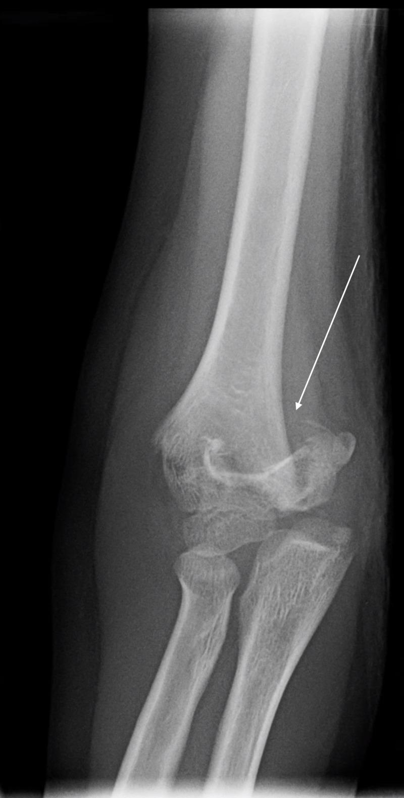
This diagnostic image is an anteroposterior (AP) radiograph of a pediatric elbow, demonstrating a supracondylar humerus fracture. A clear transverse fracture line is visible across the distal humerus, just proximal to the epicondyles. An arrow indicates the site of cortical disruption on the lateral aspect. The distal fracture fragment exhibits displacement and rotational malalignment relative to the humeral shaft, disrupting the normal anatomical relationship between the distal humerus and the articulating proximal radius and ulna. The appearance is consistent with a Gartland Type II supracondylar fracture, a common pediatric orthopedic injury typically resulting from a fall on an outstretched hand. Key educational features include the visualization of the supracondylar fracture pattern, the assessment of coronal plane displacement, and the evaluation of the elbow joint's overall alignment in a skeletally immature patient.
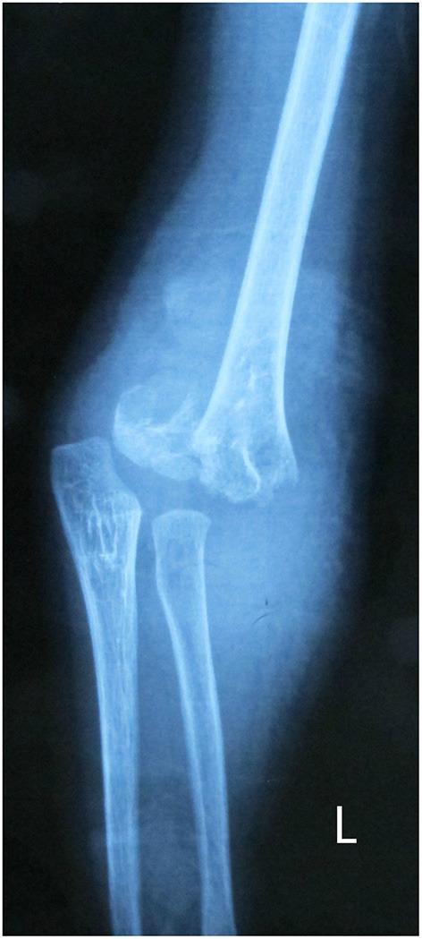
A lateral x-ray radiography of a pediatric elbow demonstrating a severe supracondylar humerus fracture, classified as a Gartland type III. The imaging reveals a complete transverse fracture line across the distal humerus, superior to the condyles. There is significant posterior displacement and angulation of the distal humeral fragment relative to the proximal humeral shaft. The anatomical alignment of the elbow joint is heavily disrupted; the normal radiocapitellar line is lost as the capitellum is displaced with the distal humeral fragment. The proximal radius and ulna remain articulated with the distal humerus but are posteriorly translated alongside it. The image serves as a clinical example of a common pediatric orthopedic emergency requiring urgent reduction to prevent neurovascular compromise or long-term deformity such as cubitus varus. This visual is typical of injuries seen in the context of a fall on an outstretched hand in children.
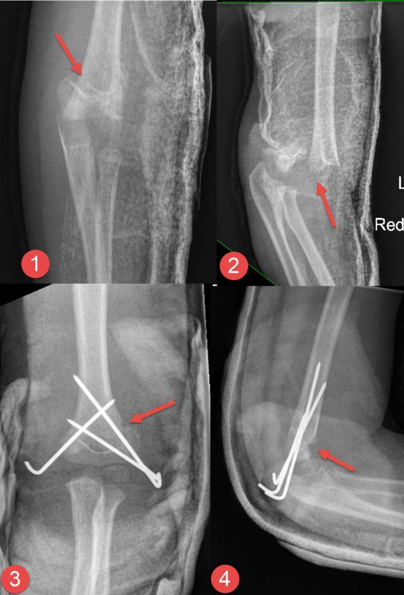
Diagnostic radiographic series of a pediatric elbow demonstrating a supracondylar humerus fracture and its subsequent surgical management. The series is divided into four panels: (1) Pre-operative anteroposterior (AP) view showing a displaced fracture of the distal humerus; (2) Pre-operative lateral view highlighting significant posterior displacement and angulation of the distal fragment; (3) Post-operative AP view showing successful closed reduction and internal fixation; (4) Post-operative lateral view confirming anatomic alignment. The fracture is stabilized using three percutaneous Kirschner wires (K-wires). Two wires are inserted from the lateral epicondyle in a divergent configuration, while a third wire is inserted from the medial epicondyle, crossing the fracture site to provide rigid stability. Red arrows in each panel point to the primary fracture line and the site of surgical reduction. The images illustrate standard orthopedic management of pediatric supracondylar fractures to prevent malunion and neurovascular complications.
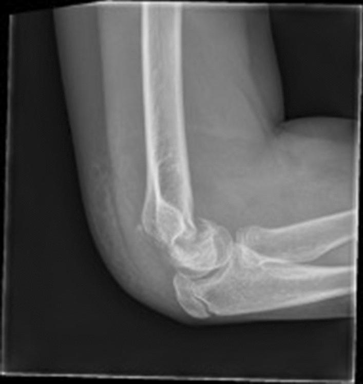
A lateral view X-ray of a pediatric elbow demonstrating a flexion-type supracondylar fracture of the humerus. The diagnostic image shows a complete transverse fracture line across the distal humerus in the supracondylar region. Key visual findings include anterior angulation and anterior displacement of the distal humeral fragment relative to the humeral shaft. Specifically, the anterior humeral line (AHL) is seen passing posterior to the capitellum, a classic radiological sign of a flexion-pattern injury. This indicates failure of the posterior cortex with the anterior periosteum potentially acting as a hinge. The joint space and proximal radius and ulna are visible, providing anatomical context for the elbow joint's sagittal alignment. This image serves as a clinical teaching aid for orthopedic traumatology, highlighting the less common supracondylar fracture variant often associated with direct trauma to the posterior elbow and a higher risk of ulnar nerve injury compared to extension-type fractures.
PT Management of Supracondylar Fracture of the Humerus
Background
Gartland Classification (Guides Treatment and PT)
| Type | Description | Treatment |
|---|---|---|
| Type I | Non-displaced / displaced ≤2 mm | Posterior splint/collar & cuff x 3 weeks, then ROM |
| Type IIA | Angulated, posterior cortex intact, stable | Closed reduction + above-elbow cast |
| Type IIB | Angulated, posterior cortex intact, unstable | CRPP (Closed Reduction + Percutaneous Pinning) |
| Type III | Completely displaced, no cortical contact | CRPP (urgent) |
| Type IV | Multi-directional instability | CRPP or open reduction |
Radiological Reference Images
PT Management: Phase by Phase
A. Conservative Management (Type I & Stable Type IIA)
- Posterior long-arm splint with elbow at 90° flexion, forearm in neutral or pronation
- Collar and cuff or above-elbow cast for 3 weeks
- Neurovascular checks (pulse, capillary refill, sensation) within the first 7-10 days
- Shoulder pendulum exercises (to prevent shoulder stiffness)
- Active ROM of fingers and wrist to reduce distal swelling
- Grip strengthening (sponge squeeze)
- Oedema management - elevation of arm above heart level
- Ice/cryotherapy for pain and swelling in early days
- Begin active elbow ROM - flexion/extension and forearm pronation/supination
- IMPORTANT: Avoid passive motion at the elbow - passive stretching predisposes to ectopic bone (myositis ossificans) formation and permanent stiffness.
"The patient should be instructed to avoid any passive motion at the elbow because it will predispose the elbow to ectopic bone formation and permanent stiffness." - Pfenninger and Fowler's Procedures for Primary Care, 3rd Ed.
- Gentle gravity-assisted active ROM is the safest approach
- Progressive active-assisted ROM once pain subsides
- Protective splint worn when not exercising until adequate callus (~6-8 weeks)
B. Post-Operative Management (CRPP - Types IIB, III, IV)
- Well-padded posterior splint with elbow at 60° flexion initially (to accommodate swelling)
- Neurovascular monitoring (Volkmann's ischemia risk)
- Elevation, ice for swelling control
- Active finger and wrist ROM
- Check at 1 week - changed to long-arm cast at 90° flexion
- Active shoulder ROM to prevent stiffness
- Active finger, hand, and wrist exercises
- Maintained in cast 2-3 more weeks (total ~3-4 weeks post-op)
- Pins removed in clinic under local anaesthetic
- Gentle active ROM exercises begin immediately
- Elbow flexion/extension - start with gravity-assisted movements
- Forearm pronation/supination
- No forced passive stretching (same principle as conservative - risk of myositis ossificans)
- Most children regain full motion without formal physiotherapy due to the natural remodeling capacity of the paediatric elbow.
"The pins are removed in 3-4 weeks, and gentle range-of-motion exercises are begun; most patients regain motion without the need for physical therapy." - Campbell's Operative Orthopaedics, 15th Ed. 2026
C. Progressive Strengthening Phase (Weeks 6-12)
| Exercise | Muscle Target |
|---|---|
| Bicep curls (light resistance) | Biceps brachii, brachialis |
| Triceps press-down | Triceps brachii |
| Forearm pronation/supination against resistance | Pronators, supinators |
| Wrist flexion/extension (resistance band) | Forearm flexors/extensors |
| Grip strengthening | Intrinsic hand muscles |
| Shoulder rotator cuff exercises | Deltoid, rotator cuff |
D. Return to Activity (Weeks 10-16)
- Sport-specific upper limb training
- Overhead activities allowed once full painless ROM achieved
- School and play activities resumed as tolerated
- Formal PT discharge when symmetrical ROM and strength achieved
Key PT Principles Specific to Supracondylar Fractures
1. The "No Passive Stretching" Rule
2. Neurovascular Monitoring During PT
- Radial pulse - brachial artery injury risk
- Capillary refill (< 2 seconds normal)
- Sensation - anterior interosseous nerve (AIN) most commonly injured
- AIN injury: inability to make "OK sign" - flexion of IP joint of thumb and DIP of index finger
- "Pointing finger sign" = inability to flex DIP of index finger
- Median nerve - thumb opposition, sensation of palm
- Radial nerve - wrist and finger extension
3. Compartment Syndrome / Volkmann's Ischemic Contracture
- Rare but most feared complication - due to excessive swelling and missed compartment syndrome
- PT must NOT force elbow into deep flexion if there is significant swelling
- Signs: pain out of proportion, pain on passive finger extension, forearm tightness, pallor, pulselessness
- If suspected: immediate referral for emergency fasciotomy
4. Established Volkmann's Ischemic Contracture (Late PT)
- Mild contracture: PT with serial splinting, stretching exercises, dynamic splints
- Moderate: May require Z-plasty + PT
- Severe: Surgical muscle slide + intensive PT post-op
Complications and PT Implications
| Complication | Incidence | PT Role |
|---|---|---|
| Cubitus varus (gun-stock deformity) | Most common angular deformity | Cosmetic mainly; monitor carrying angle; post-osteotomy PT if corrected |
| Elbow stiffness | ~5% post-CRPP | Active ROM only, no passive; may take weeks to resolve |
| AIN palsy | Most common nerve injury | Monitor recovery; function usually returns spontaneously; no specific PT for nerve |
| Radial nerve palsy | Less common | Wrist extension splint; monitor recovery |
| Ulnar nerve injury | Risk with medial pin | Monitor grip, ulnar sensation |
| Myositis ossificans | From passive stretching | Prevention: active ROM only |
| Volkmann's ischemic contracture | Rare but serious | Serial casting, intensive stretching, functional splinting |
Outcome Measures Used in PT Assessment
- Elbow ROM (flexion/extension arc, pronation/supination)
- Carrying angle (normal: 5-15° valgus) - comparing bilateral
- Grip strength dynamometry
- Flynn's criteria (loss of motion + loss of carrying angle)
- PODCI (Pediatric Outcomes Data Collection Instrument) for functional outcomes
- DASH score in older children
- Campbell's Operative Orthopaedics, 15th Ed. 2026 - CRPP technique, postoperative care protocol, complications
- Tintinalli's Emergency Medicine, 9th Ed. - Gartland classification, initial management, neurovascular assessment
- Bailey and Love's Short Practice of Surgery, 28th Ed. - conservative management, Volkmann's ischemic contracture
- Pfenninger and Fowler's Procedures for Primary Care, 3rd Ed. - no passive motion rule, ROM timeline