
Fracture of humerus
Reading File
Finding Sources
Searching PubMed
"humerus fracture"[MeSH Terms] AND management
Reading File
Reading File
Reading File
Reading File
Reading File
Finding Sources
Finding Sources
Reading File
Reading File
Reading File
Loading Image
Loading Image
I have all the information needed for a comprehensive answer. Let me compose it now.
Fracture of the Humerus
The humerus can fracture at three main regions: the proximal humerus (surgical neck, greater/lesser tuberosity, anatomical neck), the shaft (diaphysis), and the distal humerus (supracondylar and epicondylar regions). Each has distinct mechanisms, complications, and management.
1. Fractures of the Proximal Humerus
Anatomy & Mechanism
Fractures at the surgical neck are most common. The anatomical neck is rarely fractured because a fracture there would have to cross the thickest region of bone. These injuries occur predominantly in elderly women after a fall onto the shoulder, or via high-energy trauma in younger patients.
Neer Classification
The most widely used system, described in Bailey & Love's Surgery, divides the proximal humerus into four parts:
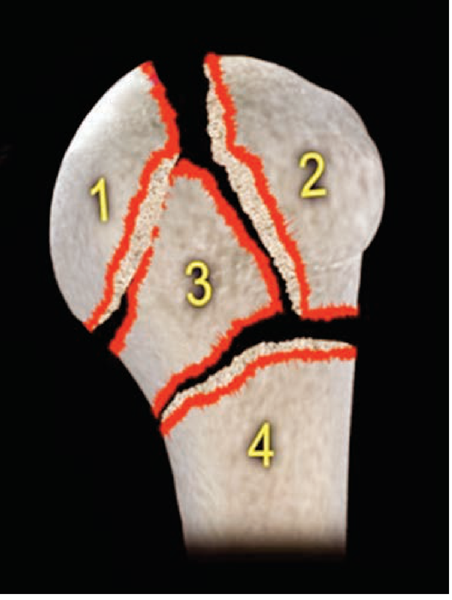
- Articular head fragment
- Greater tuberosity
- Lesser tuberosity
- Humeral shaft
A fragment is counted as a "part" only if it is displaced >1 cm or angulated >45° relative to another fragment. Based on this, fractures are classified as one-part (undisplaced), two-part, three-part, or four-part. The more parts involved, the greater the risk of avascular necrosis (AVN) of the humeral head.
Predictors of Avascular Necrosis
Three factors predict avascularity of the humeral head (Bailey & Love):
- Fracture through the anatomical neck
- Loss of the medial hinge
- Less than 8 mm of bone along the medial calcar
Neurovascular Risk
The axillary nerve and posterior circumflex humeral artery may be damaged, though this is uncommon. It is important to test the axillary nerve before any relocation attempt to document whether a deficit was pre-existing or iatrogenic.
Management
| Fracture Type | Treatment |
|---|---|
| Minimally displaced (one-part) | Sling immobilization; pendulum exercises within 2 weeks |
| Displaced (adequate bone stock) | ORIF with locking plate and screws |
| Elderly, osteoporotic, 4-part, or head-splitting | Hemiarthroplasty or reverse shoulder arthroplasty |
| Young patients | Percutaneous fixation, intramedullary nail, or plate |
Physiotherapy must begin within 2 weeks in elderly patients to prevent stiffness. Reverse shoulder arthroplasty is increasingly favored in elderly patients because it does not depend on tuberosity healing and functions under deltoid power alone. - Schwartz's Principles of Surgery, p. 1912; Bailey & Love, p. 457
2. Fractures of the Humeral Shaft (Diaphysis)
Mechanism
These occur from a direct blow or fall on the arm. Without direct violence, there is typically a rotational element producing a spiral fracture. - Pye's Surgical Handicraft, p. 186
Key Complication: Radial Nerve Injury
The radial nerve spirals around the posterior aspect of the humeral shaft in the spiral groove and is at risk with shaft fractures. It is injured in 10-12% of all closed humeral shaft fractures (Rockwood & Green, 2025).
- Clinical sign: wrist drop (inability to dorsiflex the wrist and fingers), with numbness over the dorsoradial hand
- A special pattern - the Holstein-Lewis fracture - is a spiral fracture of the distal one-third of the shaft where the distal fragment deviates radially; it has an increased risk of radial nerve entrapment
- The majority of nerve injuries are neuropraxias - expectant management is the standard, with spontaneous recovery expected within 3-4 months; waiting no longer than 6 months is advised
- Early nerve exploration is indicated for: open fractures, concomitant vascular injury, gunshot wounds, sharp/penetrating injuries, or high-energy trauma
Acceptable Alignment Criteria (Schwartz's Surgery)
- Anterior angulation: <20°
- Varus/valgus angulation: <30°
- Shortening: <3 cm
Management
- Conservative (majority of cases): Collar-and-cuff sling + coaptation splint OR a U-slab (plaster extending from axilla, under elbow, over shoulder) OR a functional brace (plastic clamshell with Velcro). Radial nerve palsy alone is NOT a contraindication to conservative management.
- Gutter splints (short medial + two anterolateral) strapped to form a triangle; reapplied weekly as swelling decreases
- Union time: approximately 12 weeks; non-union is uncommon
- Operative fixation (ORIF): indicated for polytrauma, patients confined to bed, unacceptable alignment, or failure of conservative treatment; plate fixation is standard
- Pye's, p. 186; Schwartz's, p. 1912
3. Supracondylar Fracture of the Humerus (Distal)
This is the most common elbow fracture in children, typically caused by a fall from height onto an outstretched hand. The distal fragment is displaced posteriorly (extension type, by far the most common) by the pull of the triceps.
Danger: Vascular Injury
The displaced proximal fracture fragment "bowstrings" the brachial artery, creating a risk of forearm ischemia. If unrecognized, this leads to Volkmann's ischemic contracture - a devastating outcome from anterior compartment muscle necrosis. Careful neurovascular assessment is mandatory.
- White pulseless hand = surgical emergency; requires immediate reduction; if pulse does not return, vascular exploration by a trained surgeon is required
- Pink pulseless hand = more controversial; requires early senior decision-making
- Median and ulnar nerves are particularly at risk; careful neurological examination is essential
Management
| Displacement | Treatment |
|---|---|
| Undisplaced | Collar-and-cuff or backslab for 3 weeks, then mobilize |
| Displaced, periosteal hinge intact | Closed manipulation + above-elbow cast for 3-4 weeks |
| Displaced, periosteal hinge broken | Closed manipulation + percutaneous K-wires + above-elbow cast |
| Adults (T-shaped fracture) | ORIF with Y-shaped plate (posterior surface), or lag screws; early mobilization after 6 weeks |
Important: Do NOT place the elbow in deep flexion if there is significant swelling - this risks compartment syndrome. If deep flexion is the only way to hold the reduction, K-wire fixation should be used instead.
In adults, a vertical fracture extending between the capitulum and trochlea may produce a T-shaped fracture, which may require ORIF with a Y-plate applied to the posterior humerus.
Mobilization of the elbow should begin as early as possible; 6 weeks is usually required for adequate fracture stability. - Bailey & Love, p. 462; Gray's Anatomy for Students; Pye's, p. 186
Summary Table
| Region | Common Age | Mechanism | Key Complication | First-line Treatment |
|---|---|---|---|---|
| Proximal (surgical neck) | Elderly | Low-energy fall | AVN of humeral head | Sling ± ORIF ± arthroplasty |
| Shaft | Any | Direct blow / rotational | Radial nerve palsy (wrist drop) | Conservative (functional brace) |
| Supracondylar | Children | Fall on outstretched hand | Brachial artery injury; Volkmann's contracture | Closed reduction ± K-wires |
Sources: Bailey & Love's Short Practice of Surgery (28th ed.); Schwartz's Principles of Surgery (11th ed.); Rockwood & Green's Fractures in Adults (10th ed. 2025); Pye's Surgical Handicraft (22nd ed.); Gray's Anatomy for Students
This is a shared conversation. Sign in to Orris to start your own chat.