
What is mosaic down syndrome
mosaic Down syndrome genetics features outcomes
"mosaic Down syndrome" trisomy 21 mosaicism
Mosaic Down Syndrome
What Is It?
How Does It Occur?
1. Mitotic Nondisjunction (Most Common Route)
2. Rescue of a Trisomic Conceptus
"Alternatively, nonviable cell lineages can be lost during embryogenesis, yielding, for example, viable mosaics with only the chromosome gain (e.g., 46,XY/47,XY,+21). Such a patient is a trisomy 21 mosaic with variable expression of Down syndrome."
Karyotype Notation
- 46,XX/47,XX,+21 (female) or
- 46,XY/47,XY,+21 (male)
Clinical Features
- If most cells carry trisomy 21, the individual will present very similarly to someone with full trisomy 21
- If few cells carry the trisomy, features may be quite mild and the syndrome may even go undiagnosed for years
- There is wide variability among mosaic individuals - from nearly normal appearance and cognition, to having the majority of classic Down syndrome features
"The phenotype may be milder than that of typical trisomy 21, but there is wide variability in phenotypes among mosaic patients, presumably reflecting the variable proportion of trisomy 21 cells in the embryo during early development."
Features that may be present (in varying degrees):
- Characteristic facial features (epicanthal folds, upslanting palpebral fissures, flat nasal bridge, small ears)
- Intellectual disability (often milder on average than full trisomy 21, but not always)
- Hypotonia (low muscle tone)
- Short stature
- Congenital heart defects (e.g., atrioventricular septal defects)
- Increased risk of leukemia
- Risk of early-onset Alzheimer-like dementia (due to the APP gene on chromosome 21)
- Gastrointestinal abnormalities (e.g., duodenal atresia)
- Thyroid problems
Diagnosis
- Chorionic villus sampling (CVS)
- Amniocentesis
- Cell-free fetal DNA (cfDNA/NIPT) - though sensitivity for mosaicism can be lower
Recurrence Risk
Summary Table
| Feature | Mosaic Down Syndrome | Full Trisomy 21 |
|---|---|---|
| Frequency | ~2% of Down syndrome | ~95% of Down syndrome |
| Cause | Post-zygotic mitotic error | Meiotic nondisjunction |
| Cell populations | Mixed (normal + trisomic) | All cells trisomic |
| Phenotype severity | Variable, often milder | Consistent, typically more pronounced |
| Recurrence risk | Low | Low (increases with maternal age) |
| Karyotype | 46,XX/47,XX,+21 | 47,XX,+21 |
- Thompson & Thompson Genetics and Genomics in Medicine, 9th ed.
- The Developing Human: Clinically Oriented Embryology
- Robbins, Cotran & Kumar Pathologic Basis of Disease
- Creasy & Resnik's Maternal-Fetal Medicine
- Children's Hospital of Philadelphia - Mosaic Down Syndrome
Explain the mechanism using diagram pls
A clear educational diagram showing the two mechanisms of mosaic Down syndrome: Mechanism 1 (left side): "Mitotic Nondisjunction in Normal Zygote" - Start with a normal fertilized egg (46 chromosomes, labeled "Normal Zygote 46,XX") - Arrow down to first cell division showing nondisjunction error - chromosomes 21 failing to separate - This produces two daughter cells: one with 47 chromosomes (47,XX,+21 - trisomic, shown in orange/red) and one with 45 chromosomes (shown in grey, labeled "45 - lost/nonviable") - The trisomic cell continues dividing normally - Final result shows a person with mixed cells: some normal (blue, 46,XX) and some trisomic (orange, 47,+21) - Label: "MOSAIC DOWN SYNDROME" Mechanism 2 (right side): "Trisomy Rescue" - Start with a trisomic fertilized egg (47 chromosomes, labeled "Trisomic Zygote 47,XX,+21" in orange) - Arrow down to first cell division showing one cell losing the extra chromosome 21 - One daughter cell becomes normal (46,XX, blue) and one remains trisomic (47,+21, orange) - Both lines continue dividing - Final result shows a person with mixed cells: some normal (blue) and some trisomic (orange) - Label: "MOSAIC DOWN SYNDROME" Bottom section: A comparison bar showing "% Trisomic Cells" from 0% to 100%, with labels: - 0-20%: "Very mild / possibly undiagnosed" - 20-60%: "Moderate features" - 60-100%: "Similar to full trisomy 21" Use color coding: blue for normal cells (46 chromosomes), orange/red for trisomic cells (47,+21). Include chromosome 21 illustrations as small colored dots. Clean white background, medical textbook style, clear labels and arrows.
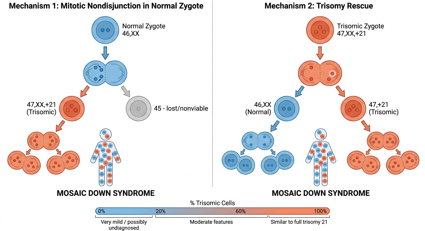
Mechanism 1 - Mitotic Nondisjunction in a Normal Zygote
- Fertilization is normal - egg and sperm fuse to produce a zygote with 46 chromosomes
- Error occurs during early mitosis - when the zygote divides, chromosome 21 fails to separate properly (nondisjunction)
- Two abnormal daughter cells result:
- One cell gets an extra chromosome 21 → 47,XX,+21 (trisomic)
- One cell loses a chromosome 21 → 45 chromosomes (monosomic, usually nonviable and lost)
- The trisomic cell line continues dividing normally from that point
- End result: a person with two cell populations - normal (46) and trisomic (47,+21)
Mechanism 2 - Trisomy Rescue
- Fertilization itself is abnormal - the original zygote already has trisomy 21 (47,XX,+21), arising from a meiotic error in the egg or sperm
- During an early mitotic division, one cell spontaneously loses the extra chromosome 21 ("rescue")
- That cell and all its descendants are now normal (46 chromosomes)
- The original trisomic line continues alongside it
- End result: again, a mosaic mix of normal and trisomic cells
Key Point - Why Severity Varies
I'm still not able to understand the mechanism
Which part is unclear?
How there are 2 cell lineage when one zygote is present it will either give a normal or a trisomic cell lineage
A simple step-by-step diagram explaining how ONE zygote produces TWO different cell lineages in mosaic Down syndrome. Layout: vertical flow, top to bottom, clean white background, large clear labels. STEP 1 (top): - Single cell labeled "Zygote" with "46 chromosomes (NORMAL)" - Show 4 pairs of colored dots inside representing chromosomes, with chromosome 21 pair shown as two small orange dots - Text below: "One normal zygote exists after fertilization" STEP 2 (middle): - The zygote is dividing into 2 cells (show a cell splitting in half) - During this split, show chromosome 21 (orange dots) failing to separate - both copies going to ONE side instead of splitting evenly - Label this error with a red "X" or "ERROR - Nondisjunction" - Text: "During the FIRST cell division, chromosome 21 fails to split equally" STEP 3 (bottom left and right - two separate daughter cells): LEFT CELL: - Show cell with 47 chromosomes - has 3 orange dots (chromosome 21 copies) - Label: "Cell A: 47 chromosomes (+21) - TRISOMIC" - Color: orange/red tint RIGHT CELL: - Show cell with 45 chromosomes - has only 1 orange dot (one chromosome 21) - Label: "Cell B: 45 chromosomes (-21) - MONOSOMIC → DIES" - Color: grey, with an X through it STEP 4 (below Cell A only, since Cell B died): - Cell A divides normally many times → show 4 orange/trisomic cells - BUT also show some normal 46-chromosome cells (blue) that came from later corrections or surviving lines - Label: "Cell A keeps dividing → ALL its children are trisomic (47,+21)" Wait - correct this: Show that the ZYGOTE itself first divides into 2 cells normally BEFORE the nondisjunction. So: REVISED FLOW: - Zygote (46) divides NORMALLY into 2 cells (Cell 1 and Cell 2), both with 46 chromosomes - Then Cell 1 divides NORMALLY → 2 normal daughters (46) shown in BLUE - Cell 2 divides with NONDISJUNCTION ERROR → one daughter gets 47 (orange, trisomic) and one gets 45 (grey, dies) - Final picture: 3 normal blue cells + 1 orange trisomic cell = MOSAIC PERSON Label at bottom: "Result: ONE person with a MIX of normal (46) and trisomic (47,+21) cells = MOSAIC DOWN SYNDROME" Use simple circles for cells, colored dots for chromosomes. Very clean, educational, easy to follow.
Simple educational diagram showing how mosaic Down syndrome produces two cell lineages from one zygote. Top: One circle labeled "Zygote (46 chromosomes, NORMAL)" with two small orange dots labeled "Chr 21 pair" First division - normal: The zygote splits into two equal cells, Cell 1 (46, blue) and Cell 2 (46, blue). Label: "1st division - normal" Second division: - Cell 1 divides normally into two blue cells both labeled 46 - Cell 2 divides but has a NONDISJUNCTION ERROR (red lightning bolt symbol). One daughter gets 3 copies of chr 21 = 47 chromosomes (orange cell, trisomic). The other daughter gets 1 copy = 45 chromosomes (grey cell, dies, marked with X). Bottom result panel showing the final body contains: BLUE cells (46, normal) AND ORANGE cells (47, trisomic). Big label: "MOSAIC = mix of two cell types in ONE person" Small note: "The error happened in Cell 2 only during the 2nd division - Cell 1 lineage stayed normal" Clean white background, simple circles, bold text labels, arrows between steps, medical illustration style.
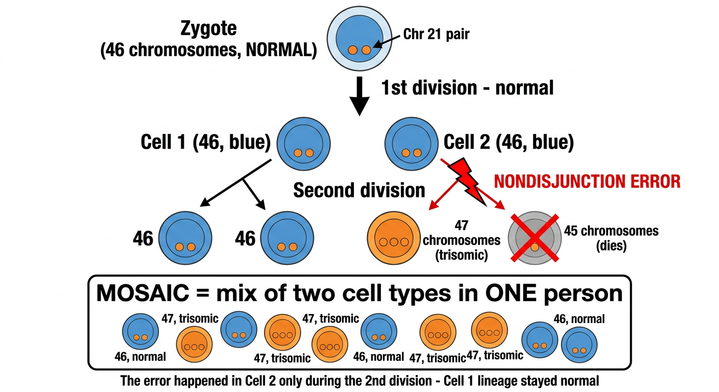
Zygote (46) — normal
     |
  1st division (normal)
  /           \
Cell 1        Cell 2
(46, normal)  (46, normal)
     |              |
 2nd division   2nd division
  (normal)      ← ERROR HERE (nondisjunction)
  /    \            /         \
46    46        47,+21       45 → DIES
(blue)(blue)   (orange)
- Cell 1's branch always divides normally → all blue (46 chromosomes)
- Cell 2's branch had the error → one daughter gets 47 (+21, trisomic), one gets 45 (dies)
- The 47 cell keeps dividing → all its children are also 47,+21
When does the nondisjunction error need to happen to affect most cells?
Educational diagram showing how timing of nondisjunction error determines what percentage of cells are trisomic in mosaic Down syndrome. Title at top: "Earlier Error = More Trisomic Cells" Show 3 scenarios side by side: SCENARIO 1 - Left column: "Error at 1st Division (2-cell stage)" - Zygote splits into 2 cells - The nondisjunction error happens RIGHT HERE at the very first division - One cell becomes trisomic (47, orange), one becomes monosomic (45, grey, dies/lost) - The trisomic cell divides into 2, then 4, then 8 orange cells - Final count: 8 orange trisomic cells out of 8 total - Big label: "~100% trisomic = Full Down Syndrome (not mosaic)" SCENARIO 2 - Middle column: "Error at 2nd Division (4-cell stage)" - Zygote divides normally into 2 cells (both blue) - Then one of those 2 cells divides with a nondisjunction error - That produces: 1 trisomic orange cell + 1 monosomic grey cell (dies) - The other normal cell divides normally into 2 blue cells - Final count shown: 1 orange + 2 blue = 3 cells, roughly 33% trisomic - Label: "~33% trisomic = Mild mosaic" SCENARIO 3 - Right column: "Error at 3rd Division (8-cell stage)" - Normal divisions until 4-cell stage (4 blue cells) - Then ONE of those 4 cells has the nondisjunction error - Produces 1 orange trisomic cell, 1 grey monosomic (dies) - Other 3 normal cells each divide into 2 = 6 blue cells - Final count: 1 orange + 6 blue = 7 cells, roughly 14% trisomic - Label: "~14% trisomic = Very mild mosaic" Bottom summary bar: "Rule: Error at division N affects roughly 1/2^(N-1) of all cells" "Earlier error → Higher % trisomic cells → More severe phenotype" Color code: Blue circles = normal 46-chromosome cells, Orange circles = trisomic 47+21 cells, Grey circles with X = monosomic cells that die. Clean white background, clear arrows, simple cell circles, bold labels.
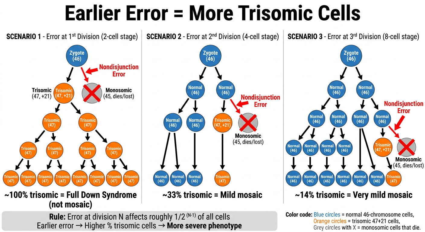
| Error happens at... | Cells at that stage | Trisomic cells produced | % of final body that is trisomic |
|---|---|---|---|
| 1st division (2-cell) | 2 | ~1 trisomic survives | ~50-100% → essentially full trisomy 21 |
| 2nd division (4-cell) | 4 | 1 out of ~3 surviving | ~33% → severe mosaic |
| 3rd division (8-cell) | 8 | 1 out of ~7 surviving | ~14% → moderate mosaic |
| Later divisions | 16, 32... | 1 out of many | <5% → very mild / may go unnoticed |
How do doctors test for mosaic Down syndrome after birth?
postnatal diagnosis mosaic Down syndrome karyotype FISH chromosomal microarray blood skin fibroblasts
https://www.imdsa.org/How-to-get-a-diagnoses
Step 1 - Clinical Suspicion
- Upslanting eyes, flat nasal bridge, single palmar crease, hypotonia
- But in mosaic cases, features may be subtle or partial - this is often why mosaic DS goes undiagnosed for longer than full trisomy 21
Step 2 - Choosing a Tissue Sample
| Tissue | How collected | Notes |
|---|---|---|
| Blood (lymphocytes) | Simple blood draw | Most common first test; may miss low-level mosaicism |
| Buccal mucosa | Cheek swab | Non-invasive; often shows higher % trisomic cells than blood |
| Skin fibroblasts | Small skin biopsy | Most sensitive; used when blood/cheek tests are negative but suspicion remains high |
Step 3 - The 3 Testing Methods
1. Conventional Karyotype (G-banding)
- Cells are grown in culture, stained, and all 46 chromosomes are photographed and arranged
- At least 20-30 cells are counted and examined
- If some show 47 chromosomes (+21) and others show 46 → mosaic confirmed
- Limitation: if mosaicism is very low (e.g., 2-3%), you might count 20 cells and happen to miss the trisomic ones - false negative is possible
2. FISH (Fluorescence In Situ Hybridization)
- Fluorescent probes that glow and attach specifically to chromosome 21 are applied to cells
- Each normal cell shows 2 glowing dots (2 copies of chr 21)
- Each trisomic cell shows 3 glowing dots
- Hundreds of cells can be scanned quickly, making it much better at detecting low-level mosaicism
- Particularly useful on buccal mucosa cells
3. Chromosomal Microarray (CMA)
- Analyzes DNA from all chromosomes at once
- Can detect extra copies of chromosome 21 across the sample
- Good for confirming findings and detecting other chromosomal issues simultaneously
- Can be done from blood, buccal swab, or skin biopsy
The Key Diagnostic Challenge - Illustrated
Same patient, different tissues:

Blood karyotype:    ██░░░░░░░░  8% trisomic  → may appear NORMAL
Buccal FISH:        ████████░░  31% trisomic → CLEARLY MOSAIC
Skin fibroblasts:   ██████████  ~50% trisomic→ MOST ACCURATE

↑ This is why a NEGATIVE blood test does NOT rule out mosaic DS
Recommendation from Experts
- Clinical features strongly suggest Down syndrome BUT the blood karyotype is normal
- A person has unexplained intellectual disability with dysmorphic features
- Blood FISH shows borderline or low-level results
Why the cell with 45 chromosome dies and explain the trisomy rescue mechanism
Two-part educational medical diagram on white background. PART 1 (top half): "Why the 45-chromosome cell dies" Show a normal cell with 46 chromosomes (represented as 23 pairs of colored bars). Then show the monosomic cell with only 45 chromosomes - chromosome 21 has only ONE copy instead of two (show one bar missing from the pair, with a red X on the gap). Explanation box next to monosomic cell: "Missing chromosome 21 = missing hundreds of essential genes → Critical proteins cannot be made → Cell cannot survive or divide → LETHAL - cell dies quickly" Contrast with trisomy 21 cell (47 chromosomes): "EXTRA chromosome 21 = excess of gene products. Cell is abnormal but can survive." Label: "Monosomy of any autosome is lethal. Extra chromosome is survivable (but causes disease)." PART 2 (bottom half): "Trisomy Rescue Mechanism" Step-by-step flow left to right: Step 1: "Fertilization goes wrong" - Show egg + sperm combining, but one sperm has an EXTRA chromosome 21 (3 dots instead of 2) - Result: Trisomic zygote 47,+21 (all orange) - Label: "Meiotic nondisjunction during egg or sperm formation → entire zygote starts with trisomy 21" Step 2: "Early cell division - rescue event" - The trisomic zygote (47) divides into 2 cells - In ONE of those cells, the extra chromosome 21 is LOST (show it floating away with an arrow, labeled "chromosome 21 lost by anaphase lag") - That cell becomes NORMAL 46 chromosomes (blue) - The other cell KEEPS all 3 copies of chromosome 21 → stays 47,+21 (orange) Step 3: "Both lineages divide and propagate" - Blue cell divides → 2 blue, then 4 blue cells (all 46, normal) - Orange cell divides → 2 orange, then 4 orange cells (all 47,+21, trisomic) - Show them mixed together in a final body outline Step 4 - BONUS box (red border): "UPD Risk!" "When rescue happens, the 2 remaining chr 21 copies may BOTH come from the same parent (uniparental disomy). This can cause imprinting disorders in 1/3 of trisomy rescue cases." Show: original 3 chr 21 copies labeled M (maternal), M, P (paternal). If P is lost → 2 M copies remain = Uniparental disomy (UPD). If one M is lost → 1M + 1P = normal biparental. Color: Blue for normal cells, Orange for trisomic, clean arrows, bold text labels, medical textbook style.
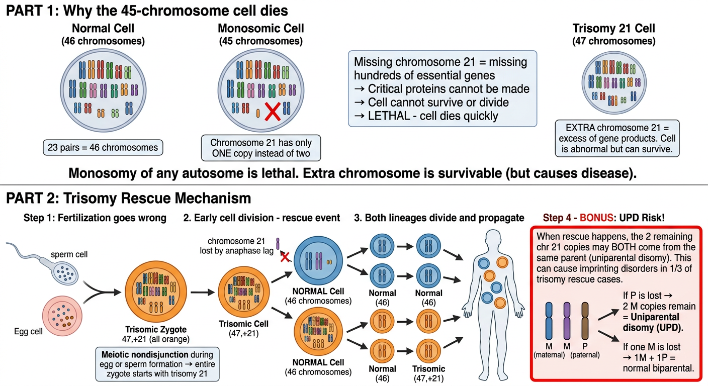
Part 1 - Why Does the 45-Chromosome Cell Die?
- Gene dosage drops by 50% for all those genes simultaneously
- Many of those genes produce proteins that are absolutely required for basic cell functions - transcription factors, metabolic enzymes, structural proteins
- The cell cannot compensate - there is no backup copy
- The cell triggers apoptosis (programmed cell death) or simply fails to divide
| State | Gene copies | Effect | Survivable? |
|---|---|---|---|
| Normal (46) | 2 copies each | Balanced | Yes |
| Trisomy (47, +21) | 3 copies of chr 21 | 50% gene overexpression - disruptive but tolerable | Yes (causes Down syndrome) |
| Monosomy (45, -21) | 1 copy of chr 21 | 50% gene underexpression - catastrophic | No - cell dies |
Part 2 - Trisomy Rescue Explained Step by Step
STEP 1 - Meiotic error during egg/sperm formation:
  Abnormal egg (has 2 copies of chr 21)  +  Normal sperm (1 copy of chr 21)
                          ↓
              Zygote: 47 chromosomes (+21)
              ALL cells start trisomic (orange)

STEP 2 - Rescue event during early mitosis:
  47-cell divides → chromosome 21 fails to be pulled into one daughter cell
  (called anaphase lag - the chromosome just "lags behind" and gets lost)
                          ↓
         One daughter: 46 chromosomes ✓ (RESCUED - now normal, blue)
         Other daughter: 47 chromosomes (still trisomic, orange)

STEP 3 - Both lineages multiply:
  Blue cell → 2 → 4 → 8 → ... normal cells
  Orange cell → 2 → 4 → 8 → ... trisomic cells
                          ↓
              MOSAIC PERSON (mix of blue + orange)
The Hidden Danger of Trisomy Rescue - UPD
- Copy M1 (from mother)
- Copy M2 (from mother, the extra one due to meiotic error in the egg)
- Copy P (from father)
- Lost P → remaining cells have M1 + M2 = both chromosomes from MOM only = Uniparental Disomy (UPD)
- Lost M1 or M2 → remaining cells have one M + one P = normal biparental inheritance