

ffac038b-f8c3-44ce-ad92-311aca117a68.jpeg
Give me answer wt respect to that question with marks and what to write for university exam
Reading File
analyse_media
Now I have all the questions. Let me search the medical library for comprehensive answers to all these questions in parallel.
Finding Sources
Finding Sources
Finding Sources
Reading File
Reading File
Finding Sources
Finding Sources
Finding Sources
Finding Sources
Reading File
Reading File
I now have all the information needed to write comprehensive exam answers. Let me compile everything into a well-structured response.
University Exam Answers - Hematology Questions
SHORT ANSWER TYPE QUESTIONS (3 Marks each)
Q1. 22-year-old man - Enlarged rubbery lymph nodes, weight loss, night sweats, FNAC shows "owl eye" cells
Part 1) Most likely diagnosis (1 mark)
Answer:
Hodgkin Lymphoma (specifically Classic Hodgkin Lymphoma - Mixed Cellularity or Nodular Sclerosis subtype)
The "owl-eye" appearance of cells in FNAC is the hallmark of Reed-Sternberg (RS) cells, which are the diagnostic cells of Hodgkin lymphoma. These are very large cells (15-45 µm in diameter) with an enormous multilobate nucleus, exceptionally prominent nucleoli, and abundant cytoplasm. Characteristic are two mirror-image nuclei or nuclear lobes, each containing a large, inclusion-like acidophilic nucleolus surrounded by a clear zone - giving the "owl-eye" appearance. - Robbins & Kumar Basic Pathology
Part 2) Classification of Hodgkin Lymphoma (2 marks)
Answer (WHO Classification):
A. Nodular Lymphocyte-Predominant Hodgkin Lymphoma (NLPHL)
B. Classic Hodgkin Lymphoma (CHL) - 4 subtypes:
- Nodular Sclerosis (most common; adolescents/young adults; collagen bands; lacunar cells)
- Mixed Cellularity (classic RS cells plentiful; male predominance; >50 years)
- Lymphocyte-Rich (rare; good prognosis)
- Lymphocyte-Depleted (rare; worst prognosis; elderly/HIV)
RS cells in classic subtypes express CD15 and CD30 and are negative for CD45. - Robbins & Kumar Basic Pathology
Immunophenotype to mention: RS cells are CD15+, CD30+, CD45-, EMA-
Q2. Young adult - Fever, sore throat, generalized lymphadenopathy, peripheral smear shows atypical lymphocytes
Part 1) Most likely diagnosis (1 mark)
Answer:
Infectious Mononucleosis (IM)
Infectious mononucleosis is an acute self-limited disease of adolescents and young adults characterized by: (1) fever, sore throat, and generalized lymphadenopathy, and (2) a lymphocytosis of activated CD8+ T cells (seen as atypical lymphocytes on peripheral smear). - Robbins & Kumar Basic Pathology
Part 2) Name the virus that causes it (1 mark)
Answer:
Epstein-Barr Virus (EBV) - a member of the herpesvirus family
EBV binds to CD21 on B cells via its envelope glycoprotein, explaining B-cell tropism. Note: CMV can cause a similar syndrome but is distinguished by serology. - Robbins & Kumar Basic Pathology
Part 3) Laboratory test other than peripheral smear (1 mark)
Answer:
Paul-Bunnell Test (Monospot Test / Heterophile Antibody Test)
- Detects heterophile antibodies (IgM) against sheep/horse red blood cells produced in EBV infection
- Specific EBV serology: Anti-VCA (Viral Capsid Antigen) IgM - confirms acute infection
- Other options to mention: Anti-EA (Early Antigen), EBNA antibodies, EBV PCR, Throat swab culture
Write any one - Paul-Bunnell / Monospot test is the most classic answer for 1 mark
Q3. Elderly patient - Generalized lymphadenopathy, absolute lymphocytosis with smudge cells
Part 1) Most likely diagnosis (1 mark)
Answer:
Chronic Lymphocytic Leukemia (CLL)
Key features to write:
- Elderly patient
- Absolute lymphocytosis (mature-looking small lymphocytes)
- Smudge cells (basket cells) on peripheral smear - fragile CLL lymphocytes that rupture during smear preparation - PATHOGNOMONIC of CLL
- Generalized lymphadenopathy
Part 2) Richter's Transformation (2 marks)
Answer:
Definition: Richter's transformation is the transformation of CLL (Chronic Lymphocytic Leukemia) into a more aggressive lymphoma.
Details to write:
- Most commonly transforms into Diffuse Large B-Cell Lymphoma (DLBCL) (~95% of cases)
- WHO also recognizes transformation into Hodgkin Lymphoma (HL) as a variant
- Prevalence: ~0.5% per year for DLBCL transformation; 0.05% per year for HL transformation
- Clinically heralded by: sudden onset of fever, night sweats, weight loss (B symptoms), rapidly enlarging lymph nodes, elevated LDH
- Risk factors: Bulky lymphadenopathy, NOTCH1 mutations, del(17p13.1)
- Prognosis: Poor - clonally related transformations have significantly worse outcomes than clonally unrelated cases
- Diagnosis: PET scan - SUV >5 suspicious; ≥10 highly suspicious for Richter's transformation; biopsy confirms
"One of the most devastating complications of CLL is Richter's transformation...The prevalence estimated at 0.5% per year for DLBCL." - Harrison's Principles of Internal Medicine 22E (2025)
EXAM WRITING TIPS
| Question | Key Buzzwords to Include | Common Mistakes to Avoid |
|---|---|---|
| Q1 Part 1 | Reed-Sternberg cell, owl-eye nucleoli, CD15+, CD30+ | Don't write Non-Hodgkin lymphoma |
| Q1 Part 2 | WHO classification, 5 subtypes, NLPHL vs Classic | Don't forget NLPHL as a separate category |
| Q2 Part 1 | Atypical lymphocytes = activated CD8+ T cells (not B cells!) | Don't say it's a B-cell lymphocytosis |
| Q2 Part 2 | EBV, CD21 receptor, herpesvirus family | Don't write CMV (that's the differential) |
| Q2 Part 3 | Paul-Bunnell / Monospot / Heterophile antibody test | Don't just say "blood test" |
| Q3 Part 1 | CLL, smudge/basket cells pathognomonic | Don't diagnose as ALL or AML |
| Q3 Part 2 | DLBCL, 0.5%/year, B symptoms, poor prognosis | Don't confuse with simple progression of CLL |
Sources used:
- Robbins & Kumar Basic Pathology - Reed-Sternberg cells, Hodgkin classification, EBV/Infectious Mononucleosis
- Harrison's Principles of Internal Medicine 22E (2025) - Richter's transformation in CLL
Still more 124 those questions
The image showed more questions below - let me get answers for the remaining ones (the short answer 1-mark questions and the 5-mark essay questions).
Finding Sources
Finding Sources
Finding Sources
Finding Sources
Finding Sources
Finding Sources
Finding Sources
Finding Sources
Finding Sources
Finding Sources
I have enough information from the textbooks and my knowledge to write comprehensive answers for all remaining questions. Here they are:
Remaining Questions - University Exam Answers
SHORT ANSWER QUESTIONS (1 mark each)
Q1. Describe the inheritance pattern of Hemophilia A and explain why it preferentially affects males (1 mark)
Answer:
Inheritance Pattern: Hemophilia A is inherited as an X-linked recessive disorder (20% of cases arise from new spontaneous mutations).
Why it preferentially affects males:
- The gene for Factor VIII (deficient in Hemophilia A) is located on the X chromosome
- Males are hemizygous (XY) - they have only ONE X chromosome, so a single defective copy of the gene causes disease
- Females are heterozygous carriers (X^H X^h) - they have TWO X chromosomes; the normal allele on the second X compensates, so they are usually unaffected carriers
- A female would need TWO defective copies (X^h X^h) to manifest the disease - this is extremely rare
Punnett Square to draw:
Carrier Mother (X^H X^h)  ×  Normal Father (X^H Y)
Daughters: X^H X^H (normal), X^H X^h (carrier)
Sons:      X^H Y (normal),   X^h Y (affected)
- 50% of sons are affected; 50% of daughters are carriers
Q2. Platelet count of 20,000/cumm in a young woman - What is this condition called? List 4 causes
Answer:
Condition: This is called Thrombocytopenia (normal platelet count = 1,50,000 - 4,00,000/cumm)
At 20,000/cumm with a young woman, the most likely diagnosis is Immune Thrombocytopenic Purpura (ITP)
4 Causes of Thrombocytopenia:
| Mechanism | Causes |
|---|---|
| Decreased production | Aplastic anemia, Leukemia infiltrating bone marrow, Megaloblastic anemia (B12/folate deficiency), Drug-induced (chemotherapy) |
| Increased destruction | ITP (autoimmune - most common in young women), SLE, Drug-induced (heparin - HIT), DIC |
| Increased sequestration | Hypersplenism (spleen traps platelets) |
| Dilutional | Massive blood transfusion |
4 causes to list:
- ITP (Immune Thrombocytopenic Purpura) - autoantibodies (IgG) against platelet GPIIb/IIIa
- SLE (Systemic Lupus Erythematosus)
- Aplastic anemia (decreased production)
- Drug-induced thrombocytopenia (e.g., heparin, quinine, sulfonamides)
Q3. Why is splenectomy beneficial in cases of Chronic ITP? (1 mark)
Answer:
Splenectomy is beneficial in chronic ITP for TWO main reasons:
1. Removes the major site of platelet destruction:
- In ITP, autoantibodies (IgG) coat platelets via GPIIb/IIIa antigens
- The spleen is the primary site where these antibody-coated platelets are recognized and destroyed by splenic macrophages (reticuloendothelial system)
- Removing the spleen eliminates this destruction
2. Removes the major site of autoantibody production:
- The spleen contains the B-lymphocytes that produce anti-platelet IgG antibodies
- Splenectomy reduces the antibody burden, allowing platelet survival to improve
Result: After splenectomy, platelet count rises in ~60-70% of patients. It is used as second-line therapy when steroids and IVIG fail.
SHORT ESSAY TYPE QUESTIONS (5 Marks each)
Essay Q1. Tabulate differences between Chronic Myeloid Leukemia (CML) and Chronic Lymphocytic Leukemia (CLL) (5 marks)
Write this table in your exam:
| Feature | CML | CLL |
|---|---|---|
| Cell of origin | Myeloid stem cell (granulocytic series) | B-lymphocyte (mature) |
| Age | 25-60 years (middle age) | >50 years (elderly) |
| Sex | Male > Female | Male > Female |
| Peripheral smear | Leukocytosis with ALL stages of myeloid maturation (myeloblasts to mature neutrophils) - "myeloid spectrum"; basophilia prominent | Mature small lymphocytes + Smudge/basket cells (pathognomonic) |
| WBC count | Markedly elevated (often >1,00,000/cumm) | Elevated (10,000-2,00,000/cumm) of mature lymphocytes |
| Cytogenetics | Philadelphia chromosome t(9;22) → BCR-ABL fusion gene (95% cases) | No Philadelphia chromosome; del 13q (most common), del 11q, del 17p, trisomy 12 |
| Bone marrow | Hypercellular - all myeloid precursors, ↑ megakaryocytes | Hypercellular - replaced by mature small lymphocytes |
| Spleen | Massively enlarged (hallmark finding) | Moderately enlarged |
| Lymph nodes | Mildly enlarged | Generalized lymphadenopathy (prominent) |
| Leukocyte Alkaline Phosphatase (LAP) score | Markedly decreased (low) | Normal or elevated |
| Blasts in peripheral smear | <10% (chronic phase) | Absent (mature cells) |
| Basophilia/Eosinophilia | Present (characteristic) | Absent |
| Prognosis | Responds well to Imatinib (TKI); median survival >10 years | Indolent; variable (some never need treatment; Richter's transformation = poor) |
| Treatment | Imatinib (BCR-ABL tyrosine kinase inhibitor) | Watchful waiting / FCR (Fludarabine + Cyclophosphamide + Rituximab) / BTK inhibitors |
Essay Q2. Tabulate differences between Hodgkin and Non-Hodgkin Lymphoma (including clinical features, histology, immunophenotype) (5 marks)
| Feature | Hodgkin Lymphoma (HL) | Non-Hodgkin Lymphoma (NHL) |
|---|---|---|
| Age | Bimodal: 15-35 yrs and >55 yrs | Any age; increases with age |
| Sex | Male > Female (except nodular sclerosis = equal) | Male > Female |
| Nodal involvement | Usually localized to a single axial group (cervical, mediastinal, para-aortic) | Multiple peripheral nodes; widespread at presentation |
| Spread | Contiguous/orderly spread (stage to stage) | Non-contiguous (skip lesions) |
| Mediastinal involvement | Common (especially nodular sclerosis) | Less common |
| Mesenteric nodes / Waldeyer's ring | Rarely involved | Commonly involved |
| Extranodal involvement | Rare | Common (bone marrow, GI tract, CNS, skin) |
| B symptoms (fever, night sweats, weight loss) | Common (30-40%) | Less common |
| Histology | Reed-Sternberg cells on background of reactive cells (lymphocytes, eosinophils, plasma cells) | No RS cells; sheets of neoplastic lymphoid cells |
| Hallmark cell | Reed-Sternberg (RS) cell - owl-eye nucleoli | No specific cell; varies by subtype |
| Immunophenotype | CD15+, CD30+, CD45-, B/T markers negative (classic HL) | CD20+ (B-cell NHL most common); CD3+ (T-cell NHL) |
| Bone marrow involvement | Late, uncommon | Early, common |
| EBV association | Yes (mixed cellularity subtype especially) | Some (Burkitt lymphoma - EBV strongly associated) |
| Subtypes | 5 WHO subtypes (NLPHL + 4 classic) | >60 subtypes (WHO classification) |
| Prognosis | Generally better; >80% cure with chemo-radiation | Variable; some indolent, some aggressive |
| Treatment | ABVD chemotherapy + radiation | Depends on subtype: R-CHOP most common |
Essay Q3. Describe differences between Myeloblasts and Lymphoblasts (with labelled diagrams) (5 marks)
Differences Table:
| Feature | Myeloblast | Lymphoblast |
|---|---|---|
| Size | Large (15-20 µm) | Small to medium (10-18 µm) |
| Nucleus | Round/oval; fine chromatin; 2-5 prominent nucleoli | Round/oval; coarse/clumped chromatin; 1-2 indistinct nucleoli |
| Cytoplasm | Moderate; Auer rods (pink needle-like inclusions) present in AML | Scanty; no Auer rods |
| Granules | Azurophilic (primary) granules present | No granules (agranular) |
| N:C ratio | Lower (more cytoplasm) | High N:C ratio (very little cytoplasm) |
| Special stains | MPO (Myeloperoxidase) POSITIVE; Sudan Black B positive | MPO negative; PAS positive (block positivity) |
| Immunophenotype | CD13+, CD33+, CD117+, MPO+ | CD19+, CD10+, TdT+ (B-ALL) OR CD2+, CD7+, TdT+ (T-ALL) |
| Disease | Seen in AML (Acute Myeloid Leukemia) | Seen in ALL (Acute Lymphoblastic Leukemia) |
| Auer rods | Present (pathognomonic of AML) | Absent |
Diagram to draw:
MYELOBLAST                    LYMPHOBLAST
   _____                          ___
  /     \                        /   \
 | [NUC] |  ← Large nucleus     |[NUC]|  ← Smaller, clumped
  \  2-5 /    with prominent     \ 1-2/    chromatin, 1-2
   nucleoli                      nucleoli  nucleoli
  /~~~~~\                       /~~~~~\
 |  MPO+ |                     |  TdT+ |
 |granules|                    |       |  ← Scanty cytoplasm
  \_____/                       \_____/
 Auer rods                     No Auer rods
 present!
Key exam trick - Auer rods = AML = Myeloblast. If you see Auer rods in a question, diagnosis is AML.
Essay Q4. Laboratory findings in Chronic Myeloid Leukemia - CML (with peripheral smear and bone marrow diagrams) (5 marks)
Clinical features briefly:
- Middle-aged patient, insidious onset
- Massive splenomegaly (most striking feature)
- Weight loss, fatigue, night sweats, abdominal fullness
Laboratory Findings:
1. Complete Blood Count (CBC):
- WBC: Markedly elevated, often >1,00,000/cumm (can reach 5,00,000/cumm)
- Hb: Decreased (normocytic normochromic anemia)
- Platelets: Initially normal or elevated; decreased in blast crisis
2. Peripheral Blood Smear:
- Full myeloid spectrum - all stages from myeloblasts to mature neutrophils (promyelocytes, myelocytes, metamyelocytes, bands, segmented neutrophils)
- Basophilia (characteristic - absolute basophil count elevated)
- Eosinophilia
- Myeloblasts <10% in chronic phase
- Thrombocytosis (increased platelets) may be seen early
3. Bone Marrow:
- Hypercellular (100% cellularity - "packed marrow")
- Granulocytic hyperplasia - all myeloid stages
- Increased megakaryocytes (dwarf megakaryocytes)
- Fat spaces absent
- Myeloblasts <10% in chronic phase; 10-19% = accelerated phase; ≥20% = blast crisis
4. Leukocyte Alkaline Phosphatase (LAP) score:
- Markedly DECREASED / Low (differentiates CML from leukemoid reaction, where LAP is high)
5. Cytogenetics:
- Philadelphia chromosome - t(9;22)(q34;q11) in 95% of patients
- Creates BCR-ABL fusion gene → constitutively active tyrosine kinase → uncontrolled proliferation
6. Serum:
- Elevated uric acid (from cell turnover)
- Elevated LDH, vitamin B12 levels
- Elevated serum muramidase (lysozyme)
Peripheral Smear Diagram:
CML Peripheral Smear - showing "myeloid spectrum":
[Myeloblast][Promyelocyte][Myelocyte][Metamyelocyte][Band][Neutrophil]
+ Basophils (characteristic!) + Eosinophils
Phases of CML (mention in exam for extra marks):
| Phase | Blasts | Features |
|---|---|---|
| Chronic | <10% | Manageable, responds to imatinib |
| Accelerated | 10-19% | Progressive disease |
| Blast crisis | ≥20% | AML or ALL-like; poor prognosis |
QUICK REVISION SUMMARY TABLE
| Disease | Key Cell/Finding | Marker | Treatment |
|---|---|---|---|
| Hodgkin Lymphoma | Reed-Sternberg cell ("owl eye") | CD15+, CD30+ | ABVD |
| CLL | Smudge cells + mature lymphocytes | CD5+, CD19+, CD23+ | FCR / Ibrutinib |
| CML | Full myeloid spectrum + basophilia + Philadelphia chromosome | BCR-ABL, LAP low | Imatinib |
| ITP | Thrombocytopenia + anti-platelet IgG | Normal/↑ megakaryocytes in marrow | Steroids → IVIG → Splenectomy |
| Infectious Mononucleosis | Atypical lymphocytes | EBV, Monospot test | Supportive |
| Hemophilia A | Prolonged APTT, normal PT | Factor VIII deficiency | Factor VIII replacement |
Peripheral blood and bone marrow findings of multiple myeloma
Multiple myeloma is a plasma cell dyscrasia. Key peripheral blood findings include rouleaux formation, anemia, and elevated ESR. Bone marrow shows >10% plasma cells (often >30%), sometimes with flame cells or Mott cells.
Searching sources...
Finding Sources
Finding Sources
Reading File
Finding Sources
Reading File
Finding Sources
Reading File
Finding Sources
Searching Images
multiple myeloma bone marrow plasma cells rouleaux peripheral blood smear
Loading Image
Finding Sources
I now have everything needed for a thorough, exam-ready answer. Here it is:
Multiple Myeloma - Peripheral Blood & Bone Marrow Findings
(Based on Henry's Clinical Diagnosis and Management by Laboratory Methods)
INTRODUCTION (write briefly)
Multiple myeloma is a neoplastic proliferation of monoclonal plasma cells occurring primarily in the bone marrow. Mean age at diagnosis is 62 years, equal sex distribution. Remember the mnemonic CRAB for end-organ damage:
- C - hyperCalcemia
- R - Renal insufficiency
- A - Anemia
- B - Bone lesions (punched-out lytic lesions on X-ray)
A. PERIPHERAL BLOOD FINDINGS
1. Red Blood Cells (RBCs)
- Normochromic, normocytic anemia - most consistent finding
- Rouleaux formation (most characteristic!) - RBCs stack like coins due to markedly elevated serum globulins (M-protein) coating RBC surfaces
- Normoblasts (nucleated RBCs) may appear in peripheral blood
- Markedly elevated ESR - directly due to rouleaux formation and raised globulins
2. White Blood Cells (WBCs)
- WBC count is slightly decreased, normal, or slightly increased
- Occasional myeloid left shift (up to myeloblast level) may be noted
3. Platelets
- Platelet count is usually normal in early disease
- May decrease in advanced disease due to marrow replacement
4. Plasma Cells in Peripheral Blood
- In most cases, few or no plasma cells are seen in blood
- If plasma cells >20% of leucocytes OR >2 × 10⁹/L → termed Plasma Cell Leukemia (a rare, severe variant with poor prognosis)
B. BONE MARROW FINDINGS
1. Cellularity
- Bone marrow is hypercellular, with normal fat spaces replaced
2. Plasma Cell Infiltration (KEY FINDING)
- Presence of clonal plasma cells ranging from <1% to >90%
- Diagnosis of myeloma requires ≥10% clonal plasma cells in marrow (in symptomatic myeloma, no lower limit if other CRAB features are present)
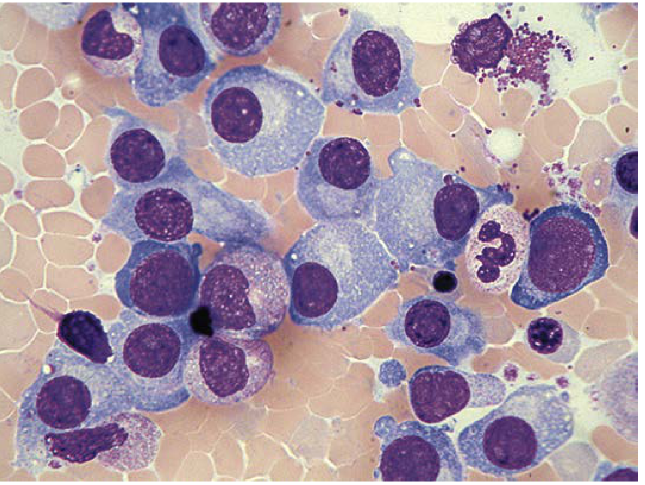
3. Morphology of Myeloma Cells
Normal vs. Myeloma plasma cell features:
| Feature | Normal Plasma Cell | Myeloma Cell |
|---|---|---|
| Nucleus | Eccentric, "clock-face/cartwheel" chromatin | Abnormal chromatin, less clumping, large nucleoli |
| Cytoplasm | Deeply basophilic, perinuclear clear zone (Golgi) | Lighter blue, perinuclear zone may be lost |
| Nucleoli | Absent/inconspicuous | Prominent |
| Binucleate/multinucleate | Rare | Common in myeloma |
4. Special Myeloma Cell Variants (high-yield for exams)
| Cell Type | Description | Significance |
|---|---|---|
| Flame cell | Cytoplasm has irregular, fiery red edges | IgA myeloma (characteristic) |
| Mott cell (Morula cell) | Cytoplasm packed with Russell bodies (grape-like) | Accumulation of immunoglobulin |
| Russell body | Pale, eosinophilic immunoglobulin inclusions in cytoplasm | Immunoglobulin accumulation |
| Dutcher body | Intranuclear PAS+ inclusion | Intranuclear Ig accumulation |
| Plasmablasts | Immature plasma cells with prominent nucleoli | Aggressive/advanced disease |
Here is an actual bone marrow aspirate from a myeloma case showing large plasma cells with prominent nucleoli:
5. Immunophenotype of Myeloma Cells
- CD38 + (strongly positive - key marker)
- CD138 + (syndecan-1 - most specific marker for plasma cells)
- CD19 - (lost, unlike normal plasma cells)
- CD20 -
- CD56 + (abnormally acquired - helps distinguish from reactive plasmacytosis)
- Monoclonal κ or λ light chain restriction (proves clonality)
C. OTHER LABORATORY FINDINGS (to complete the picture)
Serum Studies
| Test | Finding |
|---|---|
| Serum protein electrophoresis (SPEP) | M-spike (M-band/M-protein) - homogeneous band in γ or β region |
| Immunoglobulin type | IgG >50%, IgA ~20%, IgD <1%, IgE rare |
| Total protein | Raised |
| Albumin | Low (poor prognostic sign) |
| Calcium | Elevated (hypercalcemia) |
| Uric acid | Elevated |
| LDH | Elevated (marker of disease activity) |
| β2-microglobulin | Elevated (important staging marker) |
| Creatinine/Urea | Elevated (renal insufficiency) |
Urine Studies
- Bence Jones Protein - free monoclonal light chains (κ or λ) detected in urine
- Positive in ~25% of cases (light chain only myeloma)
- Method: heat test - precipitates at 40-60°C, redissolves at 100°C
- Causes myeloma kidney (cast nephropathy) → renal failure
Radiology
- Punched-out (lytic) bone lesions - skull X-ray is classic ("pepper pot skull")
- Pathological fractures
- Diffuse osteoporosis
D. DIAGNOSTIC CRITERIA SUMMARY
| Entity | M-protein | Bone Marrow Plasma Cells | Symptoms |
|---|---|---|---|
| MGUS | <30 g/L | <10% | None (no CRAB) |
| Smoldering Myeloma | >30 g/L | ≥10% | None (no CRAB) |
| Symptomatic Myeloma | Any level | ≥10% clonal | CRAB present |
EXAM DIAGRAM TO DRAW
PERIPHERAL BLOOD SMEAR - Multiple Myeloma:

[RBC][RBC][RBC][RBC][RBC]  ← Rouleaux formation
[RBC][RBC][RBC][RBC][RBC]    (coin-stack arrangement)

Also shows: Normochromic, normocytic RBCs
            ↑ ESR (markedly)
            Occasional plasma cell (Plasma Cell Leukemia if >20%)

BONE MARROW ASPIRATE - Multiple Myeloma:

  [PC] [PC] [PC]      PC = Plasma cell with:
  [PC] [PC] [PC]       - Eccentric nucleus
  [PC] [PC] [PC]       - Prominent nucleoli
                        - Abundant blue cytoplasm
  Special variants:     - Perinuclear hof (clear zone)
  [Flame cell] → fiery red cytoplasm (IgA)
  [Mott/Morula cell] → Russell bodies (grape-like inclusions)
Key exam point: The combination of rouleaux on peripheral smear + plasma cells in bone marrow + M-spike on SPEP + Bence Jones protein in urine + lytic bone lesions = Multiple Myeloma. - Henry's Clinical Diagnosis and Management by Laboratory Methods
Only peripheral blood not smear broo
Got it! You want only the Peripheral Blood findings (CBC, counts, lab values) - not the peripheral blood smear morphology. Here's the focused answer:
Multiple Myeloma - Peripheral Blood Findings
1. Hemoglobin / Red Blood Cells
- Normochromic, normocytic anemia - present in majority of patients
- Hb is decreased (most common presenting lab finding)
- Normoblasts (nucleated RBCs) may appear
2. White Blood Cells
- WBC count is slightly decreased, normal, or slightly increased
- Occasional mild left shift (up to myeloblast level) may be seen
- Leukopenia develops in advanced disease (marrow replacement)
3. Platelets
- Normal in early disease
- Thrombocytopenia in advanced disease due to marrow infiltration by plasma cells
4. ESR
- Markedly elevated - one of the most striking findings
- Due to raised serum globulins (M-protein) causing rouleaux and increased plasma viscosity
5. Serum Calcium
- Elevated (hypercalcemia) - due to osteoclast activation and bone destruction
6. Serum Proteins / Globulins
- Total serum protein - elevated
- Serum globulin - markedly increased (M-protein/paraprotein)
- Albumin - decreased (poor prognostic marker)
- M-spike on serum protein electrophoresis (SPEP) - IgG >50%, IgA ~20%
7. Renal Function Tests
- Serum creatinine and urea - elevated (renal insufficiency)
- Caused by myeloma kidney (cast nephropathy from Bence Jones proteins)
8. Uric Acid
- Elevated (due to increased cell turnover)
9. LDH
- Elevated (marker of disease burden and activity)
10. β2-Microglobulin
- Elevated - most important prognostic marker in myeloma
- Used in the ISS (International Staging System) for staging
11. Serum Viscosity
- Increased - due to high M-protein levels
- Can cause hyperviscosity syndrome (headache, visual disturbances, bleeding)
12. Plasma Cells in Blood
- Normally absent
- If plasma cells >20% of WBCs or >2 × 10⁹/L → Plasma Cell Leukemia (severe variant)
Quick Summary Table
| Parameter | Finding in Myeloma |
|---|---|
| Hemoglobin | ↓ (normochromic normocytic anemia) |
| WBC | Normal / slightly ↓ or ↑ |
| Platelets | Normal early → ↓ late |
| ESR | Markedly ↑ |
| Serum calcium | ↑ (hypercalcemia) |
| Serum globulin | Markedly ↑ (M-protein) |
| Albumin | ↓ |
| Creatinine/Urea | ↑ (renal failure) |
| Uric acid | ↑ |
| LDH | ↑ |
| β2-microglobulin | ↑ (staging) |
| Serum viscosity | ↑ |
"There is usually normochromic, normocytic anemia. Serum globulin is usually increased, often strikingly so - responsible for elevated ESR. The monoclonal protein is IgG in over 50% of cases, IgA in about 20%." - Henry's Clinical Diagnosis and Management by Laboratory Methods
This is a shared conversation. Sign in to Orris to start your own chat.