
replication of virus
Reading File
Virus replication is the process by which a virus hijacks a host cell's machinery to produce copies of itself. The general steps are: attachment to host cell receptors, entry/penetration, uncoating, replication of the viral genome, protein synthesis, assembly of new virions, and release (often by budding or cell lysis).
Searching sources...
Finding Sources
Searching the Web
virus replication cycle steps mechanism
Reading File
Reading File
Loading Image
Reading File
Reading File
Reading File
Here is a thorough breakdown of virus replication, drawn from Sherris & Ryan's Medical Microbiology (8th ed.).
Virus Replication Cycle
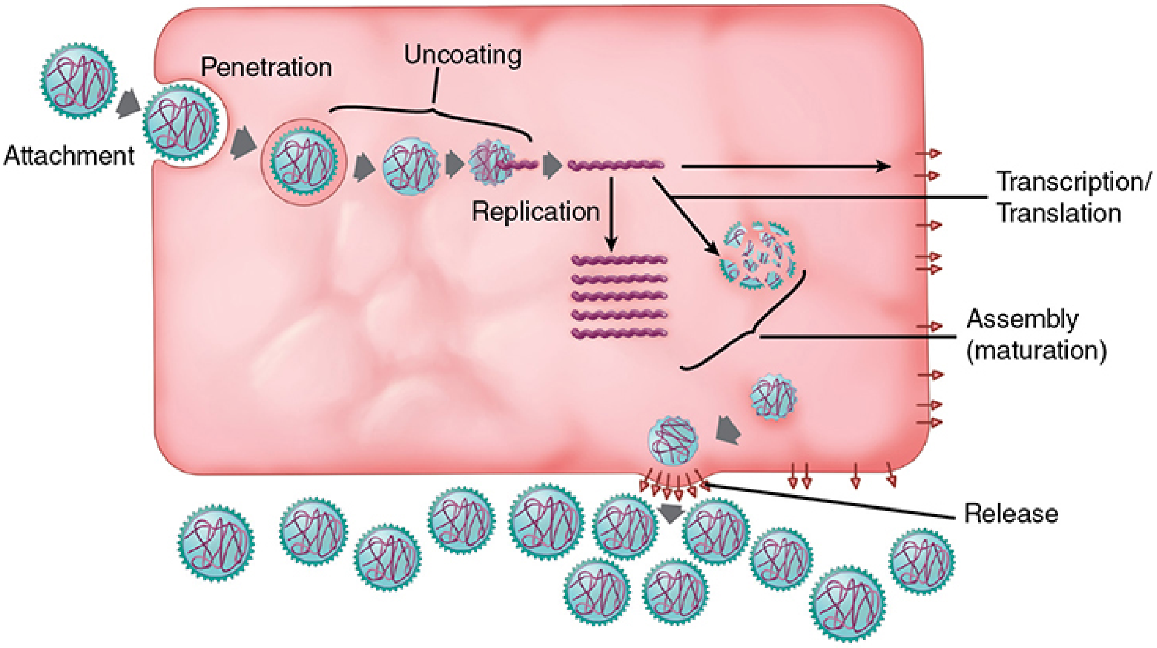
The replication cycle consists of six discrete phases:
1. Attachment (Adsorption)
Viruses have no capacity for locomotion; collision with a host cell is purely a random diffusion event. The rate of adsorption depends on the concentrations of virions and cells.
- Adsorption is a highly specific interaction between virion attachment proteins (spikes) on the virion surface and receptor molecules (usually glycoproteins) on the host cell surface.
- A typical host cell has 10^4 to 10^5 such receptors.
- Viruses have evolved to exploit a wide variety of normally functioning surface molecules - signaling proteins, immune receptors, enzymes, etc.
Examples of virus-receptor pairs:
| Virus | Receptor |
|---|---|
| SARS-CoV-2 (COVID-19) | ACE-2 |
| HIV | CD4 + CCR5/CXCR4 |
| Epstein-Barr virus | CR2 (CD21) |
| Adenoviruses | Integrins |
| MERS-CoV | Dipeptidyl peptidase 4 |
| Dengue virus | Heparin sulfate |
2. Penetration (Entry)
After attachment, the virus must enter the cell. Two main mechanisms:
a) Direct membrane fusion - The viral envelope fuses directly with the host plasma membrane (e.g., paramyxoviruses, herpesviruses, HIV). The nucleocapsid is released directly into the cytoplasm.
b) Viropexis (receptor-mediated endocytosis) - The virion is engulfed into an endosomal vesicle. The low pH of the endosome triggers a conformational change in viral spike proteins, causing membrane fusion and release of the nucleocapsid into the cytoplasm. This applies to influenza viruses, coronaviruses, rabies, and rubella viruses, as well as naked capsid viruses like poliovirus and adenovirus.
3. Uncoating
Once inside the cell, the viral capsid is removed to liberate the viral genome. This may happen in the endosome or cytoplasm, depending on the virus.
4. Synthetic Phase (Genome Replication + Protein Synthesis)
This is the most variable step and differs fundamentally between DNA viruses and RNA viruses.
DNA Viruses
- Most replicate in the nucleus using host DNA polymerases.
- Linear genomes must solve the "end problem" (inability to fully replicate chromosome ends) through various strategies:
- Phage λ: cohesive (sticky) ends that circularize the genome
- Adenovirus: protein molecules covalently attached to 5' ends serve as primers
- Parvoviruses: hairpin ends for self-priming
- Poxviruses: continuous (closed) ends
RNA Viruses
- Replicate in the cytoplasm (cells lack RNA-dependent RNA polymerases).
- Must encode their own RNA-dependent RNA polymerase (RdRP) for both transcription and replication.
- Positive-sense (+ssRNA): Genome directly serves as mRNA for immediate protein synthesis (e.g., picornaviruses, coronaviruses).
- Negative-sense (-ssRNA): Genome must first be transcribed into +ssRNA by the virion-packaged RdRP before translation can begin (e.g., influenza, rabies, Ebola).
- Double-stranded RNA (dsRNA): Transcribed within sub-viral particles (e.g., reoviruses).
- Retroviruses (HIV): +ssRNA genome is first reverse-transcribed into DNA by reverse transcriptase, integrated into the host genome as a provirus, then transcribed by host RNA polymerase.
Transcription and replication must be temporally separated in most RNA viruses so the same RNA template is not used simultaneously for both mRNA production and genome duplication.
5. Assembly
Newly synthesized viral proteins and replicated genomes are packaged together to form new virions (progeny virus). Post-translational modifications of proteins may occur at this stage. Assembly takes place in the cytoplasm, nucleus, or at the plasma membrane depending on the virus.
6. Release
Naked (non-enveloped) viruses:
- Released by cell lysis, which is often triggered by apoptosis. Viruses like picornaviruses shut off host protein synthesis early in infection; the resulting cell death lyses the cell and frees hundreds to thousands of new virions.
- Some viruses encode anti-apoptotic proteins to delay cell death until replication is complete.
Enveloped viruses - Budding:
- Viruses acquire their lipid envelope by budding through a cellular membrane.
- Viral glycoprotein spikes are inserted into the host cell membrane (via the Golgi pathway).
- A viral matrix (M) protein coats the inner face of the membrane at the budding site.
- The nucleocapsid is attracted to the M protein, triggering envelopment and pinching off of a new virion.
- Budding sites: plasma membrane (most enveloped viruses), nuclear membrane (herpesviruses), or Golgi/cytoplasmic membranes (coronaviruses, poxviruses).
- Budding does not immediately kill the cell, but progressive loss of plasma membrane integrity eventually leads to cell death.
Outcomes of Infection
| Outcome | Description |
|---|---|
| Productive (lytic) | New virions are made; host cell dies |
| Persistent (latent) | Viral genome maintained in host cells indefinitely without active replication (e.g., herpesviruses in neurons) |
| Chronic infection | Low-level ongoing virus production with minimal cell damage |
| Abortive infection | Virus enters a non-permissive cell; limited viral protein expression, no new virions, cell may die |
| Oncogenic transformation | Non-productive infection where viral DNA integrates and drives uncontrolled cell division |
Source: Sherris & Ryan's Medical Microbiology, 8th Edition, pp. 198-231
This is a shared conversation. Sign in to Orris to start your own chat.