Boil near vaginal opening

Reading File
Finding Sources
Finding Sources
Searching Images

Bartholin gland abscess vulva

**Modality:** Ultrasonography (Grayscale)
**Anatomical Region:** Vulva / Perineal soft tissue

**Description:**
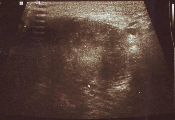
The image displays a grayscale ultrasound of the vulvar region. Centered within the field of view is a well-circumscribed, predominantly hypoechoic mass measuring approximately 2.9 × 2.5 × 2.1 cm. The internal echo texture of the lesion is heterogeneous, containing subtle internal echoes and areas of low-level echogenicity. 

**Characteristic Visual Features:**
*   **Margination:** The borders of the mass appear relatively distinct from the surrounding subcutaneous tissue.
*   **Echogenicity:** The lesion is hypoechoic compared to the adjacent fibroadipose tissue.
*   **Acoustic Features:** There is evidence of posterior acoustic enhancement, suggesting a fluid-filled or highly cellular cystic nature, often associated with lesions such as a Bartholin’s gland cyst or abscess.
*   **Artifacts:** Significant posterior acoustic shadowing is absent; however, lateral edge shadowing is visible at the margins of the mass.

**Clinical Significance:**
Findings are consistent with a localized vulvar mass or cyst. The appearance requires differentiation between inflammatory processes (e.g., abscess), benign cystic lesions (e.g., Bartholin's cyst), or solid soft tissue neoplasms.
**Modality:** Ultrasonography (Grayscale) **Anatomical Region:** Vulva / Perineal soft tissue **Description:** The image displays a grayscale ultrasound of the vulvar region. Centered within the field of view is a well-circumscribed, predominantly hypoechoic mass measuring approximately 2.9 × 2.5 × 2.1 cm. The internal echo texture of the lesion is heterogeneous, containing subtle internal echoes and areas of low-level echogenicity. **Characteristic Visual Features:** * **Margination:** The borders of the mass appear relatively distinct from the surrounding subcutaneous tissue. * **Echogenicity:** The lesion is hypoechoic compared to the adjacent fibroadipose tissue. * **Acoustic Features:** There is evidence of posterior acoustic enhancement, suggesting a fluid-filled or highly cellular cystic nature, often associated with lesions such as a Bartholin’s gland cyst or abscess. * **Artifacts:** Significant posterior acoustic shadowing is absent; however, lateral edge shadowing is visible at the margins of the mass. **Clinical Significance:** Findings are consistent with a localized vulvar mass or cyst. The appearance requires differentiation between inflammatory processes (e.g., abscess), benign cystic lesions (e.g., Bartholin's cyst), or solid soft tissue neoplasms.

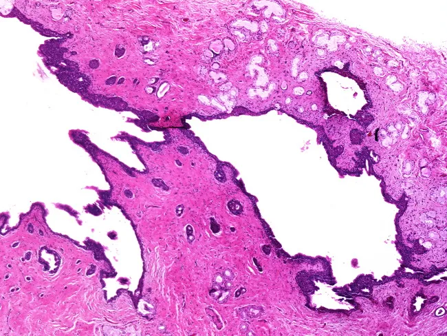
This histopathology slide depicts a Bartholin gland cyst, a common vulvar lesion resulting from cystic dilation of the Bartholin (greater vestibular) gland or its duct due to ductal obstruction, often in the setting of prior infection. In this Hematoxylin and Eosin stained preparation, low-power examination reveals a dilated duct-like structure with an anechoic to mucinous luminal content, lined by a simple to transitional-type epithelium. The cyst wall contains scattered mucous glands embedded in fibrous stroma, reflecting residual glandular architecture. The surrounding vulvar stroma displays mild chronic inflammatory infiltrate and fibrosis, compatible with retention of secretions rather than suppurative abscess. The glands are located in the posterior-lateral vestibule behind the labia minora, and drainage occurs into the postero-lateral vestibular recess. Pathology confirms a benign lesion with no dysplasia or malignancy detected in this field. Clinically, Bartholin cysts present as a unilateral vulvar mass, often fluctuating in size, and can become painful if infected (Bartholin gland abscess). This histology underscores the importance of recognizing ductal obstruction with mucus retention and glandular mucin production. Therapeutically, management may include marsupialization or gland excision depending on size, recurrence, and suspicion of neoplasm. In educational and diagnostic contexts, this image aids training in vulvar histopathology, differential with abscess or neoplasm, and correlates with clinical vulvar swelling.
This histopathology slide depicts a Bartholin gland cyst, a common vulvar lesion resulting from cystic dilation of the Bartholin (greater vestibular) gland or its duct due to ductal obstruction, often in the setting of prior infection. In this Hematoxylin and Eosin stained preparation, low-power examination reveals a dilated duct-like structure with an anechoic to mucinous luminal content, lined by a simple to transitional-type epithelium. The cyst wall contains scattered mucous glands embedded in fibrous stroma, reflecting residual glandular architecture. The surrounding vulvar stroma displays mild chronic inflammatory infiltrate and fibrosis, compatible with retention of secretions rather than suppurative abscess. The glands are located in the posterior-lateral vestibule behind the labia minora, and drainage occurs into the postero-lateral vestibular recess. Pathology confirms a benign lesion with no dysplasia or malignancy detected in this field. Clinically, Bartholin cysts present as a unilateral vulvar mass, often fluctuating in size, and can become painful if infected (Bartholin gland abscess). This histology underscores the importance of recognizing ductal obstruction with mucus retention and glandular mucin production. Therapeutically, management may include marsupialization or gland excision depending on size, recurrence, and suspicion of neoplasm. In educational and diagnostic contexts, this image aids training in vulvar histopathology, differential with abscess or neoplasm, and correlates with clinical vulvar swelling.

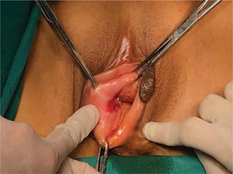
This clinical photograph displays a surgical examination of the vulva, identifying two distinct adjacent lesions classified as a collision tumor. The first lesion is located on the posterior aspect of the right labium majora, characterized by a 2.0 cm x 1.5 cm ulcerated area with significant anabrosis (surface erosion) and focal erythema, representing a poorly differentiated squamous cell carcinoma. Immediately adjacent is a second, larger mass associated with the Bartholin gland, appearing as a 3.0 cm x 2.0 cm well-circumscribed, solid, dark brown to black nodule with a smooth, expansile margin. This second component was histopathologically confirmed as an adenosquamous carcinoma. Surgical forceps and gloved hands are shown retracting the labial tissue to expose the lesions during the preoperative or intraoperative assessment. The visual highlights the morphological differences between primary vulvar squamous cell carcinoma (ulcerative) and Bartholin gland malignancies (solid, circumscribed mass), illustrating a rare clinical presentation associated with long-term high-risk HPV-16 and HPV-59 infection.

This clinical photograph displays a surgical examination of the vulva, identifying two distinct adjacent lesions classified as a collision tumor. The first lesion is located on the posterior aspect of the right labium majora, characterized by a 2.0 cm x 1.5 cm ulcerated area with significant anabrosis (surface erosion) and focal erythema, representing a poorly differentiated squamous cell carcinoma. Immediately adjacent is a second, larger mass associated with the Bartholin gland, appearing as a 3.0 cm x 2.0 cm well-circumscribed, solid, dark brown to black nodule with a smooth, expansile margin. This second component was histopathologically confirmed as an adenosquamous carcinoma. Surgical forceps and gloved hands are shown retracting the labial tissue to expose the lesions during the preoperative or intraoperative assessment. The visual highlights the morphological differences between primary vulvar squamous cell carcinoma (ulcerative) and Bartholin gland malignancies (solid, circumscribed mass), illustrating a rare clinical presentation associated with long-term high-risk HPV-16 and HPV-59 infection.

Reading File
Reading File
Loading Image
Reading File
Reading File
Here is a thorough clinical overview of a boil (abscess) near the vaginal opening:

Boil Near the Vaginal Opening - Most Likely a Bartholin's Gland Abscess

What Is It?
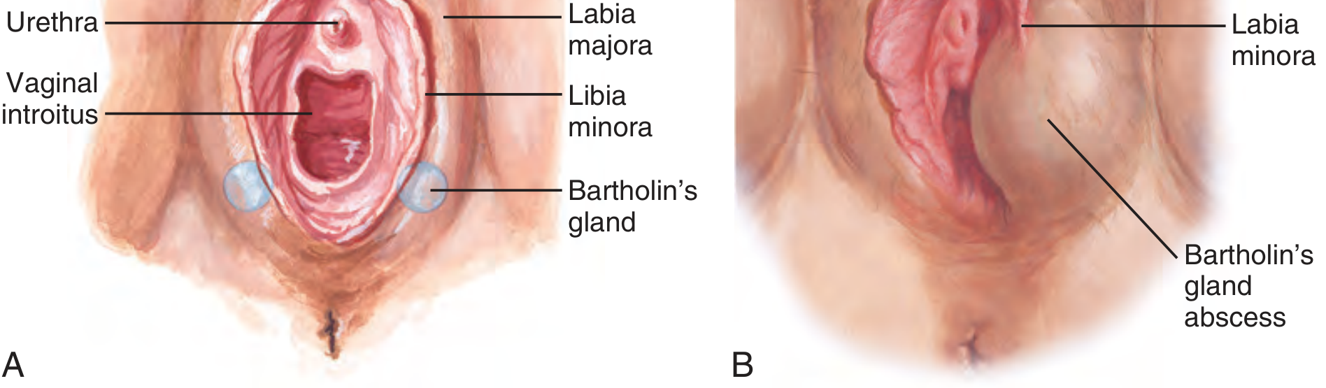
A boil near the vaginal opening in women of reproductive age is most commonly a Bartholin's gland abscess (BA). The Bartholin's glands (also called greater vestibular glands) sit at the 5 o'clock and 7 o'clock positions at the posterior vaginal introitus, and their ducts open onto the mucosa just inside the labia minora.
Anatomy of Bartholin's gland and abscess location
Left: Normal anatomy with Bartholin's glands at the lower vaginal introitus. Right: Bartholin's gland abscess as a fluctuant swelling with the labia minora transecting it.

How It Forms

  1. The Bartholin's duct becomes blocked or partially obstructed, causing secretions to accumulate.
  2. This leads to a Bartholin's duct cyst (non-infected, painless swelling).
  3. The cyst then gets secondarily infected, forming a painful abscess.
Bartholin's gland abscesses occur in about 2% of women, most commonly in reproductive-age women.
  • Roberts and Hedges' Clinical Procedures in Emergency Medicine

Bacteria Involved

The infection is usually polymicrobial - a mix of aerobic and anaerobic vaginal flora. Organisms include:
  • Vaginal anaerobes and aerobes (most common)
  • Chlamydia trachomatis or Neisseria gonorrhoeae - isolated in approximately 10% of cases
  • Staphylococcus aureus including CA-MRSA (increasingly common)
  • ROSEN's Emergency Medicine, p. 2743

Symptoms

  • Unilateral painful swelling at the posterior vaginal opening (4-5 cm is typical, can be larger)
  • Redness, warmth, and tenderness at the labial/vulvar base
  • Pain on walking, sitting, or sexual intercourse
  • Fluctuant (fluid-filled) mass when palpated
  • Rarely fever or systemic illness (unless Bartholin's abscess is very large or patient is immunocompromised)

Diagnosis

Diagnosis is clinical - by history and examination. Bedside ultrasound can help confirm if the swelling is an abscess (hypoechoic fluid collection with posterior acoustic enhancement) vs. cellulitis. An ultrasound image of a Bartholin's cyst/abscess looks like this:
Ultrasound of Bartholin's abscess

Treatment

1. Incision and Drainage (I&D) with Word Catheter - this is the standard first-line treatment in the ED.
Step-by-step:
  • Patient in lithotomy position (feet in stirrups)
  • Premedicate with IV opioids or procedural sedation as needed
  • Local anesthesia via the mucosal surface (NOT the outer skin)
  • Make a small incision with a No. 11 scalpel on the mucosal (inner) surface of the labia minora at the site of maximum fluctuance
  • Insert a Word catheter and inflate the balloon with 3-4 mL of saline
  • Leave the catheter in place for 2-4 weeks to allow a fistula tract to form - this reduces recurrence
2. Antibiotics: Antibiotics are not routinely needed after successful drainage in otherwise healthy patients. However, if there is surrounding cellulitis, signs of systemic infection, or MRSA is suspected, antibiotics are used (trimethoprim-sulfamethoxazole or clindamycin for MRSA coverage).
3. Marsupialization: If the abscess recurs, a gynecologist may perform marsupialization - an elliptical incision is made, the cyst wall is opened and sutured to the mucosa to create a permanent opening, significantly reducing recurrence.
4. For small/early abscesses: Warm sitz baths (soaking the area in warm water 3-4 times/day) can provide symptomatic relief and may help a small abscess drain on its own.
  • Roberts and Hedges' Clinical Procedures in Emergency Medicine, p. 1378-1380
  • Sabiston Textbook of Surgery, p. 2790

When to Seek Urgent/Emergency Care

Go to an emergency department or urgent care promptly if you have:
  • Rapidly enlarging or very painful swelling
  • Fever, chills, or feeling unwell
  • Difficulty walking or sitting due to pain
  • Significant redness spreading onto the inner thigh or surrounding skin
  • Diabetes, HIV, or a weakened immune system
Note: A small boil that is not near the Bartholin's gland area (e.g., on the outer labia or pubic area) could be a folliculitis (infected hair follicle) or furuncle (deep skin boil) - these are treated similarly with warm compresses, and I&D if fluctuant. Very rarely, a painful vulvar swelling in an older woman should be evaluated to rule out Bartholin's gland carcinoma, especially if there is no prior history of cysts/abscesses or if the mass is solid.

Bottom line: A boil near the vaginal opening is most often a Bartholin's gland abscess. It needs incision and drainage (Word catheter placement) in a clinic or ED - warm compresses can help in the very early stages but will not resolve a true abscess. See a doctor promptly for proper treatment.
This is a shared conversation. Sign in to Orris to start your own chat.