
Morrison pouch
Reading File
Morrison's pouch (hepatorenal recess) is the most gravity-dependent peritoneal space in the supine position, located between the liver and the right kidney. It is a key site for free fluid accumulation in trauma and peritoneal pathology.
Searching sources...
Finding Sources
Searching the Web
Morrison's pouch anatomy clinical significance FAST exam
Reading File
Reading File
Reading File
Loading Image
Reading File
Morrison's Pouch (Hepatorenal Recess)
Definition and Anatomy
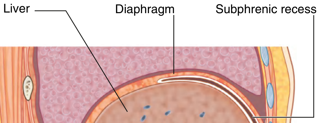
Morrison's pouch, formally called the hepatorenal recess (or hepatorenal pouch), is a peritoneal space on the right side of the abdomen between the inferior surface of the right lobe of the liver and the anterior surface of the right kidney and right suprarenal gland.
Boundaries
| Boundary | Structure |
|---|---|
| Superior | Inferior layer of the coronary ligament |
| Anterior/Superior | Inferior surface of the right lobe of the liver |
| Posterior | Anterior surface of the upper pole of the right kidney + right suprarenal gland |
| Lateral | Parietal peritoneum of the right lateral abdominal wall |
| Inferomedial | Hepatic flexure of the colon, transverse mesocolon, and 2nd part of the duodenum |
The subphrenic and hepatorenal recesses are continuous anteriorly, meaning fluid can track from one to the other.
(Gray's Anatomy for Students)
Why It Matters Clinically: Most Gravity-Dependent Space
Morrison's pouch is the most gravity-dependent peritoneal space in the supramesocolic compartment when a patient is supine. This is why free fluid accumulates here first in:
- Hemoperitoneum (trauma, ruptured ectopic, ruptured aneurysm)
- Bile leak / bowel perforation (peritonitis)
- Ascites - detected earliest here even in small amounts
- Intra-abdominal abscess - a common site after bowel surgery/appendicitis
The FAST Exam
Morrison's pouch is the right upper quadrant (RUQ) view of the FAST (Focused Assessment with Sonography in Trauma) examination. The four views of FAST are:
- RUQ - Morrison's pouch (hepatorenal recess)
- LUQ - Splenorenal recess (Koller's pouch)
- Suprapubic - Pelvis (rectovesical pouch / pouch of Douglas)
- Subxiphoid - Pericardial sac
Probe technique: Place a curvilinear probe in the posterior to mid-axillary line between ribs 8-11. On ultrasound, a normal Morrison's pouch shows the liver and kidney tightly juxtaposed with a hyperechoic line (the peritoneal interface). Free fluid appears as a hypoechoic (dark) stripe between the liver and kidney.
(Fischer's Mastery of Surgery, 8th ed.)
Intra-abdominal Abscess
The hepatorenal recess and the pelvis (recto-uterine pouch/pouch of Douglas in women) are the two most common sites for post-operative intra-abdominal abscess formation. When a supine patient has peritoneal contamination (e.g., perforated appendix), fluid and pus gravitate to this space.
A classic teaching scenario: a patient develops a right shoulder tip pain after abdominal surgery - this is referred pain from the diaphragm, as pus from the hepatorenal recess tracks superiorly to become subphrenic. The phrenic nerve (C3-C5) innervates the diaphragm; irritation refers pain to the C4 dermatome at the shoulder.
(Gray's Anatomy for Students - clinical case)
Surgical Relevance
- The right paracolic gutter communicates freely with Morrison's pouch, so blood or pus from the pelvis can track up to this space
- It is bounded superiorly by the coronary ligament, which limits upward spread; fluid then accumulates here before overflowing into the subphrenic space
- When draining a hepatorenal abscess, a subcostal (not intercostal/transpleural) route is preferred to avoid creating an empyema by passing through the pleural cavity
(Fischer's Mastery of Surgery, 8th ed.; Gray's Anatomy for Students)
This is a shared conversation. Sign in to Orris to start your own chat.