
There are 3 type of jaundice how to differentiate between all those and what are the symptoms and complains a patient have in this and give treatment too create an easy image u can create a chart too to make it remember and retain me for longer time
types of jaundice pre-hepatic hepatic post-hepatic differentiation chart
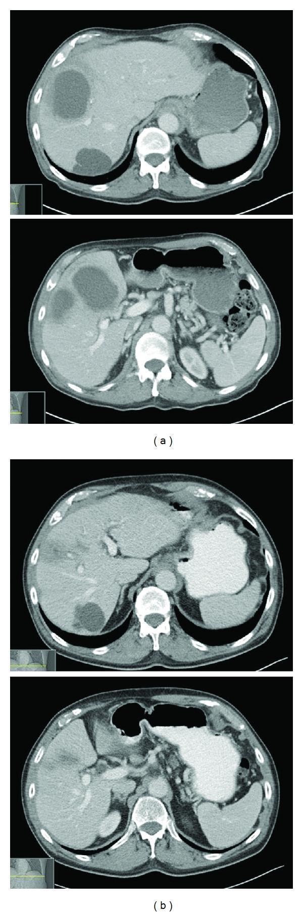
This Comparison Chart displays axial contrast-enhanced abdominal Computed Tomography (CT) scans highlighting the progression of hepatic pathology. Panel (a) illustrates pre-treatment diagnostic imaging showing multiple large, thick-walled, low-density lesions primarily localized in the right hepatic lobe. These findings are characteristic of pyogenic liver abscesses, with the largest lesion demonstrating peripheral rim enhancement and internal hypodensity. In contrast, panel (b) shows post-treatment imaging following a 6-week course of antibiotics, revealing significant radiographic resolution. The previously seen abscesses have markedly decreased in size and attenuation, with the liver parenchyma returning to a more homogeneous appearance. Notably, a simple benign liver cyst is visible in the posterior segment of the liver, which remains unchanged in size and morphology between both time points, serving as a stable anatomical landmark. The image demonstrates key concepts in hepatobiliary radiology, including the differentiation between infected fluid collections and simple cysts, as well as the radiological monitoring of treatment response in infectious diseases.
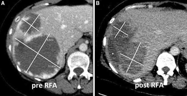
This diagnostic comparison chart consists of two side-by-side axial contrast-enhanced CT scans of the upper abdomen, labeled 'pre RFA' (A) and 'post RFA' (B), demonstrating the treatment of hepatic giant cavernous hemangiomas (GCHs). Image A shows two large, 'mating' hypodense masses in liver segments VII and VIII. The larger, inferior mass measures 10.0 x 8.8 cm, and the superior mass measures 5.7 x 5.2 cm. Both lesions exhibit characteristic peripheral nodular enhancement and heterogeneous internal consistency. Image B, taken after bipolar radiofrequency ablation (RFA), shows a significant reduction in tumor volume. The larger lesion has decreased to 6.5 x 4.6 cm, and the smaller to 3.7 x 3.2 cm. Post-treatment changes include increased internal density, slightly more irregular margins, and an asterisk marking a fluid-filled space formed by tumor retraction. The scans illustrate the efficacy of bipolar RFA in achieving tumor shrinkage and debulking for symptomatic benign liver lesions when surgical resection is high-risk.
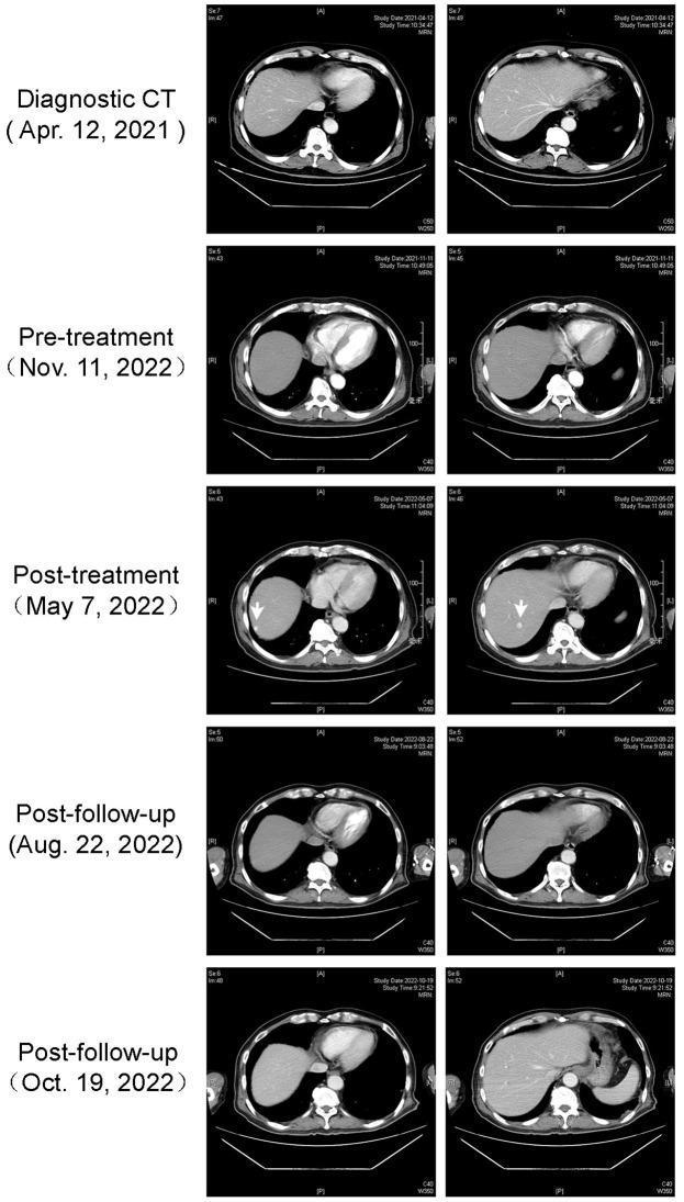
This Comparison Chart displays a series of axial contrast-enhanced CT scans of the upper abdomen over a longitudinal timeline, illustrating the progression and regression of hepatic lesions. The panels are organized into five temporal stages: 'Diagnostic CT' (Apr 2021), 'Pre-treatment' (Nov 2022), 'Post-treatment' (May 2022), and two 'Post-follow-up' sessions (Aug 2022 and Oct 2022). The 'Post-treatment' scans demonstrate two distinct, well-circumscribed, low-density (hypodense) lesions in the right liver segment S6 (indicated by white arrows), consistent with hepatic cavernous hemangiomas. Subsequent 'Post-follow-up' imaging shows a progressive reduction in the size and density of these lesions, with the final Oct 2022 scans indicating significant regression and near-disappearance of the previously visible nodules. This radiological series highlights the dynamic changes in liver pathology during and after clinical intervention, focusing on lesion morphology, margin definition, and size within the hepatic parenchyma.
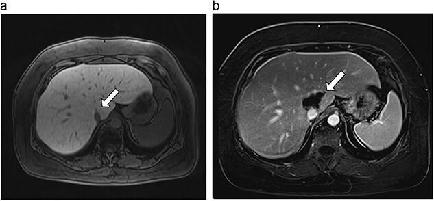
This Comparison Chart features two side-by-side axial MRI slices of the upper abdomen, illustrating a pre- and post-treatment scenario for a hepatic tumor. Image (a) is a pre-ablation MRI showing a 6 mm hypointense lesion located within the caudate lobe of the liver, indicated by a white arrow. The lesion is discrete and located near major vascular structures. Image (b) shows a post-ablation MRI taken 18 days after microwave ablation. The white arrow points to a significantly larger, 40.4 mm ablation site in the caudate lobe, characterized by altered signal intensity reflecting necrotic tissue change. Both images demonstrate the liver's anatomical relationship with the spleen (visible in the right side of the frame) and the abdominal vasculature. The educational focus is on the radiological assessment of microwave ablation success and the expected morphological changes following the procedure in the caudate lobe. This content is relevant for radiology and hepatobiliary surgical oncology training.
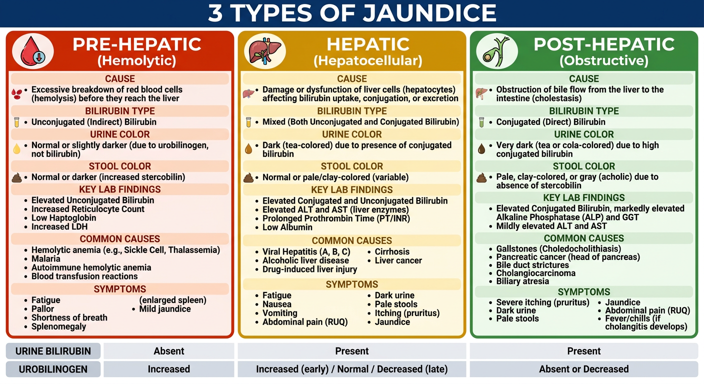
A clean, colorful medical comparison chart titled "3 Types of Jaundice" divided into 3 vertical columns with distinct colors: LEFT column (red/orange) = "Pre-Hepatic (Hemolytic)", MIDDLE column (yellow/amber) = "Hepatic (Hepatocellular)", RIGHT column (green) = "Post-Hepatic (Obstructive)". Each column has sections: Cause, Bilirubin Type, Urine Color, Stool Color, Key Lab Findings, Common Causes, Symptoms. Include small icons: blood drop for pre-hepatic, liver for hepatic, bile duct/gallstone for post-hepatic. At the bottom, a summary row showing urine bilirubin (absent/present/present) and urobilinogen (increased/increased/absent). Clean medical education style, easy to read, high contrast text.
3 Types of Jaundice - Complete Guide
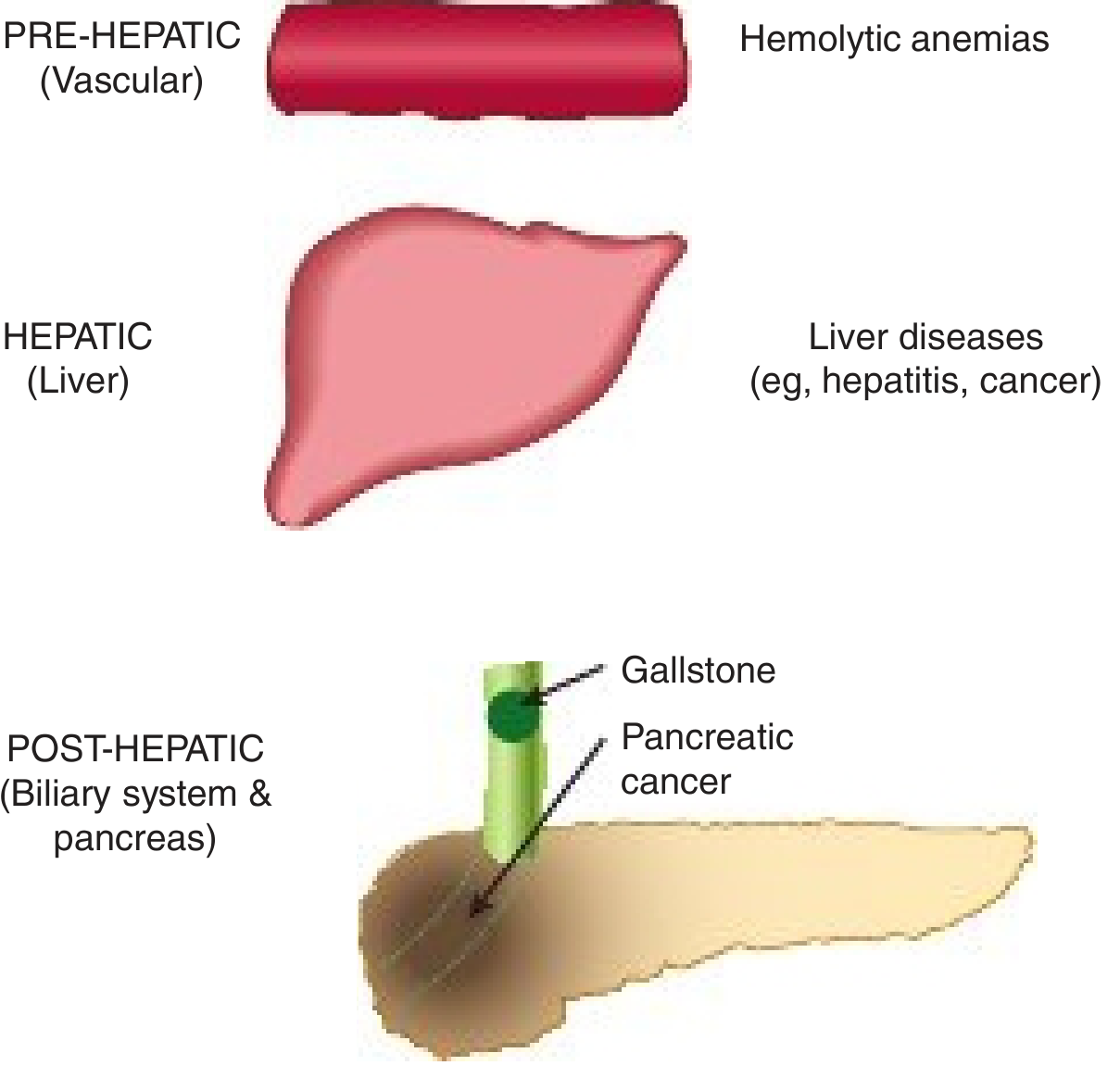
Textbook Diagram of the 3 Types
Your Memory Chart
TYPE 1 - Pre-Hepatic (Hemolytic) Jaundice
Causes
- Sickle cell anemia
- G6PD deficiency
- Pyruvate kinase deficiency
- Thalassemia
- Autoimmune hemolytic anemia
- Malaria
- Transfusion reactions
Symptoms & Patient Complaints
- Mild yellow tinge to skin and eyes (usually mild jaundice)
- Pallor (anaemia)
- Fatigue, weakness, breathlessness (from anaemia)
- Dark urine (increased urobilinogen, not bilirubin)
- Normal-colored stools (no obstruction)
- Splenomegaly (palpable spleen)
- Fever + rigors if infectious (e.g., malaria)
- No itching (pruritus absent)
Key Lab Findings
| Test | Result |
|---|---|
| Serum bilirubin | ↑ Unconjugated (indirect) only |
| Urine bilirubin | Absent (UCB not water-soluble, can't pass into urine) |
| Urine urobilinogen | Increased |
| Stool urobilinogen | Increased (dark stools) |
| ALT/AST | Normal |
| ALP | Normal |
TYPE 2 - Hepatic (Hepatocellular) Jaundice
Causes
- Viral hepatitis (A, B, C)
- Alcoholic liver disease / cirrhosis
- Drug-induced hepatitis (paracetamol overdose, isoniazid)
- Autoimmune hepatitis
- Wilson's disease
- Gilbert's syndrome (mild, benign)
- Crigler-Najjar syndrome
Symptoms & Patient Complaints
- Yellow skin and sclera (moderate-severe)
- Dark urine (tea-colored) - conjugated bilirubin in urine
- Pale/clay-colored stools (reduced stercobilin)
- Pruritus (itching) - from bile salts in skin
- Fatigue, malaise, anorexia (loss of appetite)
- Nausea, vomiting
- Right upper quadrant pain/tenderness (hepatomegaly)
- Fever (in hepatitis)
- In cirrhosis: spider naevi, palmar erythema, ascites, encephalopathy
Key Lab Findings
| Test | Result |
|---|---|
| Serum bilirubin | ↑ Both UCB and CB (direct + indirect) |
| Urine bilirubin | Present (conjugated bilirubin is water-soluble) |
| Urine urobilinogen | Increased (liver can't recycle it) |
| Stool urobilinogen | Decreased |
| ALT/AST | Markedly elevated (hepatocellular damage) |
| ALP | Mildly elevated |
TYPE 3 - Post-Hepatic (Obstructive/Cholestatic) Jaundice
Causes
- Gallstones in common bile duct (choledocholithiasis) - most common
- Cancer of the head of the pancreas
- Cholangiocarcinoma (bile duct cancer)
- Primary sclerosing cholangitis
- Stricture of the bile duct
- Pancreatitis compressing the duct
Symptoms & Patient Complaints
- Deep yellow/greenish-yellow jaundice
- Dark urine (cola-colored) - conjugated bilirubin excreted
- Pale/clay-white stools - no stercobilin reaching gut
- Severe pruritus (itching) - bile salts deposited in skin (most prominent symptom)
- Abdominal pain - right upper quadrant or epigastric (colicky if gallstones, painless if cancer)
- Nausea and vomiting
- Steatorrhoea (fatty, floating stools) - no bile for fat digestion
- Weight loss (especially in malignancy)
- Fever and rigors if secondary cholangitis (Charcot's triad: jaundice + fever + RUQ pain)
- Prolonged bleeding time (vitamin K not absorbed - fat-soluble vitamin)
Key Lab Findings
| Test | Result |
|---|---|
| Serum bilirubin | ↑ Conjugated (direct) only |
| Urine bilirubin | Present |
| Urine urobilinogen | Absent (no bile reaching gut) |
| Stool urobilinogen | Absent/trace (pale stools) |
| ALT/AST | Mildly elevated |
| ALP | Markedly elevated (hallmark) |
| GGT | Elevated |
Master Comparison Table
| Feature | Pre-Hepatic | Hepatic | Post-Hepatic |
|---|---|---|---|
| Bilirubin type raised | Unconjugated (indirect) | Both | Conjugated (direct) |
| Urine bilirubin | Absent | Present | Present |
| Urine urobilinogen | ↑ Increased | ↑ Increased | Absent |
| Stool color | Dark (normal/dark) | Pale | Pale/white |
| Urine color | Dark (urobilinogen) | Dark (bilirubin) | Dark (bilirubin) |
| Pruritus | Absent | Mild | Severe |
| ALT/AST | Normal | ↑↑ Markedly raised | Mildly raised |
| ALP | Normal | Mildly raised | ↑↑ Markedly raised |
| Key clue | Anaemia + splenomegaly | Hepatitis history + tender liver | Pale stools + severe itch |
Treatment Summary
Pre-Hepatic (Hemolytic)
- Treat the underlying cause of hemolysis
- Blood transfusion for severe anaemia
- Folic acid supplementation
- In sickle cell: hydroxyurea, exchange transfusion
- Splenectomy in some hereditary haemolytic anaemias
Hepatic (Hepatocellular)
- Treat the underlying liver disease (antiviral therapy for hepatitis B/C)
- Alcohol cessation
- Stop offending drugs (drug-induced)
- Supportive care: rest, nutrition, IV fluids
- N-acetylcysteine for paracetamol overdose
- Ursodeoxycholic acid in cholestasis
- Liver transplant for end-stage disease
Post-Hepatic (Obstructive)
- Remove the obstruction - this is primarily surgical/endoscopic
- ERCP (Endoscopic Retrograde Cholangiopancreatography) + stone extraction for gallstones
- Stenting for malignant obstruction
- Surgery: Whipple's procedure for pancreatic head cancer
- Pre-op: IV Vitamin K to correct coagulopathy, prophylactic antibiotics, IV fluids (risk of hepatorenal syndrome)
- Urinary catheter to monitor output (>40 ml/hr target)
Quick Memory Tricks
"Blood - Liver - Drain" = Pre-hepatic, Hepatic, Post-hepatic
Pale stools + severe itch = always Post-hepatic (bile can't reach gut)
Only UCB raised = Pre-hepatic (liver still working fine)
ALP markedly up = obstruction | AST/ALT markedly up = hepatitis
Urine urobilinogen absent = Post-hepatic (nothing reaching gut to form urobilinogen)
Find recent clinical guidelines on management of jaundice
jaundice management clinical guidelines
clinical guidelines management jaundice 2023 2024 2025 EASL AASLD ASGE
obstructive jaundice biliary obstruction management ERCP guidelines
cholestatic liver disease hepatic jaundice treatment guidelines
PMID: 38174913
\"biliary obstruction\"[MeSH Terms] OR \"cholangitis\" guideline management
\"neonatal jaundice\"[MeSH Terms] OR \"hyperbilirubinemia\" newborn guideline
\"hemolytic anemia\"[MeSH Terms] AND management guidelines
PMID: 35927462
PMID: 35738507
PMID: 41232665
PMID: 41400491
PMID: 39183672
PMID: 42191456
Recent Clinical Guidelines on Management of Jaundice (2022-2026)
1. POST-HEPATIC (Obstructive/Cholestatic) Jaundice
EASL Clinical Practice Guidelines on Sclerosing Cholangitis (2022)
- Diagnosis: MRI/MRCP is the primary non-invasive imaging modality; ERCP reserved for therapeutic intent
- Medical therapy: Ursodeoxycholic acid (UDCA) - evidence for biochemical improvement but no proven survival benefit in PSC; recommended in PBC
- Endoscopic management: Dominant strictures should be treated with ERCP dilation + stenting
- Monitoring: Annual liver biochemistry, 6-monthly ultrasound, colonoscopy surveillance for IBD-associated PSC (every 1-2 years)
- Liver transplant: Indicated for end-stage PSC with decompensation or recurrent cholangitis
- ⚠️ Note: An erratum was published (PMID: 37735011)
Spanish National Guidelines: Cholestatic Liver Diseases (2026)
- Incorporates next-generation genetic sequencing for genetic cholestasis diagnosis
- New targeted therapies for specific genetic cholestasis subtypes (PFIC, Alagille syndrome)
- Primary Biliary Cholangitis (PBC): UDCA first-line; obeticholic acid or fibrates (bezafibrate, fenofibrate) as second-line in UDCA non-responders
- Intrahepatic cholestasis of pregnancy (ICP): UDCA is the treatment of choice; early delivery if bile acids >100 µmol/L
- Multidisciplinary approach strongly recommended
EASL-AASLD Delphi Consensus on Primary Biliary Cholangitis (2026)
- Three second-line PBC therapies now conditionally approved: obeticholic acid, bezafibrate, elafibranor
- Recommends integration of real-world evidence and patient-reported outcomes (PROs) into PBC management decisions
- Framework for future drug development and regulatory approval in PBC
Clinical Practice Guidelines for Peroral Cholangioscopy (2026)
- Indications: Indeterminate biliary strictures, papillary cholangiocarcinoma, difficult bile duct stones
- Antibiotic prophylaxis: Recommended before POCS to prevent cholangitis
- Lithotripsy: POCS-guided electrohydraulic or laser lithotripsy for stones resistant to standard ERCP
- Biopsy: POCS-guided biopsy improves diagnostic yield for malignant strictures
2. HEPATIC (Hepatocellular) Jaundice
ACG Clinical Guideline: Alcohol-Associated Liver Disease (2024)
- Severity scoring: MELD score >20 = severe disease (1-month mortality 20-50%)
- Corticosteroids: Prednisolone 40 mg/day for 28 days - only therapy with proven efficacy for severe AH; benefit at 1 month in 50-60% of patients
- Lille Model score at Day 7: If score >0.45, corticosteroids should be stopped (non-response)
- Alcohol cessation: Crucial for long-term outcomes; integrated addiction medicine referral required
- Liver transplant: Now considered in selected severe AH patients unresponsive to steroids with low relapse risk
- Multidisciplinary care: Hepatology + addiction medicine + social work model strongly recommended
AGA Clinical Practice Update: Pregnancy-Related Liver Disease (2024)
- HELLP syndrome / Acute fatty liver of pregnancy (AFLP): Urgent delivery is the definitive treatment
- Intrahepatic cholestasis of pregnancy: UDCA first-line; fetal monitoring required
- Hepatitis B in pregnancy: Antiviral therapy (tenofovir) in third trimester if HBV DNA >200,000 IU/mL to prevent vertical transmission
- Pre-existing liver disease: Multidisciplinary management with hepatology + obstetrics
3. PRE-HEPATIC (Neonatal Hyperbilirubinemia - Special Population)
AAP Clinical Practice Guideline: Neonatal Hyperbilirubinemia (2022, revised)
- Applies to newborns ≥35 weeks gestation
- Risk stratification: Universal pre-discharge bilirubin screening (TcB or TSB) + hour-specific nomogram
- Phototherapy thresholds: Now lower than previous 2004 guidelines - accounts for gestational age, age in hours, and neurotoxicity risk factors
- Escalation thresholds: Exchange transfusion threshold if bilirubin rises beyond defined safety limits
- Outpatient follow-up: All newborns discharged <48 hours must be seen within 2 days
AAP Technical Report: Phototherapy for Neonates (2024)
- Optimal wavelength: Narrow-band blue-to-green LED (460-490 nm; 478 nm optimal) - best matches bilirubin absorption spectrum
- Minimum irradiance: ≥30 µW/cm²/nm for term infants
- Body surface area: Maximize exposure (35-80%)
- Efficacy: With effective phototherapy and no concurrent hemolysis, bilirubin falls within 4-6 hours
- Device variability: Wide variation in commercial devices - institutions should verify irradiance measurement accuracy
Chinese National Guidelines: Neonatal Jaundice in Primary Care (2025)
Summary Table: Which Guideline for Which Type?
| Jaundice Type | Condition | Guideline | Year | PMID |
|---|---|---|---|---|
| Post-Hepatic | Sclerosing cholangitis | EASL CPG | 2022 | 35738507 |
| Post-Hepatic | Cholestatic liver diseases (full spectrum) | AEEH/SEGHNP | 2026 | 41232665 |
| Post-Hepatic | Primary biliary cholangitis | EASL-AASLD Delphi | 2026 | 42191456 |
| Post-Hepatic | Biliary endoscopy (POCS) | Japan Biliary Assoc. | 2026 | 41400491 |
| Hepatic | Alcohol-associated hepatitis (jaundice) | ACG | 2024 | 38174913 |
| Hepatic | Pregnancy-related liver disease | AGA | 2024 | 39140906 |
| Pre-Hepatic/Neonatal | Neonatal hyperbilirubinemia | AAP revised CPG | 2022 | 35927462 |
| Pre-Hepatic/Neonatal | Phototherapy technique | AAP Technical Report | 2024 | 39183672 |
| Pre-Hepatic/Neonatal | Neonatal jaundice primary care | Chinese Med. Assoc. | 2025 | 41401948 |
Key Clinical Takeaways from the Guidelines
- Obstructive jaundice workup - MRCP first (non-invasive), then ERCP only if intervention needed (stone extraction, stenting, biopsy via cholangioscopy)
- PBC management - UDCA remains the cornerstone first-line; obeticholic acid and fibrates are now established second-line options (EASL-AASLD 2026)
- Severe alcoholic hepatitis - Corticosteroids are the only evidence-based treatment; use Lille score at Day 7 to decide whether to continue
- Neonatal jaundice - The AAP 2022 revision lowered phototherapy thresholds and mandates universal pre-discharge screening; all neonatal jaundice is not the same - hemolysis changes the threshold
- Pregnancy jaundice - AFLP and HELLP require urgent delivery; ICP is managed with UDCA + fetal monitoring