
Give me histological labeled diagram of renal medulla
renal medulla histology labeled diagram collecting ducts thin loop of Henle
kidney medulla histology
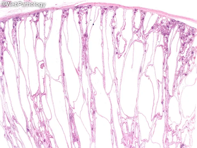
Imaging modality and technique: Light microscopy histology of renal tissue stained with Hematoxylin and Eosin (H&E). Specimen: Formalin-fixed paraffin-embedded kidney biopsy displaying medullary to cortico-medullary architecture. Anatomical region: Renal cortex and medulla with prominent dilated collecting ducts in the medulla. Visual features: Fusiform dilation of collecting tubules with long axis oriented perpendicular to the renal cortical surface, producing radiating arrays from medulla toward cortex. Cyst lining is a uniform cuboidal epithelium; cysts are irregularly distributed with thin fibrous septa containing normal glomeruli and tubules. Interstitium shows little or no fibrosis or inflammatory infiltrate in early disease; in longstanding disease, interstitial fibrosis and glomerulosclerosis may develop. Medullary cysts tend to be rounded; kidneys may show variable size and appearance in less severe cases, with less dramatic enlargement. In all ARPKD, additional liver pathology is present, including irregularly branching and anastomosing bile ductules and portal fibrosis, aiding distinction from infantile-adult polycystic kidney disease spectrum. Diagnostic relevance: The observed histology supports autosomal recessive polycystic kidney disease. Differential diagnoses include autosomal dominant polycystic kidney disease (ADPKD) and nephronophthisis; correlation with clinical history, age of onset, and hepatic involvement is essential. Potential clinical applications: confirm diagnosis in infants or children, guide genetic testing, inform prognosis, and stage hepatic involvement in congenital hepatic fibrosis.
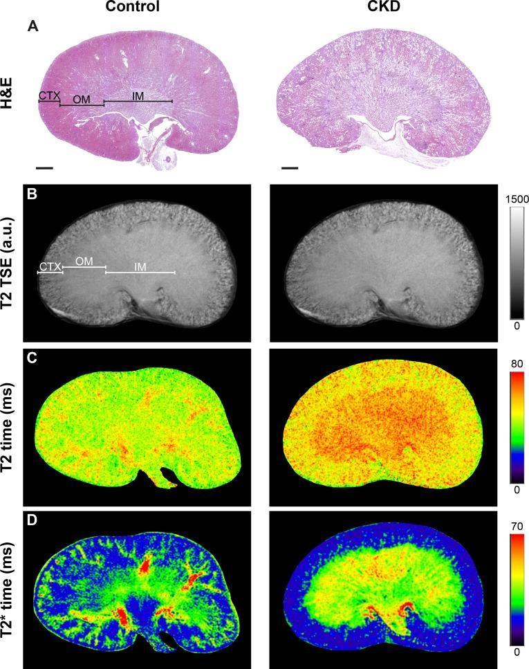
This multi-panel figure compares renal histology and magnetic resonance imaging (MRI) between a control kidney and a Chronic Kidney Disease (CKD) model. Row A presents Hematoxylin & Eosin (H&E) stained sections, highlighting the renal cortex (CTX), outer medulla (OM), and inner medulla (IM). In the CKD kidney, there is visible cortical thinning and loss of clear structural demarcation between medullary layers. Row B displays T2-weighted Turbo Spin Echo (TSE) images, showing morphological changes and signal intensity variations corresponding to the tissue layers. Rows C and D provide functional mapping through T2 and T2* relaxation time maps using pseudo-color scales (0-80ms and 0-70ms respectively). These maps demonstrate significantly elevated relaxation times in the CKD kidney, particularly within the outer medulla, visualized by a shift from cooler green/blue tones to warmer yellow/red tones. The composite image illustrates the correlation between histological tissue remodeling and functional MRI biomarkers in renal pathology.

This composite figure illustrates a multimodal imaging study of a kidney, integrating radiological, molecular, and histological data. Panel (a) presents a semi-transparent 3D MRI volume rendering fused with molecular clusters derived from 3D MALDI Mass Spectrometry Imaging (MSI). The spatial distribution of renal compartments is color-coded: the renal cortex is shown in blue forming the outer parenchyma, the renal medulla in green, and the renal pelvis as a central branching red structure. Panel (b) shows the spatial localization of a specific ion species (m/z 4808) overlaid on the MRI volume, demonstrating molecular mapping within the anatomical framework. Panel (c) displays an H&E-stained histological cross-section of the inner kidney co-registered with a 3D segmentation map. The histology reveals distinct tissue architecture including the cortex (purple/blue), medulla (green), and hilar structures. This visualization demonstrates the clinical relevance of combining molecular MSI data with traditional diagnostic imaging and pathology for enhanced tissue characterization and anatomical-molecular correlation.
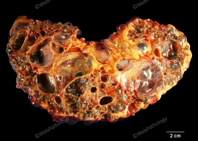
This is a gross pathology photograph of an adult human kidney affected by autosomal dominant polycystic kidney disease (ADPKD). The organ is markedly enlarged (reniform in outline) yet preserves its overall bean‑shaped kidney morphology. On the cut surface, innumerable cysts of variable size disrupt the renal parenchyma, with some cysts less than 0.5 cm and others exceeding 6 cm in diameter. The cysts are fluid‑filled and appear transparent to reddish, cumulatively replacing much of the normal renal cortex and medulla, leaving only scant residual parenchyma between cavities. The external cortex is irregular, with a patchy, nodular feel, and the cortex–medulla junction is distorted by multilocular dilatations. This specimen derives from an autopsy of an adult male who had end‑stage renal disease from ADPKD and died of an unrelated cause; the kidneys reflect chronic, progressive cystic degeneration rather than an acute process. Clinically, ADPKD is a common hereditary cause of renal failure, characterized by bilateral renal enlargement, hypertension, and progressive decline in function; pathologically, multiple cysts originate from tubular epithelium and communicate loosely with collecting system. This image illustrates the phenotype, useful for education, differential diagnosis with other cystic nephropathies, and correlation with microscopic histology showing cystic dilation and fibrotic parenchyma.
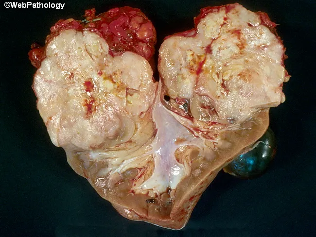
Gross pathology photograph of a nephrectomy kidney displaying an exophytic yellow-tan tumor with focal hemorrhage and necrosis. The cut surface reveals a large mass intermingled with hemorrhagic and necrotic zones within the renal parenchyma. The exterior of the kidney shows irregular borders. Microscopically, the lesion comprises predominantly malignant fibrous histology–like sarcomatous areas characterized by highly pleomorphic spindle- to epithelioid-shaped cells, marked mitotic activity, and a storiform-to-haphazard architecture. Intermixed are small foci of conventional clear cell renal cell carcinoma, confirming histologic heterogeneity and suggesting a biphasic neoplasm or collision/dedifferentiation. Areas of extensive necrosis and hemorrhage contribute to the gross appearance and may correlate with aggressive behavior. The tumor appears intraparenchymal, infiltrating renal cortex and medulla, with potential involvement of surrounding parenchyma. Clinically, such renal tumors portend poorer prognosis due to sarcomatoid transformation and RCC admixture. Immunohistochemical and molecular studies are often required to confirm lineage and distinguish from primary renal sarcomas. This image provides a reference for educational pathology, aiding recognition of mixed histology renal tumors, differentiation from pure sarcomas, and correlation with histology slides, differential diagnoses, and treatment decision-making in nephrectomy specimens, with implications for prognosis, therapy planning, and follow-up."
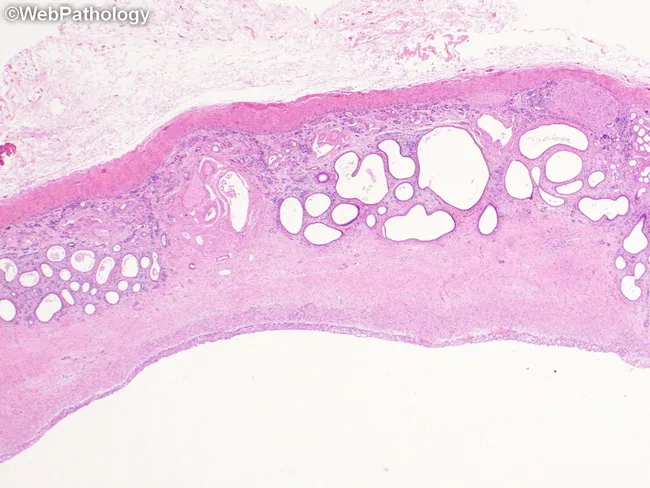
Histopathology slide of renal tissue from a case of multicystic dysplastic kidney (MKD). The section shows a large cyst with cyst wall lined by flattened to cuboidal epithelium and adjacent dysplastic parenchyma. Immature ducts and tubules are embedded in fibromuscular stroma, forming collarettes of primitive smooth muscle around these immature elements. Normal cortical architecture is disrupted with poor corticomedullary differentiation and sparse or absent mature glomeruli. The overall renal architecture is disorganized, reflecting dysplastic transformation rather than normal nephrogenesis. This pattern distinguishes MKD from pure hydronephrosis, which exhibits extreme thinning of the renal medulla and cortex without dysplastic features such as primitive ductal collars. Differential considerations include cystic Wilms tumor or mesoblastic nephroma, sporadic glomerulocystic kidney disease, tuberous sclerosis–associated renal lesions, and polycystic kidney disease. Clinically, these findings indicate a nonfunctional or poorly functioning renal unit and guide surgical planning, prognosis, and genetic counseling within the CAKUT spectrum, highlighting the importance of histology for accurate diagnosis and differential exclusion of neoplastic or other cystic kidney diseases.
normal kidney histology cortex medulla labeled nephron diagram collecting duct loop of Henle
renal medulla histology labeled diagram collecting duct thin loop of Henle vasa recta

- Outer stripe of outer medulla: proximal straight tubules (thick, eosinophilic cuboidal cells with brush border) and collecting ducts.
- Inner stripe of outer medulla: thick ascending limbs of the loop of Henle plus collecting ducts.
- Inner medulla: thin limbs of the loop of Henle (small, flattened simple squamous epithelium, easily confused with capillaries) and larger collecting ducts lined by pale cuboidal "clear" cells with distinct cell borders - these converge near the papilla into papillary ducts (ducts of Bellini).
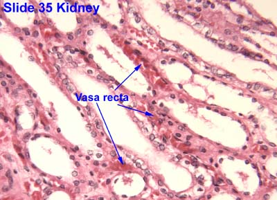
- No glomeruli (unlike cortex) - the medulla is purely tubular and vascular.
- Vertically-running (striated) parallel tubules: thin/thick limbs of loop of Henle, collecting ducts, and vasa recta, all oriented toward the papilla.
- Collecting ducts are the largest tubules, lined by pale cuboidal "clear" cells with sharp, distinct lateral cell borders (less eosinophilic than proximal tubule cells).
- Thin limbs of the loop of Henle are small caliber with flattened simple squamous epithelium, similar in appearance to capillaries - vasa recta help distinguish true vessels from thin tubule segments in this region.
- Interstitial connective tissue between tubules is scant in the outer medulla but increases toward the inner medulla/papilla.