
Hi
Hey! How can I help you today?
Can u explain me about mitochondria
Mitochondria are membrane-bound organelles found in most eukaryotic cells, often called the "powerhouse of the cell" because they generate most of the cell's ATP through a process called cellular respiration. They have a double membrane structure and contain their own DNA, suggesting they originated from an ancient bacterial endosymbiosis.
Searching sources...
Reading File
Finding Sources
Reading File
Reading File
Reading File
Loading Image
Loading Image
Here is a thorough explanation of mitochondria, drawn from Guyton & Hall Textbook of Medical Physiology, Junqueira's Basic Histology, and Basic Medical Biochemistry:
Mitochondria - The Powerhouse of the Cell
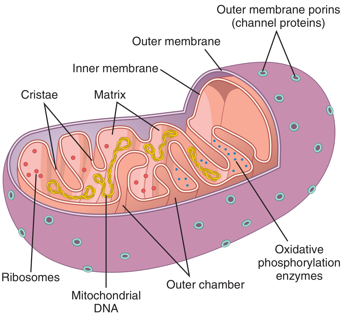
What Are Mitochondria?
Mitochondria are membrane-enclosed organelles found in the cytoplasm of nearly all eukaryotic cells. Their primary role is aerobic respiration and the production of ATP (adenosine triphosphate) - the main energy currency of the cell. Without mitochondria, cells cannot extract sufficient energy from nutrients, and essentially all cellular functions would cease. - Guyton and Hall Textbook of Medical Physiology
Structure
Each mitochondrion has two distinct membranes, creating two separate compartments:
1. Outer Membrane
- Contains channel proteins called porins
- These porins regulate the flow of ions and small metabolites (like pyruvate) from the cytoplasm into the intermembrane space
- Permeable to molecules up to ~1,000 Da
2. Inner Membrane
- Highly impermeable - this is critical for energy production
- Forms deep infoldings called cristae, which dramatically increase the surface area for chemical reactions
- Houses the electron transport chain and ATP synthase (oxidative phosphorylation enzymes)
3. Matrix (Inner Compartment)
- The innermost space, enclosed by the inner membrane
- Contains:
- Large quantities of dissolved enzymes for the Krebs (TCA) cycle
- Mitochondrial DNA (mtDNA) - 37 genes
- Ribosomes for protein synthesis
- Enzymes for fuel oxidation pathways
4. Intermembrane Space
- The narrow space between the outer and inner membranes
- A proton gradient is built up here during oxidative phosphorylation, which drives ATP synthesis
Size and Number
- Size: typically 0.5-1 µm in diameter, up to 10 µm long - can be globular or elongated
- Number per cell: varies from fewer than 100 to several thousand, depending on energy demands
- High-energy cells (cardiac muscle, kidney tubule cells) have abundant mitochondria
- Low-energy cells (fat cells/adipocytes) have few mitochondria
- Mitochondria concentrate in the most energy-demanding parts of the cell
How They Generate Energy
- Glycolysis (in the cytoplasm) - converts glucose to pyruvate, releasing a small amount of ATP
- Pyruvate is imported into the mitochondrial matrix
- The Krebs/TCA cycle oxidizes pyruvate to CO₂ and H₂O, generating electron carriers (NADH, FADH₂)
- The electron transport chain on the cristae uses these carriers to build a proton gradient
- ATP synthase uses this proton gradient to synthesize ATP
Result: Mitochondrial metabolism yields 15 times more ATP than glycolysis alone. - Junqueira's Basic Histology
A small portion of the released energy escapes as heat, helping maintain body temperature.
Mitochondrial DNA (mtDNA)
- Contains 37 genes essential for mitochondrial function
- Primarily maternally inherited (from the egg, not the sperm - sperm mitochondria are typically destroyed after fertilization)
- Controls self-replication of mitochondria
- When cells face increased energy demands (e.g., chronic exercise training), mitochondria can replicate to supply additional ATP
Dynamic Behavior
Mitochondria are not static - they are highly plastic organelles that:
- Continuously change shape
- Fuse with one another
- Divide (fission)
- Are transported through the cytoplasm along microtubules
Clinical Relevance
| Condition | Mechanism |
|---|---|
| MERRF (Myoclonic Epilepsy with Ragged-Red Fibers) | Mutation in mtDNA gene for lysine-tRNA → defective respiratory chain proteins → structural abnormality in muscle fibers |
| Mitochondria and Apoptosis | Mitochondria release cytochrome c, triggering programmed cell death |
| Pompe Disease | Deficiency of lysosomal enzyme (acid maltase) leading to glycogen accumulation; linked to mitochondrial energy metabolism |
Summary Table
| Feature | Detail |
|---|---|
| Main function | ATP production via oxidative phosphorylation |
| Membranes | Two (outer + inner) |
| Key structure | Cristae (inner membrane folds) |
| DNA | Yes - 37 genes, maternally inherited |
| Self-replication | Yes |
| Number per cell | <100 to several thousand |
| Key enzymes | TCA cycle enzymes, electron transport chain, ATP synthase |
Sources: Guyton and Hall Textbook of Medical Physiology; Junqueira's Basic Histology, 17e; Basic Medical Biochemistry, 6e
What is the use of cristae
Cristae are the infoldings of the inner mitochondrial membrane. Their main purpose is to dramatically increase the surface area available for the electron transport chain and ATP synthase, which are the protein complexes that generate ATP. More surface area means more ATP can be produced per mitochondrion.
Searching sources...
Finding Sources
Finding Sources
Reading File
Reading File
Loading Image
Loading Image
Here is a detailed explanation of cristae and their functions, backed by authoritative histology and physiology textbooks:
The Role of Cristae in Mitochondria
What Are Cristae?
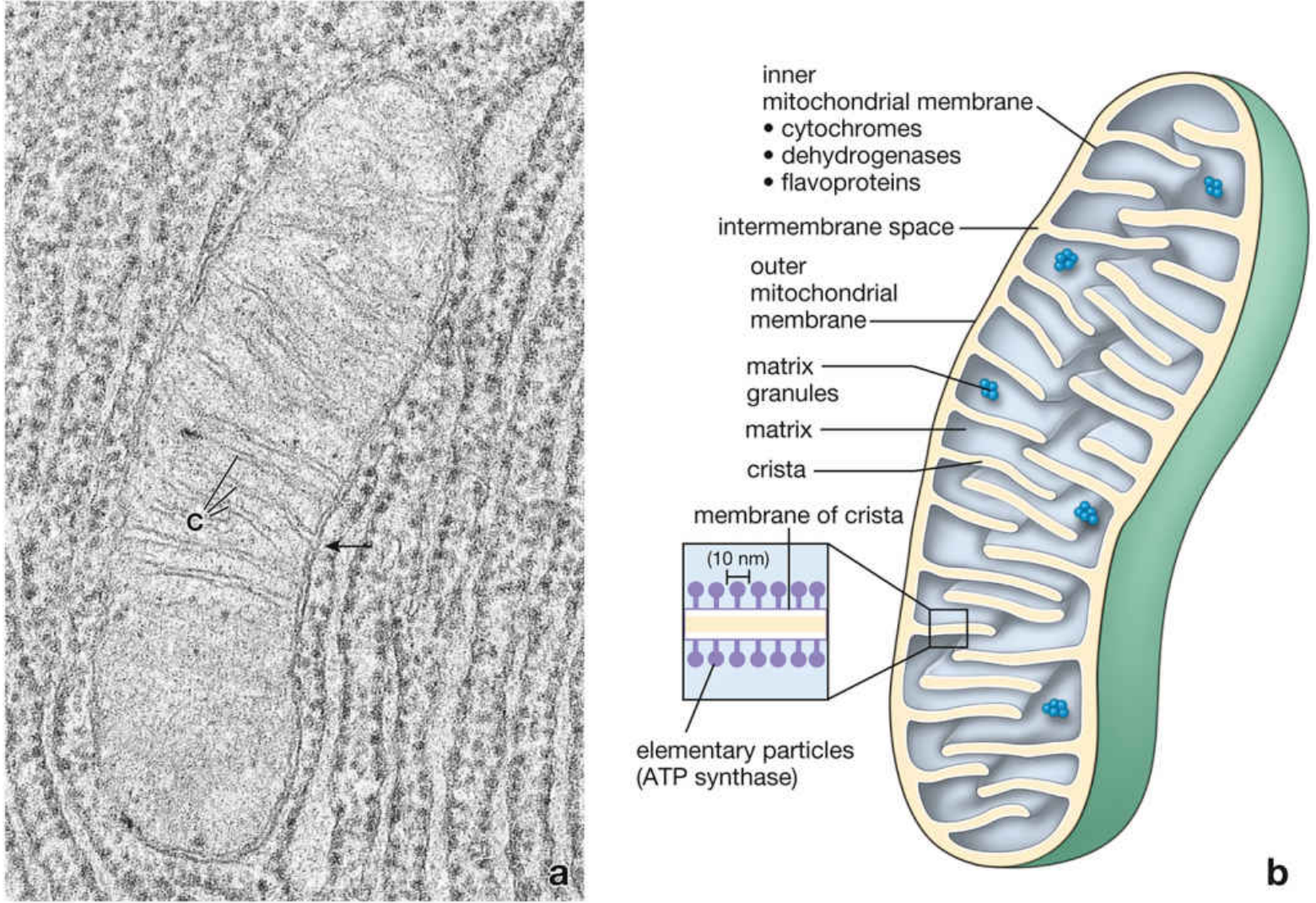
Cristae (singular: crista) are the sharp inward folds of the inner mitochondrial membrane that project into the matrix. The word comes from Latin, meaning "crests" or "ridges." Under the electron microscope, they appear as shelf-like or finger-like projections extending deep into the interior of the mitochondrion.
Functions of Cristae
1. Massively Increase Surface Area
The primary purpose of cristae is to greatly increase the inner membrane's surface area. The inner membrane by itself would be too small to accommodate all the protein complexes needed for energy production. By folding inward repeatedly, cristae multiply the available surface area many times over.
"The inner membrane has many long folds called cristae, which project into the matrix and greatly increase the membrane's surface area." - Junqueira's Basic Histology, 17e
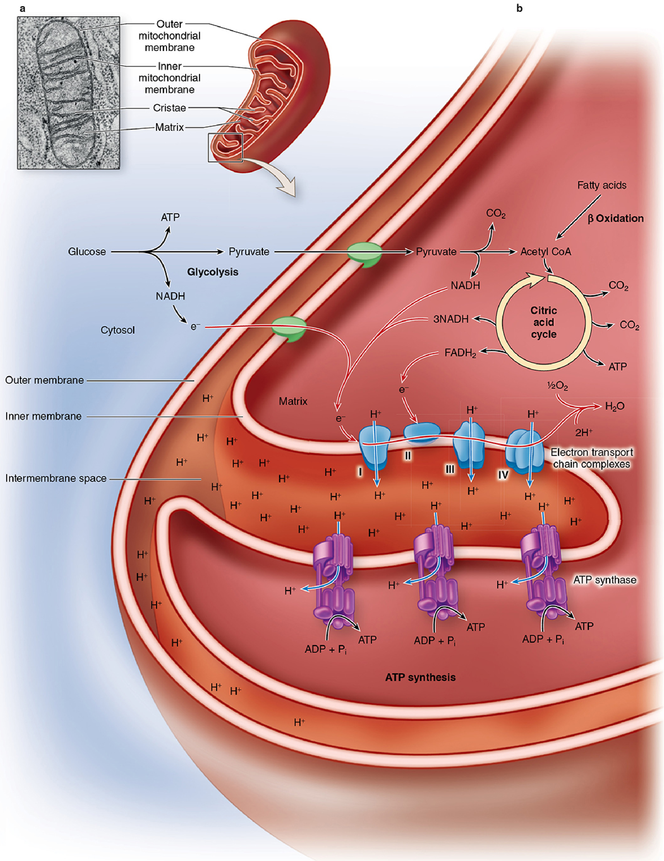
2. House the Electron Transport Chain (Complexes I-IV)
The membrane forming the cristae contains the respiratory electron transport chain - four large protein complexes (I, II, III, IV) that pass electrons along in a relay. As electrons move through these complexes:
- Protons (H⁺) are pumped from the matrix into the intermembrane space
- This builds up an electrochemical gradient (proton-motive force)
3. Host ATP Synthase ("Elementary Particles")
Cristae are studded with ATP synthase complexes, visible in electron microscopy as tennis racquet-shaped structures called elementary particles (~10 nm in diameter). Their heads project into the matrix. Protons flow back down through these complexes (from intermembrane space into matrix), and this flow drives the rotation of ATP synthase, converting ADP + phosphate into ATP - a process called oxidative phosphorylation (chemiosmosis).
4. Serve as Docking Sites for Electron Transport Supercomplexes
Cristae are enriched with the phospholipid cardiolipin, which helps maintain the ultrastructure of cristae and acts as the docking site for mitochondrial electron transport supercomplexes - organized assemblies of multiple respiratory chain complexes that improve efficiency of electron transfer. - Fuster & Hurst's The Heart, 15th ed.
5. Number Reflects Energy Demand
The number of cristae directly correlates with a cell's energy needs:
- High-energy cells (cardiac muscle, kidney tubule cells) → many, densely packed cristae
- Low-energy cells (fat cells) → few, poorly developed cristae
- In steroid-synthesizing cells (adrenal cortex), cristae can be tubular rather than shelf-like, to accommodate cholesterol-converting enzymes alongside ATP production
Step-by-Step: How Cristae Generate ATP
| Step | Location | What Happens |
|---|---|---|
| 1. Fuel entry | Outer membrane porins | Pyruvate and fatty acids enter the intermembrane space |
| 2. Matrix reactions | Matrix | Citric acid (Krebs) cycle produces NADH and FADH₂ |
| 3. Electron transfer | Cristae membrane (ETC Complexes I-IV) | Electrons from NADH/FADH₂ pass along the chain; H⁺ pumped into intermembrane space |
| 4. Proton gradient | Intermembrane space | H⁺ accumulates, creating electrochemical gradient |
| 5. ATP synthesis | Cristae membrane (ATP synthase) | H⁺ flows back into matrix through ATP synthase → ATP is produced |
Clinical Relevance: What Happens When Cristae Are Disrupted?
- Aging: Mitochondria may become swollen with disrupted cristae, leading to impaired ATP production and increased electron leakage. - Harrison's Principles of Internal Medicine, 22e
- Heart disease: Cardiomyocyte mitochondria show morphological changes including cristae abnormalities, reducing cardiac energy supply
- Cardiolipin deficiency: Cardiolipin is essential for maintaining cristae ultrastructure; its loss destabilizes the electron transport supercomplexes, reducing ATP output
In Simple Terms
Think of cristae like the folds inside a radiator - the more folds, the greater the surface area, and the more heat (or in this case, ATP) that can be produced. Without cristae, the mitochondrion would be far too small to pack in all the machinery needed to power the cell.
Sources: Junqueira's Basic Histology, 17e; Histology: A Text and Atlas with Correlated Cell and Molecular Biology; Fuster & Hurst's The Heart, 15th ed.; Harrison's Principles of Internal Medicine, 22e
This is a shared conversation. Sign in to Orris to start your own chat.