
Macular sparring
Reading File
Macular sparing refers to the preservation of central (macular) vision when there is a lesion affecting the visual pathway, most notably in the occipital cortex. It occurs because the macula has dual blood supply (posterior cerebral artery + middle cerebral artery), so strokes affecting one vessel may spare the macular representation.
Searching sources...
Finding Sources
Searching the Web
macular sparing visual field defect mechanism occipital lobe
Now let me read the most detailed sections from the textbooks to give a thorough answer:
Reading File
Reading File
Reading File
Now let me fetch the key visual diagram and also read the Neuroanatomy discussion section for clinical context:
Loading Image
Reading File
Excellent - I now have thorough information to compile a comprehensive answer. Here is the complete explanation:
Macular Sparing
Definition
Macular sparing is a visual field defect pattern in which the central (macular) region of vision is preserved despite significant peripheral or hemianopic loss. It is the opposite of macular splitting, where the vertical midline cuts precisely through the central field.
Anatomy Recap: Why the Macula Is Special
The macula (central ~5 degrees of the visual field) has a disproportionately large cortical representation at the occipital pole (tip of the occipital lobe). This area receives blood supply from two sources:
- The posterior cerebral artery (PCA) - supplies most of the visual cortex
- The middle cerebral artery (MCA) - gives collaterals to the occipital pole
This dual supply is the key to understanding macular sparing.
Mechanisms
1. Dual Vascular Supply (most accepted)
When a PCA stroke infarcts the occipital cortex, the MCA collaterals can still perfuse the occipital pole (macular cortex). Peripheral field cortex - further from the pole - loses its only PCA supply and infarcts. Central vision is thus spared.
2. Bilateral Macular Representation
The macula projects to both hemispheres to some degree (overlap around the vertical meridian), meaning a unilateral lesion may spare macular input from the contralateral hemisphere.
3. Cortical Magnification
The macular representation is so large relative to peripheral fields that a single lesion must extend a considerable distance to destroy all macular neurons. As Ganong states: "occipital lesions must extend considerable distances to destroy both macular and peripheral vision." - Ganong's Review of Medical Physiology, 26th Ed.
Classic Patterns
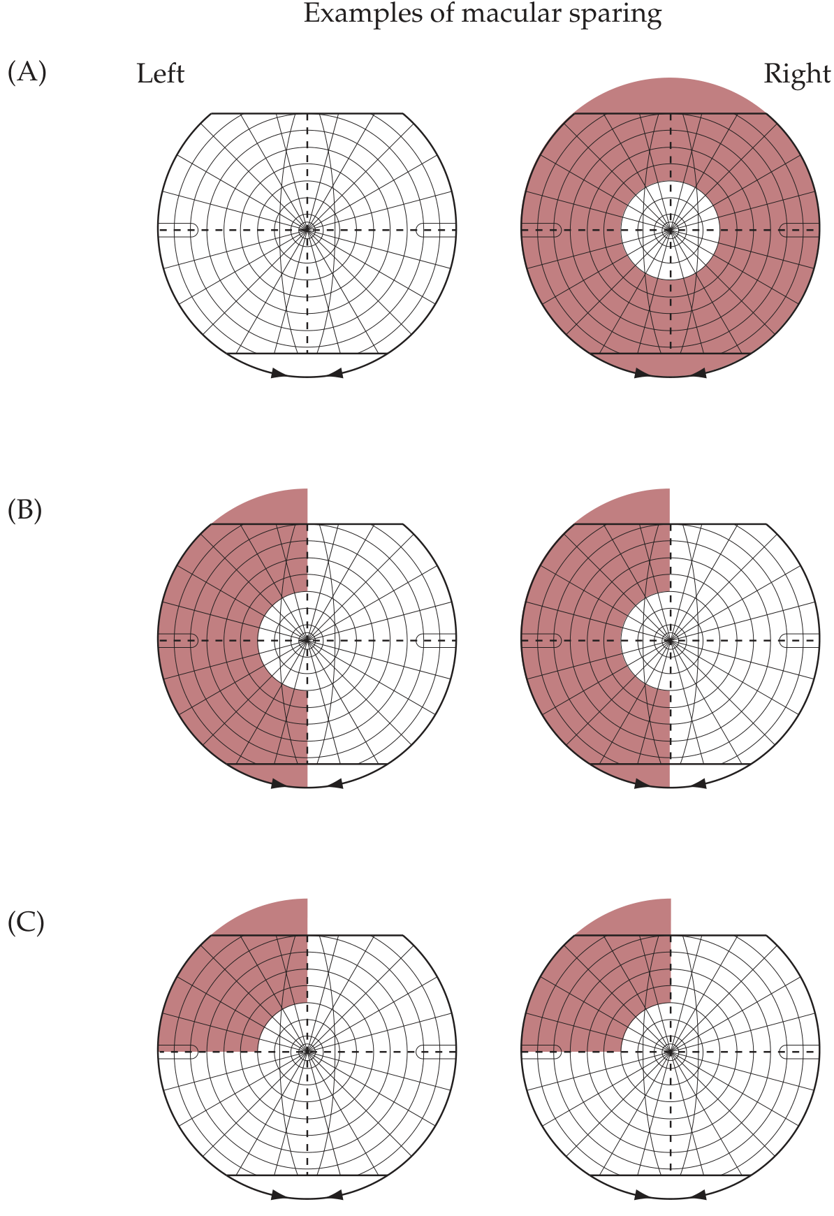
The diagram above (from Neuroanatomy through Clinical Cases) shows three classic patterns:
| Pattern | Description | Typical Cause |
|---|---|---|
| (A) Monocular concentric loss | Peripheral loss with central sparing in one eye | Chronic elevated ICP, retinitis pigmentosa |
| (B) Homonymous hemianopia with macular sparing | Left field lost in both eyes, central circle intact | Right PCA infarct sparing occipital pole |
| (C) Superior quadrantanopia with macular sparing | Left upper quadrant lost, macula intact | Lesion of inferior bank of right calcarine fissure |
Where in the Visual Pathway Does It Occur?
Macular sparing is characteristic of lesions at or behind the geniculocalcarine tract (optic radiations) / visual cortex.
- Optic nerve, chiasm, or optic tract lesions do NOT produce macular sparing - they split the macula cleanly
- Geniculocalcarine tract lesions: cause homonymous hemianopia with macular sparing
- Primary visual cortex (V1/calcarine cortex) lesions: most common cause; sparing of the occipital pole = sparing of macula
Key rule: Macular sparing suggests an occipital (cortical) origin of the hemianopia, rather than a tract or radiation lesion. - Neuroanatomy through Clinical Cases, 3rd Ed.
Bilateral Occipital Lesions
Bilateral occipital infarcts can cause bilateral homonymous hemianopia (cortical blindness) but may leave a small "keyhole" of central vision intact - this is called keyhole vision or tunnel of central macular sparing. Rarely, patients with cortical blindness deny their defect (Anton syndrome). - Localization in Clinical Neurology, 8th Ed.
Clinical Significance
- Lesion localization - Macular sparing points to an occipital cortex lesion (typically PCA territory infarct), distinguishing it from optic tract or LGN lesions, which produce macular splitting.
- Preserved reading ability - Because the macula handles fine detail/central vision, macular sparing can preserve the ability to read, recognize faces, and perform tasks requiring central acuity, even with large peripheral field loss.
- Differential diagnosis - Not all central sparing is macular sparing. Tunnel vision (e.g., glaucoma, retinitis pigmentosa) is monocular peripheral loss; true macular sparing is binocular and homonymous.
Macular Sparing vs. Macular Splitting
| Feature | Macular Sparing | Macular Splitting |
|---|---|---|
| Central field | Intact | Lost (bisected by midline) |
| Typical location | Occipital cortex (PCA territory) | Optic tract, LGN, optic radiation |
| Vascular cause | PCA infarct with MCA collaterals | MCA territory infarct |
| Reading ability | Usually preserved | Often impaired |
Sources:
- Neuroanatomy through Clinical Cases, 3rd Ed. (Blumenfeld), Figures 11.16, p. 501
- Costanzo Physiology, 7th Ed., Optic Pathways section
- Ganong's Review of Medical Physiology, 26th Ed., Effect of Lesions in the Optic Pathways
- Localization in Clinical Neurology, 8th Ed., Chiasmal Syndrome chapter
Astigmation
Astigmatism is a refractive error caused by an unequal curvature of the cornea (or less commonly the lens), so light rays in different meridians focus at different points rather than a single point on the retina, producing blurred or distorted vision at all distances.
Searching sources...
Finding Sources
Searching the Web
astigmatism types classification corneal lenticular treatment
The index results weren't useful - let me search with better targeted queries in ophthalmology textbooks:
Finding Sources
Reading File
Reading File
Now let me search for more detailed clinical content on astigmatism types, optics, and the Wills Eye Manual section:
Finding Sources
Finding Sources
Finding Sources
Finding Sources
Finding Sources
Reading Web Page
https://www.ncbi.nlm.nih.gov/books/NBK582142
I now have comprehensive information to compile a thorough answer. Here it is:
Astigmatism
Definition
Astigmatism is a refractive error caused by unequal curvature of the cornea (most common) or lens, resulting in different focal points for light rays entering in different meridians. Instead of converging at a single point on the retina, light forms two focal lines separated by a region called the interval of Sturm (Sturm's conoid).
Optics - Sturm's Conoid
In a normal (spherical) eye, all meridians have equal refracting power and light converges to a single point. In astigmatism:
- The two principal meridians are perpendicular to each other (in regular astigmatism)
- Each meridian has a different focal power - the steeper meridian focuses closer, the flatter meridian focuses farther
- Between the two focal lines lies an interval of Sturm - the "circle of least confusion" lies at its midpoint and represents the best blur circle
Classification
1. By Anatomical Origin
| Type | Cause |
|---|---|
| Corneal astigmatism | Unequal corneal curvature (most common) |
| Lenticular astigmatism | Unequal curvature or tilting of the crystalline lens |
| Retinal astigmatism | Oblique placement of the macula or tilted optic disc |
2. Regular vs. Irregular
Regular astigmatism
- The two principal meridians are always 90° apart
- There is a smooth, consistent variation in refractive power from one meridian to another
- Usually present from birth; may be hereditary
- Correctable with cylindrical spectacle lenses or toric contact lenses
Irregular astigmatism
- The principal meridians are not 90° apart, or curvature varies across the same meridian
- Usually acquired - from corneal scarring (trauma, infection), keratoconus, pterygia, post-surgical changes, or pellucid marginal degeneration
- Cannot be corrected with spectacle lenses - requires rigid gas-permeable (RGP) contact lenses
3. By Axis of the Principal Meridian (Regular Only)
| Type | Description |
|---|---|
| With-the-rule (WTR) | Vertical meridian is steeper (corrected with + cylinder at 90°). Common in children/young adults |
| Against-the-rule (ATR) | Horizontal meridian is steeper (corrected with + cylinder at 180°). More common in the elderly |
| Oblique | Principal meridians lie between 30-60° and 120-150° |
| Bi-oblique | The two principal meridians are perpendicular but both oblique (not horizontal/vertical) |
Age trend: Infants (0-4 yrs) tend to have ATR astigmatism. Ages 4-18 shift toward WTR. In older adults, WTR gradually converts to ATR.
4. By Relationship of Focal Lines to Retina
| Type | Description |
|---|---|
| Simple myopic astigmatism | One focal line on the retina, the other in front of it |
| Simple hyperopic astigmatism | One focal line on the retina, the other behind it |
| Compound myopic astigmatism | Both focal lines in front of the retina |
| Compound hyperopic astigmatism | Both focal lines behind the retina |
| Mixed astigmatism | One focal line in front, one behind the retina |
Symptoms
- Blurred or distorted vision at all distances
- "Shadowing" of letters or images (ghosting)
- Difficulty seeing fine detail
- Asthenopic symptoms - eyestrain, headaches, squinting
- In children: amblyopia (if uncorrected during critical period)
Diagnosis
| Investigation | Purpose |
|---|---|
| Visual acuity | Baseline assessment |
| Retinoscopy | Objective refraction; characteristic "scissor reflex" in keratoconus |
| Subjective refraction (Jackson cross-cylinder) | Refines cylinder power and axis |
| Astigmatic fan test | Identifies the principal meridians |
| Keratometry / Topography | Measures corneal curvature meridians |
| Scheimpflug imaging (Pentacam) | Detailed anterior segment mapping |
| Slit lamp | Detects irregular astigmatism causes (scars, keratoconus, pterygium) |
| Pachymetry | Corneal thickness (rule out keratoconus) |
Treatment
Optical Correction
| Option | Details |
|---|---|
| Cylindrical spectacles | Standard correction for regular astigmatism |
| Soft toric contact lenses | For up to ~2-3 D of regular astigmatism |
| Rigid gas-permeable (RGP) contact lenses | Corrects both regular and irregular astigmatism by replacing the irregular corneal surface with a smooth tear lens interface |
| Hybrid contact lenses | Hard centre + soft periphery; useful for irregular astigmatism with comfort issues |
Surgical Correction
| Procedure | Details |
|---|---|
| Limbal relaxing incisions / Arcuate keratotomy | Paired arcuate cuts in the steep meridian to flatten it; useful for low-moderate astigmatism and post-keratoplasty astigmatism |
| PRK / LASEK | Corrects up to 3 D of astigmatism |
| LASIK | Corrects up to 5 D of astigmatism |
| Toric IOL implantation | Used at time of cataract surgery or as phakic IOL; risk of axis rotation post-op |
| Conductive keratoplasty (CK) | Radiofrequency energy in corneal periphery for low-moderate astigmatism; regression is a drawback |
| Repeat optical keratoplasty | Reserved for very high astigmatism (>20 D) |
Special Condition: Keratoconus
Keratoconus is the most important cause of progressive irregular astigmatism:
- Acquired corneal thinning causing protrusion, typically inferiorly/centrally
- Onset in adolescence; progressive over 5-10 years
- Produces high irregular astigmatism (often ATR)
- Managed with RGP lenses, corneal collagen cross-linking (CXL) to halt progression, or corneal transplant (PK/DALK) for severe cases
Prognosis
Prognosis is generally good with timely correction. Untreated astigmatism in children can cause amblyopia (lazy eye) with permanent vision reduction. Regular astigmatism is often stable; irregular astigmatism (e.g., keratoconus) may progress and require escalating intervention.
Sources:
- Goldman-Cecil Medicine, 2-vol ed., Astigmatism section
- Kanski's Clinical Ophthalmology, 10th Ed., Correction of astigmatism
- StatPearls - Astigmatism (NCBI)
Whatt do you mean by meridian in this
In the context of the eye, a meridian is simply an imaginary line that passes through the centre (optical axis) of the cornea - like lines of longitude on a globe, but drawn across the front surface of the eye.
Visualising It
Imagine looking straight at someone's eye from the front. The cornea (the clear front surface) is a dome. Now draw lines across that dome, all passing through the very centre:
- The vertical meridian runs 12 o'clock to 6 o'clock (90°)
- The horizontal meridian runs 9 o'clock to 3 o'clock (180°)
- You can draw a meridian at any angle - 45°, 60°, 135°, etc.
By convention, meridians are measured in degrees 0° to 180° (not 0-360°), since a line at 0° and 180° is the same line.
Why It Matters in Astigmatism
A perfectly spherical cornea has the same curvature (steepness) in every meridian - it curves equally whether you measure along the vertical, horizontal, or any other line.
In astigmatism, different meridians have different curvatures:
- One meridian is steeper (more curved, bends light more strongly)
- The perpendicular meridian is flatter (less curved, bends light less strongly)
Think of it like a rugby ball vs. a football:
- A football = same curve in all directions = normal spherical cornea
- A rugby ball = steeper along one axis, flatter along the other = astigmatic cornea
Because the steep and flat meridians bend light by different amounts, they focus light at two different distances instead of one point - and that is what causes the blur.
The Two Principal Meridians
In regular astigmatism, there are always two principal meridians - the steepest and the flattest - and they are always 90° apart (perpendicular to each other). Everything in between gradually transitions from one to the other.
When an optometrist writes a glasses prescription like -1.00 / -0.75 × 180, the "× 180" tells you which meridian the cylindrical correction is aligned to.
This is a shared conversation. Sign in to Orris to start your own chat.