
Dysphagia Approach
dysphagia approach classification oropharyngeal esophageal
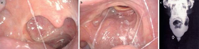
This composite educational clinical figure illustrates oropharyngeal and esophageal pathologies contributing to dysphagia. Image (a) is a transoral endoscopic view of an 87-year-old patient showcasing post-surgical changes following an extended partial resection of the right oropharynx and neck dissection. Visible features include a disrupted pharyngeal wall structure on the right and distorted mucosa consistent with radiotherapy and chemotherapy effects. Image (b) presents a transnasal endoscopic view of the velum and posterior oropharyngeal wall. It demonstrates significant scarring and asymmetrical contraction of the right-sided velum, with visible scar tissue extending across the posterior pharyngeal wall. Prominent white, viscous saliva is seen spanning the lumen, indicating impaired clearance and a transportation disorder. Image (c) is a contrast radiography (barium swallow) from a different patient, identifying tissue formations (indicated by an arrow) in the pharynx or esophagus that create mechanical obstacles. Together, these images teach how oncological treatments, scarring, and structural abnormalities disrupt the swallowing mechanism.
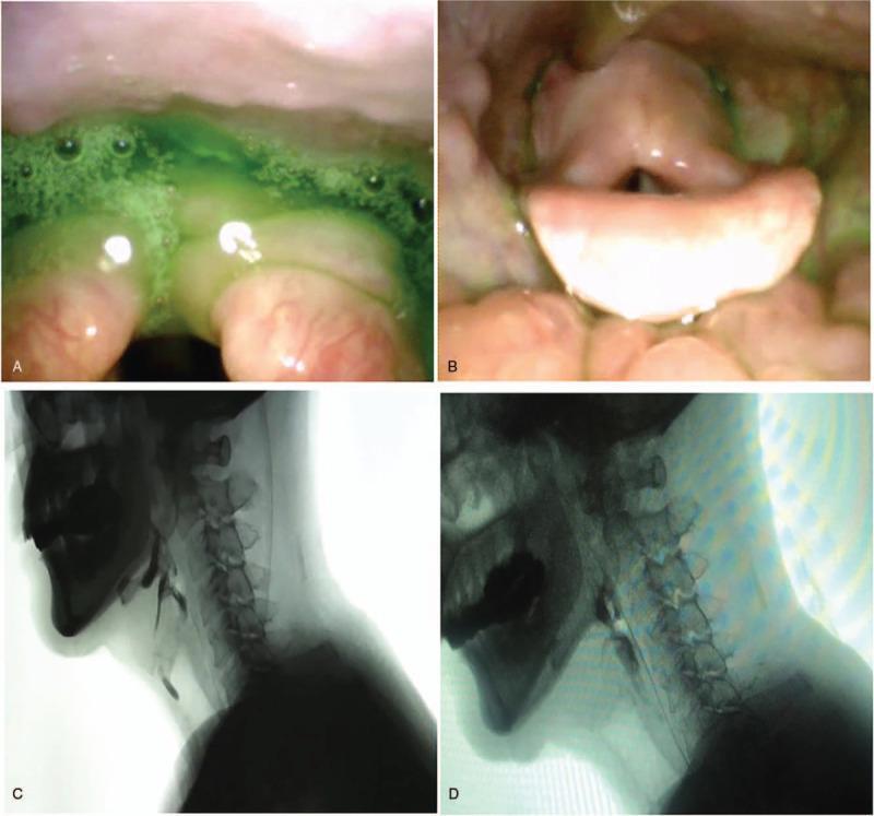
This composite figure illustrates a comparative evaluation of oropharyngeal dysphagia before and after treatment for cricopharyngeal (CP) muscle dysfunction. Panels A and B are Fiberoptic Endoscopic Evaluation of Swallowing (FEES) stills. Panel A (Pre-treatment) shows massive green-dyed mucilage secretions and pooling in the postcricoid region, indicating penetration and impaired clearance. Panel B (Post-treatment) shows a significant reduction in pharyngeal residue and clearer visualization of the laryngeal inlet. Panels C and D are lateral Video Fluoroscopic Swallowing Study (VFSS) frames. Panel C (Pre-treatment) demonstrates limited epiglottic folding and a persistent posterior indentation at the level of the CP muscle, signifying non-relaxation during a swallow attempt. Panel D (Post-treatment) reveals improved CP muscle opening, successful bolus passage through the upper esophageal sphincter, and decreased residue in the vallecula and pyriform sinuses. The images collectively demonstrate the therapeutic efficacy of botulinum toxin injection in improving upper esophageal sphincter dynamics and reducing aspiration risk in patients with neurogenic dysphagia.
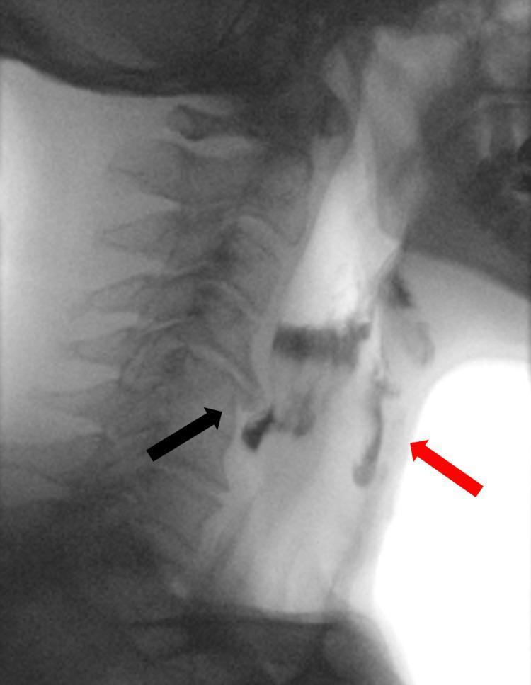
A lateral view videofluoroscopic swallowing study (VFSS) image demonstrating oropharyngeal dysphagia. The image captures the cervical spine and the pharyngeal/esophageal soft tissue structures during the transit of a radiopaque contrast bolus. A prominent anterior osteophyte at the C5 vertebral body level (indicated by a black arrow) causes focal extrinsic compression and narrowing of the proximal cervical esophagus. This mechanical obstruction leads to the stasis and pooling of the contrast material above the upper esophageal sphincter. Significantly, contrast is seen misdirected into the laryngeal vestibule and trachea (indicated by a red arrow), confirming laryngeal penetration and tracheal aspiration. This diagnostic image illustrates the clinical relationship between cervical spondylosis (DISH or severe osteophytosis) and mechanical dysphagia, highlighting the risk of respiratory complications due to impaired bolus clearance and subsequent airway entry.
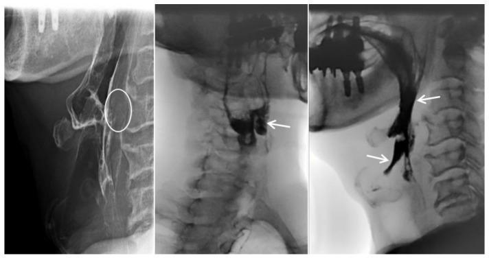
This diagnostic image consists of three lateral-view cervical esophagrams (fluoroscopic barium swallow study) demonstrating oropharyngeal dysphagia and mechanical obstruction. The first panel highlights prevertebral soft tissue swelling at the C3–C4 level (white circle), which causes extrinsic compression and anterior displacement of the esophagus. The subsequent panels illustrate functional swallowing impairment: radiopaque contrast material is seen abnormally pooling within the laryngeal vestibule and epiglottic space. White arrows indicate laryngeal penetration and aspiration, where contrast moves anteriorly into the airway rather than proceeding through the upper esophageal sphincter. These findings are clinically significant for diagnosing mechanical dysphagia, in this case secondary to an anteriorly extruded cervical disc. Key educational concepts include the radiographic identification of aspiration, the assessment of prevertebral soft tissue thickness, and the recognition of extrinsic esophageal compression in the context of cervical spine pathology.
dysphagia evaluation management approach
Dysphagia: A Systematic Approach
Step 1: Classify the Type of Dysphagia
| Feature | Oropharyngeal (Transfer) | Esophageal |
|---|---|---|
| Timing | Difficulty initiating swallow; symptoms within 1 second | Food sticks after swallowing |
| Site perception | Cervical/throat region | Lower sternum, epigastrium |
| Associated symptoms | Nasal regurgitation, coughing/choking on swallowing, drooling, dysarthria, hoarseness | Heartburn, bland regurgitation, chest pain |
| Aspiration | Common | Less common |
Note: Some patients with distal esophageal disease (e.g., achalasia) may point to the upper esophageal region as the site of hold-up -- this can mislead toward an ENT referral when the real problem is a motility disorder. - Yamada's Textbook of Gastroenterology, 7th ed.
Step 2: History - The Three Key Questions for Esophageal Dysphagia
- What type of food causes symptoms? (solids only vs. solids AND liquids)
- Is the dysphagia intermittent or progressive?
- Is there associated heartburn?
Diagnostic Algorithm
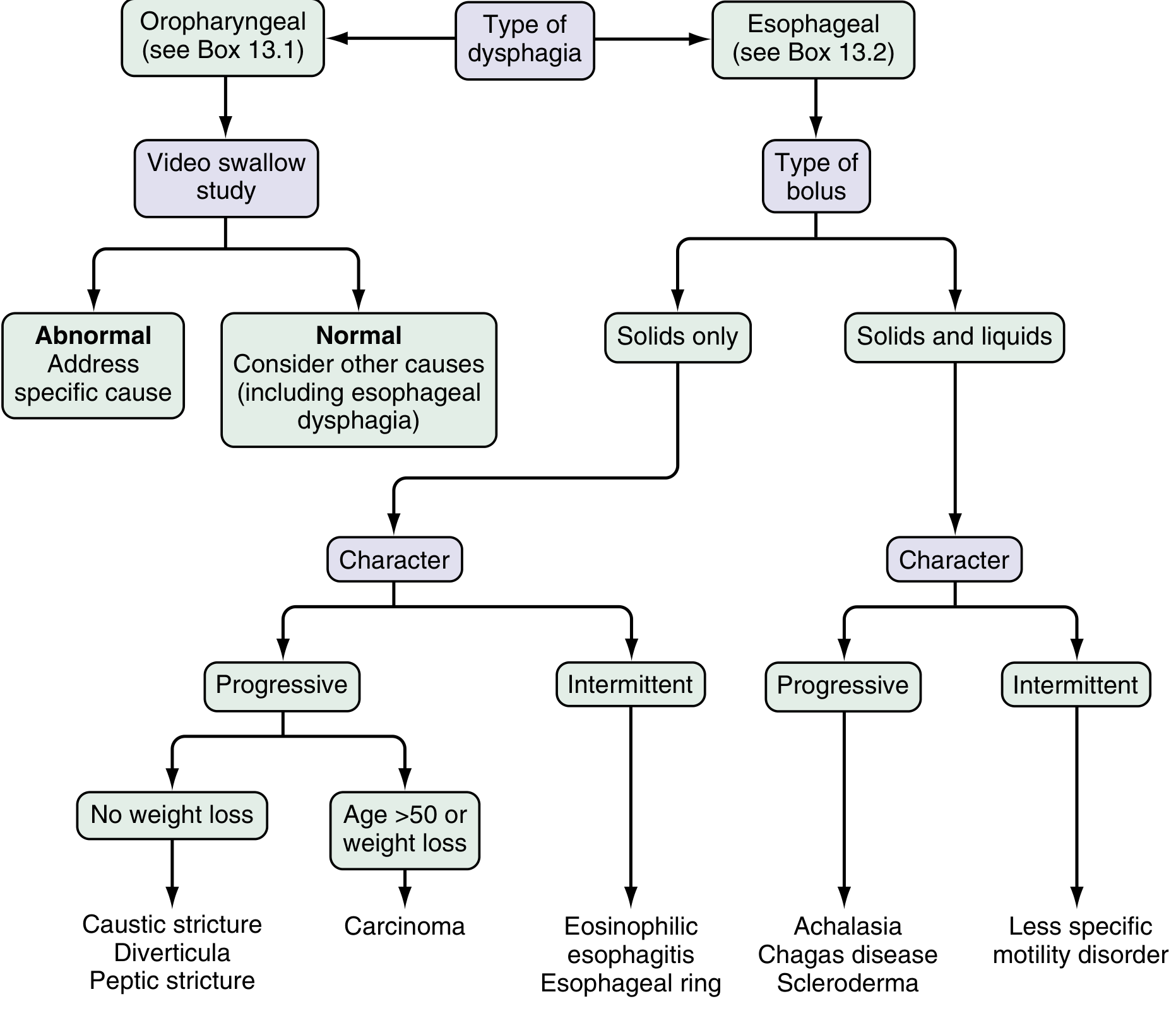
Step 3: Differential Diagnosis
A. Oropharyngeal Dysphagia - Causes
- Stroke (most common)
- Parkinson disease
- ALS (amyotrophic lateral sclerosis)
- Multiple sclerosis
- Myasthenia gravis
- Polymyositis/dermatomyositis
- Muscular dystrophy (oculopharyngeal type)
- Postpolio syndrome
- Thyroid dysfunction
- Zenker's diverticulum (pharyngeal pseudodiverticulum at Killian's triangle)
- Carcinoma
- Cervical osteophytes (e.g., DISH - most common at C5-6)
- Proximal esophageal web
- Prior surgery or radiation therapy
- Infections of pharynx/neck
- Thyromegaly
B. Esophageal Dysphagia - Causes
Motility (Neuromuscular) Disorders
- Achalasia - dysphagia to solids AND liquids, progressive; nocturnal regurgitation of undigested food; bird's-beak appearance on barium swallow
- Diffuse esophageal spasm - dysphagia + chest pain; corkscrew appearance on barium; triggered by hot/cold liquids
- Hypercontractile (jackhammer) esophagus
- Hypertensive LES / Nutcracker esophagus
- Scleroderma (systemic sclerosis) - Raynaud's phenomenon + heartburn + regurgitation
- Chagas disease
- Reflux-related dysmotility
Structural (Mechanical) Disorders
- Peptic stricture - progressive dysphagia for solids; history of GERD
- Carcinoma - progressive, with weight loss; age >50 is a red flag
- Schatzki's ring - episodic, intermittent dysphagia for solids; occurs at start of meal; no weight loss
- Eosinophilic esophagitis (EoE) - intermittent dysphagia/food impaction; young men; linear furrows + corrugated rings on endoscopy
- Medication-induced stricture
- Esophageal webs, diverticula, foreign body
- Mediastinal mass (lymphoma, lung cancer)
- Vascular compression (dysphagia lusoria)
- Spinal osteophytes
Step 4: Red Flag Symptoms (Alarm Features)
- Progressive dysphagia (especially solids -> liquids)
- Unintentional weight loss
- Age >50 with new-onset dysphagia
- Odynophagia (pain on swallowing)
- Hematemesis or melena
- Hoarseness (recurrent laryngeal nerve involvement)
Step 5: Investigation
For Oropharyngeal Dysphagia:
- Modified Barium Swallow (MBS) - first-line; performed with a speech-language pathologist; assesses all phases; different bolus consistencies; can test compensatory maneuvers
- FEES (Fiberoptic Endoscopic Evaluation of Swallowing) - direct pharyngeal visualization before and after swallow; detects penetration, aspiration, pooling; good for bedside/non-ambulatory patients; cannot visualize oral phase or events during the swallow itself
- FEESST - FEES + sensory testing of laryngeal adduction reflex
- Manometry - evaluates UES, LES, and peristaltic coordination
- EAT-10 questionnaire - validated symptom severity and quality-of-life tool
For Esophageal Dysphagia:
- Upper endoscopy (EGD) - first-line when mechanical obstruction is suspected; allows biopsy and immediate dilation; can miss subtle rings
- Barium esophagram - useful for rings, webs, achalasia (bird's beak), diffuse spasm (corkscrew); complementary to endoscopy
- High-resolution manometry (HRM) - essential for motility disorders; detects peristaltic gaps, achalasia subtypes (Chicago Classification), spasm
- CT chest/mediastinum - if extrinsic compression suspected
Key point: When mechanical obstruction is suspected, endoscopy is the preferred initial test because it permits biopsy AND dilation simultaneously. When transfer dysphagia or motility disorder is suspected, start with modified barium swallow or manometry. - Harrison's Principles of Internal Medicine 22E
Step 6: Management Overview
Oropharyngeal Dysphagia
| Approach | Technique |
|---|---|
| Diet modification | Thickened liquids, soft/pureed diet |
| Supraglottic swallow | Inhale and hold breath, swallow, then cough before inhaling; used for vocal cord paresis |
| Effortful swallow | Squeeze hard with all muscles; propels bolus |
| Mendelsohn maneuver | Lift and hold larynx during swallow to extend UES opening duration |
| Shaker head-lift exercise | Strengthens suprahyoid muscles (geniohyoid, thyrohyoid, digastric); improves UES opening |
| Head tilt | Tilt toward paralyzed vocal cord to direct food away from airway |
| Thermal-tactile stimulation | Cold stimulus to anterior tonsillar pillars; reduces pharyngeal delay |
| Zenker's diverticulum | Diverticulectomy + cricopharyngeal myotomy (open or endoscopic) |
Esophageal Dysphagia
| Condition | Treatment |
|---|---|
| Peptic stricture | PPI therapy + endoscopic dilation |
| Schatzki's ring | Endoscopic dilation (balloon or Savary); acid suppression may reduce recurrence |
| Eosinophilic esophagitis | Dietary elimination + topical corticosteroids + dilation if needed |
| Achalasia | Pneumatic dilation; Heller myotomy (± fundoplication, 70-90% success); POEM (per-oral endoscopic myotomy); botulinum toxin into LES (temporary) |
| Diffuse esophageal spasm | Smooth muscle relaxants (CCBs, anticholinergics); botulinum toxin injections |
| Malignant stricture | Self-expanding metal stent (palliation); surgical/oncologic referral |
| Scleroderma esophagus | PPI, prokinetics; treat underlying disease |
Quick Summary Mnemonic: DYSPHAGIA
| Letter | Key Point |
|---|---|
| D | Distinguish: oropharyngeal vs. esophageal |
| Y | Years: progressive over months/years = malignancy until proven otherwise |
| S | Solids only = mechanical; Solids + liquids = motility |
| P | Progressive vs. intermittent character |
| H | Heartburn history (suggests GERD-related peptic stricture or EoE) |
| A | Alarm features (weight loss, age >50, odynophagia) = urgent endoscopy |
| G | Go-to tests: MBS for oropharyngeal; EGD + manometry for esophageal |
| I | Initiate treatment: dilation, dietary modification, or surgery as indicated |
| A | Aspiration risk: always assess pulmonary consequences |
Key Imaging Examples