
Anatomy and Physiology of the eye in detail
Reading File
“Anatomy and physiology of the eye in detail” is a large topic. It can be covered systematically as:
- Gross anatomy and protective structures - orbit, eyelids, conjunctiva, lacrimal apparatus, extraocular muscles
- Eyeball structure - fibrous, vascular, and neural coats; anterior and posterior segments
- Optical apparatus - cornea, aqueous humour, lens, vitreous, accommodation and refraction
- Retina and visual transduction - photoreceptors, retinal layers, macula, optic disc, light-to-nerve signalling
- Visual pathways - optic nerve to visual cortex, pupillary light reflexes, visual fields
- Physiology of vision - colour, dark adaptation, acuity, depth perception, eye movements
- Applied anatomy - glaucoma, cataract, retinal detachment, refractive errors, common examination findings
At a high level, the eye focuses incoming light through the cornea and lens onto the retina. Retinal photoreceptors convert light into electrical signals, which travel through the optic nerve and visual pathways to the occipital cortex, where vision is interpreted. Aqueous humour maintains pressure and nourishes anterior structures, while the lacrimal system, lids, and ocular muscles protect, lubricate, and position the globe.
Asking for Preferences
Which section should I explain first in detailed study-note format?
“Anatomy and physiology of the eye in detail” is a large topic. It can be covered systematically as:
- Gross anatomy and protective structures - orbit, eyelids, conjunctiva, lacrimal apparatus, extraocular muscles
- Eyeball structure - fibrous, vascular, and neural coats; anterior and posterior segments
- Optical apparatus - cornea, aqueous humour, lens, vitreous, accommodation and refraction
- Retina and visual transduction - photoreceptors, retinal layers, macula, optic disc, light-to-nerve signalling
- Visual pathways - optic nerve to visual cortex, pupillary light reflexes, visual fields
- Physiology of vision - colour, dark adaptation, acuity, depth perception, eye movements
- Applied anatomy - glaucoma, cataract, retinal detachment, refractive errors, common examination findings
At a high level, the eye focuses incoming light through the cornea and lens onto the retina. Retinal photoreceptors convert light into electrical signals, which travel through the optic nerve and visual pathways to the occipital cortex, where vision is interpreted. Aqueous humour maintains pressure and nourishes anterior structures, while the lacrimal system, lids, and ocular muscles protect, lubricate, and position the globe.
Please choose one section for detailed notes:
- Eyeball anatomy
- Optics and accommodation
- Retina and physiology of vision
- Visual pathways and reflexes
- Full exam-focused overview
Give me a full exam-focused overview of the eye
Reading File
Finding Sources
Finding Sources
Searching Images
labeled diagram anatomy of the human eye cornea lens retina optic nerve
Searching PubMed
eye anatomy physiology
Searching the Web
human eye anatomy physiology overview NEI
Finding Sources
Finding Sources
Reading File
Reading File
Reading File
Reading Web Page
https://www.nei.nih.gov/eye-health-information/healthy-visio…
Anatomy and Physiology of the Eye: Exam-Focused Overview
1. Basic organization of the eye
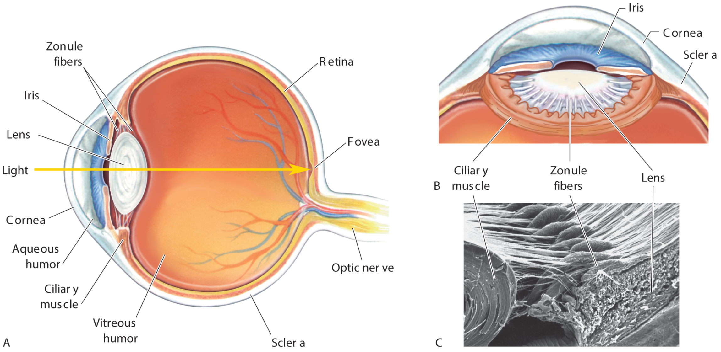
The eyeball is approximately spherical, about 24 mm in diameter, and lies in the orbit. Its function is to:
- Receive light
- Refract and focus it on the retina
- Convert light into electrical signals
- Transmit those signals to the visual cortex for interpretation
Components
| Part | Main function |
|---|---|
| Accessory structures | Protect, lubricate, and move the globe |
| Eyeball | Optical focusing and photoreception |
| Optic nerve and visual pathway | Carry and process visual signals |
The eyeball has three coats:
- Fibrous coat: sclera and cornea
- Vascular coat / uvea: choroid, ciliary body, iris
- Neural coat: retina
2. Accessory structures
A. Orbit
The orbit is a pyramidal bony cavity that contains the globe, extraocular muscles, lacrimal gland, nerves, vessels, orbital fat, and connective tissue.
- Apex: posterior, directed toward the optic canal.
- Base: anterior orbital opening.
- The optic nerve enters the orbit through the optic canal with the ophthalmic artery.
B. Eyelids
The eyelids protect the eye, spread the tear film, and prevent excessive light exposure.
Layers of eyelid, anterior to posterior
- Skin
- Subcutaneous tissue
- Orbicularis oculi muscle
- Tarsal plate
- Palpebral conjunctiva
Important glands
- Meibomian (tarsal) glands: modified sebaceous glands within tarsal plates. Their oily secretion reduces tear evaporation.
- Glands of Zeis: sebaceous glands associated with eyelashes.
- Glands of Moll: modified sweat glands.
Muscles
- Orbicularis oculi: closes the eye. Facial nerve, CN VII.
- Levator palpebrae superioris: elevates upper eyelid. Oculomotor nerve, CN III.
- Superior tarsal muscle (Müller muscle): assists eyelid elevation. Sympathetic supply.
Clinical correlation
- CN III palsy: ptosis due to levator weakness.
- Horner syndrome: mild ptosis due to loss of superior tarsal muscle activity.
- Facial palsy: inability to close the eye, risking corneal exposure.
C. Conjunctiva
A thin, transparent mucous membrane covering:
- Posterior surface of eyelids: palpebral conjunctiva
- Anterior sclera: bulbar conjunctiva
It does not cover the cornea. The space between palpebral and bulbar conjunctiva is the conjunctival sac.
Functions:
- Lubrication
- Protection against pathogens
- Allows smooth movement of lid over globe
D. Lacrimal apparatus
Components:
- Lacrimal gland
- Excretory ducts
- Lacrimal puncta
- Lacrimal canaliculi
- Lacrimal sac
- Nasolacrimal duct
Flow of tears
Lacrimal gland → conjunctival sac → lacrimal puncta → canaliculi → lacrimal sac → nasolacrimal duct → inferior meatus of nose
Tears contain water, electrolytes, mucin, lipids, lysozyme, lactoferrin, and immunoglobulins. They lubricate the cornea, wash out debris, and contribute to the eye's refractive surface.
3. Extraocular muscles and movements
There are six muscles moving each globe:
| Muscle | Primary action | Nerve |
|---|---|---|
| Medial rectus | Adduction | CN III |
| Lateral rectus | Abduction | CN VI |
| Superior rectus | Elevation, adduction, intorsion | CN III |
| Inferior rectus | Depression, adduction, extorsion | CN III |
| Superior oblique | Intorsion, depression, abduction | CN IV |
| Inferior oblique | Extorsion, elevation, abduction | CN III |
Nerve mnemonic
LR6 SO4, all others 3
- Lateral rectus: CN VI
- Superior oblique: CN IV
- All remaining extraocular muscles: CN III
Actions tested in examination
- Superior rectus elevates best when the eye is abducted.
- Inferior oblique elevates best when the eye is adducted.
- Inferior rectus depresses best when the eye is abducted.
- Superior oblique depresses best when the eye is adducted.
4. Coats of the eyeball
A. Fibrous coat
1. Sclera
The sclera is the opaque, white posterior five-sixths of the fibrous coat.
Functions:
- Maintains globe shape
- Protects intraocular structures
- Provides attachment for extraocular muscles
- Resists raised intraocular pressure
2. Cornea
The cornea forms the transparent anterior one-sixth of the fibrous coat.
Functions:
- Main refractive surface of the eye
- Protects anterior structures
- Permits entry of light
The cornea is avascular, so it receives nutrients and oxygen from tears, aqueous humour, limbal blood vessels, and atmospheric oxygen. It is highly sensitive because it receives sensory innervation mainly from the ophthalmic division of trigeminal nerve, CN V1.
The cornea is the eye's major refractive element. Neuroscience: Exploring the Brain, 5th ed., p. 831.
Corneal layers, anterior to posterior
- Epithelium
- Bowman layer
- Stroma
- Descemet membrane
- Endothelium
The corneal endothelium maintains relative dehydration of the stroma. Failure of this pump function causes corneal edema and loss of transparency.
B. Vascular coat: uvea
The uvea comprises the choroid, ciliary body, and iris.
1. Choroid
A pigmented, vascular layer between sclera and retina.
Functions:
- Supplies outer retina, especially photoreceptors
- Absorbs stray light and reduces internal reflection
- Helps regulate retinal temperature
2. Ciliary body
The ciliary body has two major functions:
- Production of aqueous humour
- Control of lens shape for accommodation
It contains:
- Ciliary processes, which produce aqueous humour
- Ciliary muscle, which alters tension on zonular fibers
3. Iris
The iris is the colored, anterior part of the uvea. Its central opening is the pupil.
Muscles of the iris:
| Muscle | Action | Nerve supply |
|---|---|---|
| Sphincter pupillae | Constricts pupil, miosis | Parasympathetic, CN III |
| Dilator pupillae | Dilates pupil, mydriasis | Sympathetic |
5. Chambers and fluids of the eye
The eye has three chambers:
| Chamber | Boundaries | Contents |
|---|---|---|
| Anterior chamber | Cornea anteriorly, iris posteriorly | Aqueous humour |
| Posterior chamber | Iris anteriorly, lens and zonules posteriorly | Aqueous humour |
| Vitreous chamber | Lens anteriorly, retina posteriorly | Vitreous body |
Histology: A Text and Atlas with Correlated Cell and Molecular Biology, 8th ed., p. 2372.
A. Aqueous humour
Aqueous humour is a clear fluid produced mainly by the ciliary processes.
Flow of aqueous humour
Ciliary processes → posterior chamber → pupil → anterior chamber → trabecular meshwork at iridocorneal angle → canal of Schlemm → episcleral veins
Functions:
- Nourishes avascular cornea and lens
- Removes metabolic waste
- Maintains intraocular pressure
- Helps preserve globe shape
Glaucoma
Glaucoma is optic neuropathy usually associated with raised intraocular pressure. It can result from:
- Reduced aqueous outflow through the trabecular meshwork: open-angle glaucoma
- Obstruction of the iridocorneal angle by the iris: angle-closure glaucoma
B. Vitreous body
The vitreous body is a transparent gel filling the vitreous chamber. It is approximately 99% water and helps:
- Maintain shape of the eye
- Support the retina
- Cushion the eye during movement
Histology: A Text and Atlas with Correlated Cell and Molecular Biology, 8th ed., pp. 2372-2374.
6. Lens and accommodation
The lens is a transparent, biconvex, avascular structure behind the iris. It is suspended by zonular fibers attached to the ciliary body.
Functions:
- Fine focusing of light on retina
- Accommodation for near vision
Accommodation for near vision
For a near object:
- Parasympathetic fibers travel in CN III to the ciliary muscle.
- Ciliary muscle contracts.
- Tension on zonular fibers decreases.
- Lens becomes more rounded and convex.
- Refractive power increases.
- Near image is focused on the retina.
The near response consists of the near triad:
- Accommodation
- Convergence of eyes
- Pupillary constriction
For distant vision:
- Ciliary muscle relaxes.
- Zonular tension increases.
- Lens becomes flatter.
- Refractive power decreases.
Presbyopia
With age, the lens becomes less elastic and accommodation decreases. This causes difficulty focusing on near objects, called presbyopia.
7. Refraction and refractive errors
The eye's refractive media are:
- Cornea
- Aqueous humour
- Lens
- Vitreous humour
The cornea provides most of the total refractive power; the lens provides adjustable focusing. The National Eye Institute overview describes the sequence as cornea, pupil, lens, retina, and optic nerve.
Refractive errors
| Condition | Optical problem | Image focus |
|---|---|---|
| Emmetropia | Normal eye | On retina |
| Myopia | Eye too long or refractive power too strong | In front of retina |
| Hypermetropia | Eye too short or refractive power too weak | Behind retina |
| Astigmatism | Unequal curvature of cornea or lens | Different focal points |
| Presbyopia | Reduced accommodation | Near objects cannot be focused clearly |
- Myopia is corrected by a concave lens.
- Hypermetropia is corrected by a convex lens.
- Astigmatism is corrected by a cylindrical lens.
8. Retina
The retina is the neural, light-sensitive inner coat of the eye. It extends from the optic disc posteriorly to the ora serrata anteriorly.
It has two parts:
- Optic retina: photosensitive posterior part
- Nonvisual retina: anterior continuation over ciliary body and posterior iris
Important retinal landmarks
Optic disc
- Site where optic nerve exits the eye
- Contains retinal blood vessels
- Has no rods or cones
- Produces the physiological blind spot
Macula lutea
- Yellowish central retinal area
- Responsible for central vision
Fovea centralis
- Central depression in macula
- Site of highest visual acuity
- Contains densely packed cones
- Avascular region
The fovea is the primary visual axis and site of maximal visual acuity. Gray's Anatomy for Students, 5th ed., p. 1345.
Retinal cells and signal flow
Main neural sequence:
Photoreceptors → bipolar cells → ganglion cells → optic nerve
Supporting interneurons:
- Horizontal cells: integrate signals laterally in the outer retina.
- Amacrine cells: modify signaling between bipolar and ganglion cells.
Ganglion-cell axons form the optic nerve.
Photoreceptors
| Feature | Rods | Cones |
|---|---|---|
| Number | More numerous | Fewer |
| Main location | Peripheral retina | Macula and fovea |
| Light sensitivity | Very high | Lower |
| Function | Dim-light vision, night vision | Daylight and color vision |
| Visual acuity | Lower | High |
| Color discrimination | Absent | Present |
Rods
- Mediate scotopic vision: vision in dim light.
- Contain rhodopsin.
- More sensitive but less precise due to convergence of many rods onto fewer ganglion cells.
Cones
- Mediate photopic vision: daylight vision.
- Provide high acuity and color vision.
- Three classes respond preferentially to short, medium, or long wavelengths, often simplified as blue, green, and red sensitivity.
9. Phototransduction
Phototransduction is conversion of light energy into an electrical neural signal.
In darkness
- cGMP concentration is high.
- cGMP-gated sodium channels remain open.
- Sodium and calcium enter the photoreceptor.
- Cell remains relatively depolarized.
- Glutamate is continuously released.
In light
- Photon activates rhodopsin in rods or photopsin in cones.
- Activated pigment stimulates the G-protein transducin.
- Transducin activates phosphodiesterase.
- Phosphodiesterase lowers intracellular cGMP.
- cGMP-gated sodium channels close.
- Photoreceptor hyperpolarizes.
- Glutamate release decreases.
- Bipolar and ganglion-cell signaling changes, transmitting visual information to the brain.
This light-induced reduction in cGMP closes cation channels and hyperpolarizes photoreceptors. Ganong's Review of Medical Physiology, 26th ed., p. 196.
Dark adaptation
Dark adaptation is progressive improvement in sensitivity after moving from bright light to darkness.
- Initial phase: cones adapt quickly.
- Later, more marked phase: rods adapt more slowly but become much more sensitive.
- Rhodopsin regeneration contributes to rod dark adaptation.
10. Visual pathway
Main pathway
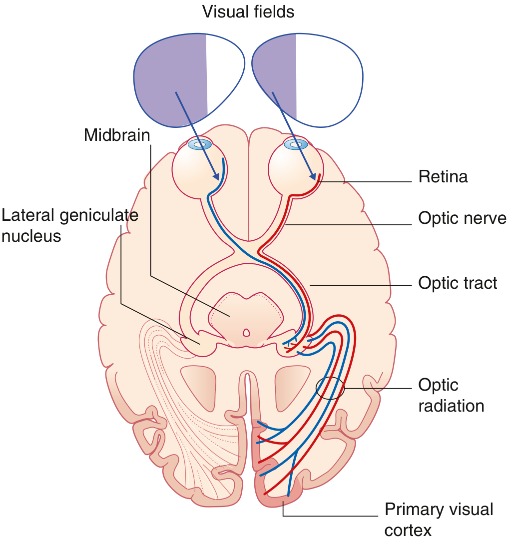
Retina → optic nerve → optic chiasma → optic tract → lateral geniculate nucleus → optic radiation → primary visual cortex
At the optic chiasma
- Fibers from the nasal retina cross to the opposite side.
- Fibers from the temporal retina remain uncrossed.
Therefore, each optic tract carries information from the contralateral visual field:
- Right optic tract carries left visual field information.
- Left optic tract carries right visual field information.
Cortical destination
Fibers synapse in the lateral geniculate nucleus (LGN) of the thalamus. From there, optic radiations pass to the primary visual cortex around the calcarine sulcus in the occipital lobe.
- Upper visual field projects to lower bank of calcarine sulcus.
- Lower visual field projects to upper bank.
- Macular vision has a disproportionately large cortical representation.
The optic nerve is part of the central nervous system, and nasal retinal fibers decussate at the optic chiasm. Gray's Anatomy for Students, 5th ed., pp. 1345-1346.
Visual field defects
| Site of lesion | Typical visual defect |
|---|---|
| Optic nerve | Ipsilateral monocular blindness |
| Optic chiasma | Bitemporal hemianopia |
| Optic tract | Contralateral homonymous hemianopia |
| Temporal optic radiation, Meyer loop | Contralateral superior quadrantanopia |
| Parietal optic radiation | Contralateral inferior quadrantanopia |
| Occipital cortex | Contralateral homonymous hemianopia, often with macular sparing |
11. Pupillary reflexes
Pupillary light reflex
Afferent limb
Retina → optic nerve → optic tract → pretectal nucleus
Efferent limb
Pretectal nucleus → Edinger-Westphal nucleus → CN III → ciliary ganglion → short ciliary nerves → sphincter pupillae
Light in one eye normally produces:
- Direct light reflex: constriction of illuminated pupil
- Consensual light reflex: constriction of opposite pupil
Accommodation reflex
Near viewing produces:
- Lens accommodation
- Pupillary constriction
- Convergence of both eyes
12. Blood supply
Arterial supply
The ophthalmic artery is a branch of the internal carotid artery.
Major branches relevant to the eye:
- Central retinal artery: supplies inner retina
- Short posterior ciliary arteries: supply choroid and optic nerve head
- Long posterior ciliary arteries: supply anterior uvea
- Anterior ciliary arteries: contribute to anterior segment supply
Venous drainage
- Central retinal vein
- Vorticose veins draining the choroid
- Ophthalmic veins draining into cavernous sinus
Clinical point: The central retinal artery is an end artery. Its sudden occlusion can cause abrupt, severe, painless monocular visual loss.
13. Common high-yield clinical correlations
Cataract
Opacity of the crystalline lens.
- Painless progressive decrease in vision
- Commonly age-related
- Causes glare and reduced visual clarity
Glaucoma
Progressive optic neuropathy often associated with increased intraocular pressure.
- Open-angle glaucoma: gradual peripheral visual-field loss
- Acute angle-closure glaucoma: painful red eye, halos around lights, headache, nausea, fixed mid-dilated pupil
Retinal detachment
Separation of the neurosensory retina from the retinal pigment epithelium.
Typical symptoms:
- Flashes of light
- Floaters
- Painless curtain-like loss of vision
Macular degeneration
Disease involving the macula, producing central visual loss while peripheral vision may initially remain relatively preserved.
Papilledema
Optic-disc swelling due to raised intracranial pressure.
Optic neuritis
Inflammation of optic nerve, often presenting with:
- Pain on eye movement
- Reduced visual acuity
- Reduced color vision
- Relative afferent pupillary defect in unilateral disease
14. Rapid revision points
- The cornea is the chief refracting structure.
- The lens fine-tunes focus and becomes more convex during near accommodation.
- Aqueous humour flows from ciliary processes to posterior chamber, through pupil to anterior chamber, then through trabecular meshwork and canal of Schlemm.
- Rods mediate dim-light vision; cones mediate color and high-acuity vision.
- The fovea has the highest visual acuity and is cone-rich.
- The optic disc is the blind spot because it lacks photoreceptors.
- Nasal retinal fibers cross in the optic chiasma.
- Each occipital cortex receives information from the opposite visual field.
- Parasympathetic fibers constrict the pupil; sympathetic fibers dilate it.
- LR6 SO4, all others 3 summarizes extraocular-muscle innervation.
For a plain-language confirmation of light's path through the eye, see the National Eye Institute guide.
This is a shared conversation. Sign in to Orris to start your own chat.