
Levels of neck lymph nodes , the content which drains into it . And anatomically covered this topic
Reading File
Finding Sources
Finding Sources
Searching PubMed
"cervical lymph nodes" AND anatomy
Searching the Web
cervical lymph node levels anatomical boundaries drainage official head neck cancer staging
Searching Images
anatomical diagram cervical neck lymph node levels I II III IV V VI boundaries
Reading File
Reading File
Reading Web Page
https://radiopaedia.org/articles/lymph-node-levels-of-the-ne…
Reading File
Reading File
Cervical lymph-node levels: anatomy and drainage
The neck-node level system is used in clinical examination, imaging, head-and-neck cancer staging, and neck dissection. It divides cervical nodes into Levels I to VI, with Level VII sometimes used for the upper mediastinum.
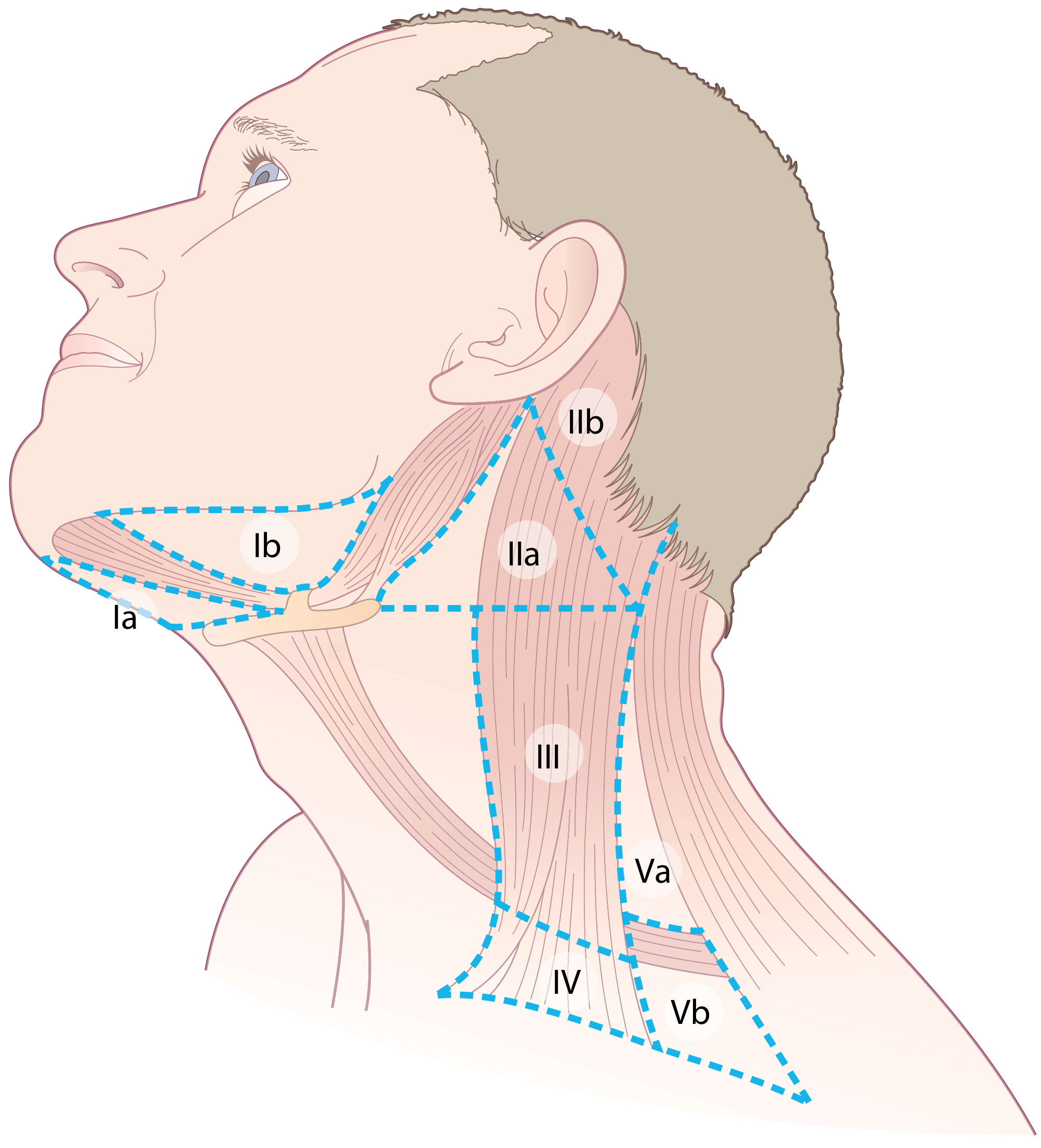
Overall lymphatic pathway
Superficial head and neck nodes
(occipital, mastoid/post-auricular, parotid/pre-auricular, facial, submental, submandibular)
→ superficial cervical or deep cervical chain
→ jugular lymphatic trunk
→ right lymphatic duct on the right, or thoracic duct on the left.
(occipital, mastoid/post-auricular, parotid/pre-auricular, facial, submental, submandibular)
→ superficial cervical or deep cervical chain
→ jugular lymphatic trunk
→ right lymphatic duct on the right, or thoracic duct on the left.
The deep cervical chain lies along the internal jugular vein. It is the final major cervical nodal drainage pathway. - Gray's Anatomy for Students, p. 1178
Table: Levels, anatomical boundaries, nodes, and drainage
| Level | Name / principal nodes | Anatomical location and boundaries | Main structures draining to it |
|---|---|---|---|
| IA | Submental nodes | Midline submental triangle, between the right and left anterior bellies of digastric; from mandibular symphysis region to hyoid | Central lower lip, chin, anterior floor of mouth, tip/ventral anterior tongue, lower incisors |
| IB | Submandibular nodes | Submandibular triangle. Lateral to anterior belly of digastric and beneath mandible; related to submandibular gland and facial vessels | Cheek, lateral lower lip, gums, teeth, lateral anterior tongue, lateral floor of mouth, submandibular gland; much of face drains here |
| II | Upper jugular chain, especially jugulodigastric node | Skull base to inferior border of hyoid. Along upper internal jugular vein, deep to upper sternocleidomastoid | Palatine tonsil and oropharynx, nasopharynx, oral cavity, parotid, hypopharynx, supraglottic larynx |
| IIA | Upper jugular nodes anterior/medial to spinal accessory nerve | Same vertical limits as Level II; generally anterior or medial to CN XI | Same drainage territory as Level II, especially oropharynx and tonsil |
| IIB | Upper jugular nodes posterior/lateral to spinal accessory nerve | Posterior to CN XI, bounded by posterior belly of digastric superiorly and SCM posteriorly | Usually receives less direct mucosal drainage; can receive drainage from posterior scalp/upper neck and from adjacent Level II nodes |
| III | Middle jugular chain | Inferior border of hyoid to inferior border of cricoid cartilage, along middle internal jugular vein | Lower oropharynx, oral tongue, hypopharynx, and larynx |
| IV | Lower jugular chain | Inferior border of cricoid cartilage to clavicle, along lower internal jugular vein | Hypopharynx, larynx, cervical oesophagus, thyroid. Left lower Level IV can contain Virchow node, which may reflect thoracic or abdominal malignancy |
| VA | Upper posterior-triangle nodes, including spinal accessory chain | Posterior triangle above inferior border of cricoid. Between posterior border of SCM anteriorly and anterior border of trapezius posteriorly | Posterior scalp and neck, and may receive spread from nasopharynx and oropharynx |
| VB | Lower posterior-triangle and supraclavicular nodes | Posterior triangle below inferior border of cricoid to clavicle | Posterior scalp/neck and lower neck drainage; receives lymph from other cervical nodal groups. Supraclavicular nodes may receive thoracic and abdominal lymphatic drainage, especially on the left |
| VI | Central/anterior compartment: prelaryngeal (Delphian), pretracheal, paratracheal, perithyroid nodes | Midline compartment. Superior: hyoid; inferior: suprasternal notch or upper mediastinal continuation; lateral: common carotid arteries | Thyroid, parathyroids, glottic/subglottic larynx, cervical trachea, cervical oesophagus, apex of piriform sinus |
Scott-Brown's Otorhinolaryngology describes IA as draining the anterior floor of mouth, lower lip and ventral tongue; IB as receiving drainage from lateral tongue, lateral floor of mouth and buccal cavity; and Levels II to IV as the upper, middle, and lower jugular chains. - Scott-Brown's Otorhinolaryngology Head & Neck Surgery, p. 599
Anatomical divisions to remember
Level I: submental and submandibular
- IA = central, under the chin.
- IB = beneath the mandible, lateral to IA.
- Easy memory:
- IA drains the midline anterior oral cavity.
- IB drains the lateral oral cavity and face.
Levels II, III, IV: deep jugular chain
These are arranged vertically along the internal jugular vein:
- Level II: skull base → hyoid
- Level III: hyoid → cricoid
- Level IV: cricoid → clavicle
Easy memory: Upper, middle, lower jugular nodes.
A clinically important Level II node is the jugulodigastric node, near the posterior belly of digastric and internal jugular vein. It classically enlarges in tonsillitis because it receives drainage from the palatine tonsil. - Gray's Anatomy for Students, p. 1178
Level V: posterior triangle
- Anterior boundary: posterior border of sternocleidomastoid.
- Posterior boundary: anterior border of trapezius.
- Inferior boundary: clavicle.
- VA: above cricoid.
- VB: below cricoid, including supraclavicular nodes.
Level VI: central compartment
- Lies between the carotid arteries.
- Contains prelaryngeal, pretracheal, paratracheal, and perithyroid nodes.
- Important in thyroid carcinoma and cancers of the larynx, trachea, cervical oesophagus, and hypopharynx.
The reference boundaries on cross-sectional imaging differ slightly among surgical and radiology conventions, but the core division is consistent. Radiopaedia’s level reference summarizes the commonly used imaging boundaries.
Other named cervical nodes not routinely included in Levels I-VI
| Node group | Location | Main drainage |
|---|---|---|
| Occipital | At skull base near trapezius attachment | Posterior scalp and upper posterior neck |
| Mastoid / posterior auricular | Behind auricle | Posterolateral scalp |
| Parotid / pre-auricular | In and anterior to parotid gland | Eyelids, lateral face, anterior auricle, anterolateral scalp |
| Retropharyngeal | Behind pharynx | Nasopharynx, posterior nasal cavity, soft palate, posterior pharyngeal wall |
| Facial / buccal | Along facial vessels | Cheek and adjacent face |
| Supraclavicular | Above clavicle, chiefly lower Level IV/VB region | Lower neck, thorax; left-sided node can receive lymph via thoracic duct |
High-yield clinical correlations
- Tonsillitis / oropharyngeal cancer: commonly Level II, especially jugulodigastric nodes.
- Oral cavity cancers: usually Level I first, then Levels II and III.
- Nasopharyngeal cancer: retropharyngeal nodes and Level II, sometimes Level V.
- Laryngeal and hypopharyngeal cancers: Levels II, III, IV.
- Thyroid cancer: first involves Level VI, then Levels II to IV and sometimes Level VII.
- Left supraclavicular node (Virchow node): may signal malignancy from stomach or another thoracic/abdominal site.
- Posterior scalp lesions: commonly drain to occipital/posterior auricular nodes, then superficial cervical and deep cervical nodes.
One-line exam summary
Level I drains oral cavity and face; Levels II-IV are the upper-to-lower jugular chain draining pharynx and larynx; Level V is the posterior triangle draining scalp/posterior neck; Level VI is the central compartment draining thyroid, larynx, trachea, and cervical oesophagus.
This is a shared conversation. Sign in to Orris to start your own chat.