
nagler's reaction
Nagler's reaction Clostridium perfringens egg yolk agar
Nagler reaction egg yolk agar Clostridium perfringens lecithinase
Nagler's Reaction
What It Is
Principle
Medium Used
Procedure
- The egg yolk agar plate is divided into two halves.
- One half is smeared with specific anti-alpha toxin (antitoxin/antiserum against C. perfringens alpha-toxin).
- The test organism is inoculated across both halves.
- Incubated anaerobically at 37°C for 24-48 hours.
Result Interpretation
| Half of Plate | Antitoxin Applied | Result |
|---|---|---|
| Without antitoxin | No | Opaque white halo around colonies (positive - lecithinase active) |
| With antitoxin | Yes | No halo - reaction inhibited (Nagler inhibition) |
- Positive Nagler reaction: Pearly white/opaque zone of precipitation around colonies on the untreated half, with inhibition on the antitoxin-treated half - confirms C. perfringens.
- If both halves show opacity (no inhibition), the organism produces a non-specific lecithinase and is NOT C. perfringens.
Significance of Alpha-Toxin
- Damages cell membranes (lecithinase activity)
- Causes hemolysis (responsible for the double zone of hemolysis on blood agar)
- Key pathogenic factor in gas gangrene (clostridial myonecrosis)
- Also contributes to food poisoning
- Identified in clinical settings using the Nagler reaction
Key Points to Remember
- Organism identified: Clostridium perfringens (also called Cl. welchii)
- Toxin detected: Alpha-toxin (phospholipase C / lecithinase)
- Medium: Egg yolk agar
- Hallmark: Inhibition of the opaque halo by specific antitoxin on one half of the plate
- C. perfringens also shows "boxcar" (short, fat) Gram-positive rods and double zone of hemolysis on blood agar
- The antitoxin-based inhibition makes the test specific - it rules out other lecithinase-producing organisms (e.g., Bacillus cereus, C. bifermentans)
laboratory diagnosis of drug resistant tuberculosis
laboratory diagnosis drug resistant tuberculosis WHO 2025 methods
Laboratory Diagnosis of Drug-Resistant Tuberculosis (DR-TB)
Classification of Drug Resistance (WHO Definitions)
| Type | Definition |
|---|---|
| Monoresistance | Resistance to one first-line drug only |
| Polydrug resistance (PDR) | Resistance to >1 first-line drug, but NOT both INH + RIF |
| MDR-TB | Resistance to at least both isoniazid (INH) AND rifampicin (RIF) |
| Pre-XDR-TB | MDR/RR-TB + resistance to any fluoroquinolone |
| XDR-TB | MDR/RR-TB + fluoroquinolone resistance + resistance to bedaquiline or linezolid |
| RR-TB (Rifampicin-resistant) | Resistance to RIF by any method, with or without other resistance |
Two Major Approaches to DST
1. Phenotypic (Culture-Based) Methods
2. Genotypic (Molecular) Methods
A. PHENOTYPIC METHODS
1. Proportion Method (Reference Standard)
- Cultures inoculated on drug-free media and drug-containing media in parallel.
- Resistance defined as: >1% of colonies grow on drug-containing medium compared to drug-free control.
- Can be done on solid (Lowenstein-Jensen agar) or liquid (broth) media.
- Results on liquid media: ~7-14 days; solid media: up to 21 days or longer.
- (Murray & Nadel's Respiratory Medicine; Harrison's 22e)
2. Automated Broth-Based Systems (MGIT, BACTEC)
- Gold standard liquid culture using fluorometric/radiometric growth detection.
- First-line drugs (INH, RIF, PZA, EMB) tested using a critical concentration.
- Sensititre MycoTB plate can determine MICs for multiple drug concentrations.
- WHO recommends automated liquid culture for all Group A drugs (moxifloxacin, levofloxacin, bedaquiline, linezolid), Group B (clofazimine), and Group C (amikacin, delamanid, pyrazinamide).
- (Tietz Textbook of Laboratory Medicine 7e; Murray & Nadel)
3. Colorimetric Redox Methods
- AlamarBlue / Resazurin assay: Viable bacteria reduce the indicator, causing a color change (blue to pink = resistant). Sensitivity 98% / Specificity 99% for RIF; 97% / 98% for INH.
- Nitrate Reductase Assay (NRA): Based on M. tuberculosis reducing nitrate to nitrite. Sensitivity 97%, specificity 100% for RIF resistance.
- (Murray & Nadel's Respiratory Medicine)
4. MODS (Microscopic Observation Drug Susceptibility) Assay
- A liquid broth culture method in a multi-well plate WITH and WITHOUT antibiotics.
- Microscopy used to identify characteristic cording of M. tuberculosis complex.
- Detects resistance to RIF and INH simultaneously.
- Inexpensive, suitable for resource-limited settings.
- (Jawetz Melnick & Adelberg's Medical Microbiology 28e; Murray & Nadel)
B. GENOTYPIC (MOLECULAR) METHODS
1. GeneXpert MTB/RIF & MTB/RIF Ultra (CB-NAAT)
- The most widely deployed rapid test worldwide.
- Detects M. tuberculosis DNA AND rifampicin resistance (mutations in rpoB gene) simultaneously in unprocessed sputum in ~90 minutes.
- Uses molecular beacons (fluorescent probes) in a real-time PCR format.
- Sensitivity ~83%, Specificity ~98% for TB detection; excellent for RIF resistance.
- Xpert Ultra has higher sensitivity for paucibacillary disease (smear-negative TB, children, PLHIV).
- Rifampicin resistance on Xpert is used as a proxy marker for MDR-TB.
- (Park's PSM; Campbell's Operative Orthopaedics 15e; Comprehensive Clinical Nephrology 7e)
2. Line Probe Assays (LPA) - MTBDRplus, MTBDRsl
- Detects resistance to Rifampicin (rpoB gene) and Isoniazid (katG and inhA genes).
- Applied directly to smear-positive sputum or culture isolates.
- For smear-negative specimens: culture first, then LPA on isolate.
- PCR amplification followed by reverse hybridization onto nitrocellulose strips.
- Results in 24-48 hours from isolate.
- Detects fluoroquinolone resistance (gyrA, gyrB genes) and second-line injectable resistance (rrs, eis genes).
- WHO recommends as the initial test for rapid detection of FQ and injectable resistance in confirmed MDR-TB isolates.
- (Park's PSM; Harrison's Principles 22e)
| Gene | Drug resistance detected | Detection rate |
|---|---|---|
| rpoB | Rifampicin | ~95-96% of RIF-resistant strains |
| katG | Isoniazid | 50-95% of INH-resistant strains |
| inhA promoter | Isoniazid, ethionamide | Up to 45% of INH resistance |
| gyrA, gyrB | Fluoroquinolones | - |
| rrs, eis | Aminoglycosides (amikacin, kanamycin) | - |
| pncA | Pyrazinamide | - |
| tlyA | Capreomycin | - |
3. Targeted Next-Generation Sequencing (tNGS)
- Amplification and sequencing of key drug resistance gene regions directly from sputum.
- Now WHO-recommended after TB diagnosis to guide comprehensive DST and treatment decisions.
- Faster than phenotypic DST; especially useful when comprehensive resistance profiling is needed.
- Commercial assays: Deeplex Myc-TB (GenoScreen) - targets 18 resistance-associated gene loci for 13 anti-TB drugs; DeepChek-TB (ABL).
- Results within 48 hours from clinical specimens.
- Still requires phenotypic confirmation for some newer drugs (bedaquiline, linezolid, pretomanid).
- (Murray & Nadel; Harrison's 22e)
4. Whole Genome Sequencing (WGS)
- The most comprehensive method - sequences the entire M. tuberculosis genome.
- Provides a complete drug resistance profile, strain typing for epidemiology, and transmission cluster analysis.
- England implemented nationwide WGS of all M. tuberculosis clinical isolates in 2017 as a public health initiative.
- Currently requires culture before processing (limitation); direct-from-sputum approaches under development.
- Best suited for reference laboratory settings.
- (Tietz Textbook of Laboratory Medicine 7e)
5. Sanger Sequencing / Pyrosequencing
- Used by reference laboratories (e.g., CDC) to predict resistance to first- and second-line drugs.
- High sensitivity and specificity compared to phenotypic DST.
- (Henry's Clinical Diagnosis 23e)
Summary: Comparison of Methods
| Method | Time to Result | Drugs Covered | Best Use |
|---|---|---|---|
| Solid proportion (LJ) | 4-8 weeks | All 1st/2nd line | Reference standard |
| Liquid culture (MGIT) | 7-21 days | All 1st/2nd line + Group A | Routine DST |
| Colorimetric (resazurin/NRA) | Days-weeks | RIF, INH | Low-resource settings |
| MODS | Days | RIF, INH | Low-resource settings |
| Xpert MTB/RIF / Ultra | 90 minutes | RIF resistance (rpoB) | First rapid screen |
| LPA (1st line) | 24-48 hrs (from isolate) | RIF (rpoB) + INH (katG, inhA) | Rapid initial DST |
| LPA (2nd line) | 24-48 hrs | FQ (gyrA/B) + injectables (rrs/eis) | Confirmed MDR-TB |
| tNGS | 48 hrs | 13 drug classes | Comprehensive DST |
| WGS | Days (from culture) | All genes - complete profile | Reference labs, epidemiology |
Important Limitations of Molecular Methods
- Failure to detect a mutation does not exclude resistance - not all resistance mechanisms are known.
- Phenotypic DST remains essential to detect resistance caused by novel or unknown mutations.
- Molecular methods are adjuncts to, not replacements for, phenotypic DST.
- Testing for PZA resistance has a high false-positive rate without proper quality assurance.
- (Henry's Clinical Diagnosis; Murray & Nadel; Harrison's 22e)
WHO Recommended Diagnostic Cascade (Current Standard)
- Universal DST for all TB patients - at minimum, test for RIF resistance.
- Rapid molecular test (Xpert MTB/RIF) as initial test for all presumptive TB patients.
- If RIF resistance detected - confirm MDR-TB; test 2nd-line LPA or tNGS for FQ and injectable resistance.
- Culture-based DST for all second-line drugs in confirmed MDR-TB.
- tNGS or WGS at reference laboratories for comprehensive resistance profiling and epidemiology.
reason out staphylooocus infections are localised in contrast to the spreading nature of streptococcus infections
why staphylococcus infections localized streptococcus spreading enzymes mechanism
Why Staphylococcal Infections Are Localized vs. Streptococcal Infections Spreading
Part 1: STAPHYLOCOCCUS - The "Fortress Builder"
1. Coagulase - The Key Factor
- S. aureus produces two forms of coagulase:
- Bound coagulase (clumping factor): on the bacterial surface; directly converts fibrinogen to fibrin, coating the bacterial cell in fibrin.
- Free coagulase: secreted enzyme; reacts with a plasma factor (coagulase-reacting factor) to form a staphylothrombin complex, which then converts fibrinogen to fibrin in the surrounding environment.
- Result: A thick fibrin wall is laid down around the infection site, physically preventing the bacteria from spreading into surrounding tissue and simultaneously blocking host phagocytes from penetrating the abscess core.
- This fibrin cocoon is the structural basis of a boil, furuncle, or abscess - the hallmark of staphylococcal disease.
2. Protein A
- A surface protein of S. aureus that binds the Fc region of IgG antibodies backwards (Fc end instead of Fab).
- This prevents opsonization - antibodies coat the bacterium the wrong way, so neutrophils cannot phagocytose it efficiently.
- The organism survives inside the fibrin wall, protected from humoral immunity.
3. Leukocidin (Panton-Valentine Leukocidin / PVL)
- Kills neutrophils and macrophages that do manage to enter the abscess zone.
- Contributes to the pus formation (dead neutrophils = pus) that characterizes staphylococcal abscesses.
- The liquefied center of the abscess (necrotic tissue + dead leukocytes) further consolidates the "local" nature of the infection.
4. What Staphylococcus Lacks
- S. aureus does not produce hyaluronidase in clinically significant quantities as a spreading factor (unlike streptococci).
- It does not produce streptokinase - in fact, it produces the opposite: coagulase (clot-forming, not clot-lysing).
- It does not elaborate the M protein that resists phagocytosis in the way streptococci use to traverse tissue.
Net Result for Staphylococcus
Fibrin wall formed → bacteria trapped inside → neutrophils killed by leukocidin → pus accumulates → localized abscess
Part 2: STREPTOCOCCUS - The "Tissue Destroyer and Spreader"
1. Streptokinase (Fibrinolysin) - The Counter to Coagulase
- Converts plasminogen to plasmin, an enzyme that digests fibrin clots.
- This is the direct biochemical opposite of coagulase - instead of forming a fibrin wall, streptococcus dissolves any fibrin barriers.
- Any attempt by the host to wall off the infection is actively degraded.
- Clinical consequence: no abscess formation - instead, the infection spreads along tissue planes.
2. Hyaluronidase ("Spreading Factor")
- Degrades hyaluronic acid, the main cement substance of connective tissue ground substance.
- This breaks down the intercellular matrix that physically holds tissue together.
- Creates "lanes" through which the organism can dissect along fascial planes.
- Leads to the spreading, flat, non-fluctuant inflammation of erysipelas and cellulitis.
- (Henry's Clinical Diagnosis 23e: "hyaluronidase may enhance the spread of the organism through connective tissue")
3. Deoxyribonucleases (DNases A, B, C, D) and NADase
- DNases liquefy the thick, viscous pus (which is largely composed of DNA from dead neutrophils).
- Thin, liquid pus spreads more easily through tissue than viscous pus.
- This prevents the formation of a thick, walled-off abscess.
- DNase B (streptodomase) is used clinically as an anti-DNase B titer in diagnosing post-streptococcal sequelae.
4. M Protein - Anti-phagocytic Surface Protein
- The M protein on the streptococcal surface binds fibrinogen and serum factor H, disrupting opsonization by complement (C3b cannot bind effectively).
- Bacteria resist phagocytosis and are free to keep dividing and spreading.
- Over 60 M protein types exist - immunity is type-specific, allowing re-infection with different M types.
- (Henry's Clinical Diagnosis 23e)
5. Streptolysins (O and S)
- Streptolysin O (oxygen-labile, antigenic) and Streptolysin S (oxygen-stable, non-antigenic) lyse red blood cells, platelets, and leukocytes.
- Kill host immune cells attempting to contain the infection, allowing spread.
- ASO (anti-streptolysin O) titer is the basis of serological diagnosis of streptococcal infection.
6. Pyrogenic Exotoxins (Erythrogenic toxins A, B, C)
- Superantigens - activate enormous numbers of T cells non-specifically.
- Drive systemic inflammation, contributing to scarlet fever, toxic shock-like syndrome.
- Facilitate tissue destruction and spread.
Net Result for Streptococcus
No fibrin wall (streptokinase dissolves it) → hyaluronic acid matrix degraded → pus liquefied by DNases → bacteria spread freely along tissue planes → cellulitis, erysipelas, necrotizing fasciitis
Summary Comparison Table
| Feature | Staphylococcus aureus | Streptococcus pyogenes |
|---|---|---|
| Key enzyme | Coagulase (clot-FORMING) | Streptokinase (clot-DISSOLVING) |
| Effect on fibrin | Forms fibrin wall around bacteria | Dissolves fibrin barriers |
| Connective tissue | Not significantly degraded | Hyaluronidase breaks it down |
| Pus character | Thick, viscous, localized | Thin, fluid, spreads easily |
| DNase production | Yes (helps evade NETs) | Yes (liquefies pus for spreading) |
| Typical lesion | Abscess, boil, furuncle, carbuncle | Cellulitis, erysipelas, necrotizing fasciitis |
| Abscess formation | YES - characteristic | Rare (pus doesn't wall off) |
| Anti-phagocytic factor | Protein A (Fc receptor block) | M protein (anti-complement) |
The One-Line Mechanistic Summary
Staphylococcus builds a fibrin fortress around itself with coagulase, trapping the infection locally into an abscess; Streptococcus tears down every barrier with streptokinase, hyaluronidase, and DNase, allowing it to spread freely through tissue planes.
rapid plasma reagin test replaced vdrl test in many labs
RPR replaced VDRL syphilis test advantages differences laboratory
RPR vs. VDRL: Why RPR Has Replaced VDRL in Most Labs
Background: What Both Tests Share
- Non-treponemal (reaginic) tests for syphilis
- They detect IgG and IgM reagin antibodies - antibodies directed against a cardiolipin-cholesterol-lecithin antigen derived from beef heart
- These antibodies arise because Treponema pallidum damages host cells, releasing membrane lipids (including cardiolipin) that stimulate antibody production
- Both are flocculation tests - the antibody-antigen interaction forms visible clumps (flocculation/agglutination)
- Both are used for screening and for quantitative monitoring of treatment response (four-fold titer fall = adequate treatment)
- Neither is specific for syphilis - both give biological false positives
The Key Differences That Make RPR Superior for Routine Use
1. Antigen Formulation and Visualization Method
| Feature | VDRL | RPR |
|---|---|---|
| Antigen | Cardiolipin-cholesterol-lecithin in aqueous suspension | Same antigen + choline chloride + charcoal (carbon) particles |
| Reaction product | Microscopic floccules | Macroscopic black clumps visible to naked eye |
| Reading method | Darkfield/light microscope required | Read with naked eye - no microscope needed |
| Reaction vessel | Glass slide | Disposable cardboard card (circle card test) |
2. Specimen Requirements
| Feature | VDRL | RPR |
|---|---|---|
| Specimen for serum | Requires heated (inactivated) serum (56°C for 30 min) | Uses unheated plasma or serum (hence "Unheated" in related TRUST test) |
| CSF testing | YES - VDRL is the ONLY validated test for CSF | Cannot reliably be used on CSF |
| Automation | Difficult to automate | Can be semi-automated |
3. Practical Advantages of RPR Over VDRL
| Advantage | Explanation |
|---|---|
| No microscope needed | VDRL requires reading under a microscope at 100x; RPR is read with the naked eye |
| Faster | RPR results in minutes; no need to heat serum beforehand |
| Simpler to perform | Fewer steps, disposable cards, no glass slide preparation |
| Stable reagents | RPR reagents have a longer shelf life and more stable antigen suspension |
| Suitable for field/peripheral use | Can be performed in low-resource settings without laboratory infrastructure |
| Less technical expertise needed | No microscopy training required for result reading |
| Higher throughput | Disposable cards allow many tests to be run simultaneously |
4. Sensitivity and Specificity - Comparable to VDRL
| Stage | Sensitivity |
|---|---|
| Primary syphilis | ~70-78% (both tests) |
| Secondary syphilis | 99-100% |
| Latent/Late syphilis | ~70% |
- Specificity: 98-99% for both tests
- False-positive rate in the general population: ~1-2%
- (Goldman-Cecil Medicine; Medical Microbiology 9e)
The One Area Where VDRL Cannot Be Replaced: Neurosyphilis
"Only the VDRL test should be used to test CSF from patients with suspected neurosyphilis." (Medical Microbiology 9e)
- The RPR uses a modified antigen suspension optimized for serum/plasma - it is not validated for CSF and gives unacceptably high false-negative rates in CSF.
- The VDRL antigen for CSF is specially diluted (to account for the much lower immunoglobulin concentration in CSF compared to blood) and must be freshly prepared within 2 hours of use.
- CSF-VDRL: highly specific for neurosyphilis but only 30-70% sensitive in late neurosyphilis (tabes dorsalis, general paresis).
- Serum RPR titers are used in parallel to support the diagnosis when CSF-VDRL is negative but suspicion remains high.
False Positives: Both Tests Share This Limitation
- Autoimmune diseases (SLE, antiphospholipid syndrome)
- Pregnancy
- Viral infections (EBV, hepatitis, HIV, COVID-19)
- Intravenous drug use
- Advanced age
- Malaria, leprosy, tuberculosis
The Prozone Phenomenon (Affects Both Tests)
Summary
| Feature | VDRL | RPR |
|---|---|---|
| Reading method | Microscope (100x) | Naked eye |
| Serum preparation | Must heat to 56°C | Unheated |
| Specimen | Serum or CSF | Serum/plasma only |
| CSF (neurosyphilis) | YES - gold standard | NO - not validated |
| Lab infrastructure | Microscope required | Minimal |
| Practical use | Reference labs, neurosyphilis | Routine screening, treatment monitoring |
| Sensitivity/specificity | ~70-99% / 98-99% | ~70-99% / 98-99% |
reason out proteus strains are used to detect rickettsial antibodies in weil felix test
Weil Felix test Proteus strains OX19 OX2 OXK rickettsial antibodies cross reaction antigen basis
https://www.ncbi.nlm.nih.gov/books/NBK559225?report=printabl…
The Weil-Felix Test: Why Proteus Strains Are Used to Detect Rickettsial Antibodies
The Core Concept: A Fortunate Coincidence of Nature
Proteus bacteria and Rickettsia species share structurally similar surface antigens - specifically, the O-polysaccharide chains of their outer membrane lipopolysaccharide (LPS).
Step-by-Step Reasoning
1. Why Not Just Use Rickettsial Antigen Directly?
- They are extremely difficult and dangerous to culture in a standard laboratory.
- Preparing rickettsial antigen suspensions requires BSL-3 biosafety infrastructure.
- Antigen suspensions would be unstable, expensive, and hazardous.
2. What Is the Shared Antigen? The Molecular Basis
- Amano et al. demonstrated chemical and structural similarities between the LPS O-polysaccharides of Proteus vulgaris OX-19 and those of the typhus group rickettsiae (R. prowazekii, R. typhi).
- The target of the Weil-Felix antibodies in patients was recently confirmed to be the rickettsial LPS O-antigen, which is highly conserved across rickettsial groups.
- This O-antigen "mimicry" between Proteus and Rickettsia is the molecular explanation for the cross-reaction.
- (StatPearls - NBK559225)
3. The Three Proteus Strains Used and What They Represent
| Proteus Strain | Species | Cross-reacts with |
|---|---|---|
| OX-19 | P. vulgaris | Typhus group (R. prowazekii, R. typhi) - strongly |
| OX-2 | P. vulgaris | Spotted fever group (R. conorii, R. rickettsii) - with OX-19 |
| OX-K | P. mirabilis | Scrub typhus (Orientia tsutsugamushi) - alone |
4. Interpretation Pattern - How the Test Identifies the Disease Group
| Disease | OX-19 | OX-2 | OX-K |
|---|---|---|---|
| Epidemic typhus (R. prowazekii) | +++ | ± | - |
| Murine typhus (R. typhi) | +++ | ± | - |
| Scrub typhus (O. tsutsugamushi) | - | - | +++ |
| Rocky Mountain Spotted Fever (R. rickettsii) | ++ | ++ | - |
| Indian Tick Typhus (R. conorii) | ++ | ++ | - |
| Q fever (Coxiella burnetii) | - | - | - |
| Rickettsialpox (R. akari) | - | - | - |
- Q fever and rickettsialpox are both negative across all strains - these organisms do not share the cross-reactive O-antigen, making them exceptions.
- The scrub typhus agent (Orientia tsutsugamushi) uniquely cross-reacts only with OX-K.
- (Park's Textbook of Preventive & Social Medicine; StatPearls)
5. The Antibody Being Detected
- The test detects predominantly IgM anti-rickettsial antibodies.
- These cross-reactive IgM antibodies appear 5-10 days after onset of illness - so a test done in the first week may be falsely negative.
- Diagnostic criterion: A single titer of ≥1:160 to 1:320 with a compatible clinical picture is suggestive. A four-fold rise between acute and convalescent serum samples (taken 2-4 weeks apart) is the most reliable evidence.
Why Isn't This Test Considered Specific?
| Limitation | Explanation |
|---|---|
| False positives from Proteus infections | A patient with a Proteus urinary tract infection will have antibodies against Proteus O-antigens - giving a positive test without any rickettsial infection |
| False positives in other febrile illnesses | Leptospirosis, Borrelia, severe liver disease can give non-specific positive results |
| Negative in Q fever and rickettsialpox | These lack the shared OPS antigen |
| Poor sensitivity in early disease | IgM takes 5-10 days to appear |
| OX-K non-specificity | Titers up to 1:160 have been observed in completely healthy individuals for OX-K |
| Does not detect rickettsiae directly | Only detects host antibody cross-reaction |
One-Line Summary of the Reasoning
Rickettsia and Proteus OX strains share structurally similar LPS O-polysaccharide antigens - so anti-rickettsial antibodies raised during infection cross-agglutinate Proteus bacteria, which are safe and easy to culture, making them practical surrogates in place of the dangerous, unculturable rickettsiae. This is a heterophile agglutination reaction.
bacterial conjugation from Apurba shastri textbook of microbiology 5 marks answer mbbs
bacterial conjugation Apurba Sastri microbiology F factor Hfr F prime mechanism
bacterial conjugation F factor sex pilus F+ Hfr diagram gene transfer
Bacterial Conjugation
Definition
The F Factor (Fertility Factor)
-
The F factor contains:
- tra genes (transfer genes): encode all functions needed for conjugation, including sex pilus synthesis
- oriT (origin of transfer): the site where DNA strand nicking initiates transfer
- IS elements (insertion sequences): allow integration into the bacterial chromosome
-
Cells containing the F factor = F+ (male/donor)
-
Cells lacking the F factor = F− (female/recipient)
Mechanism of Conjugation
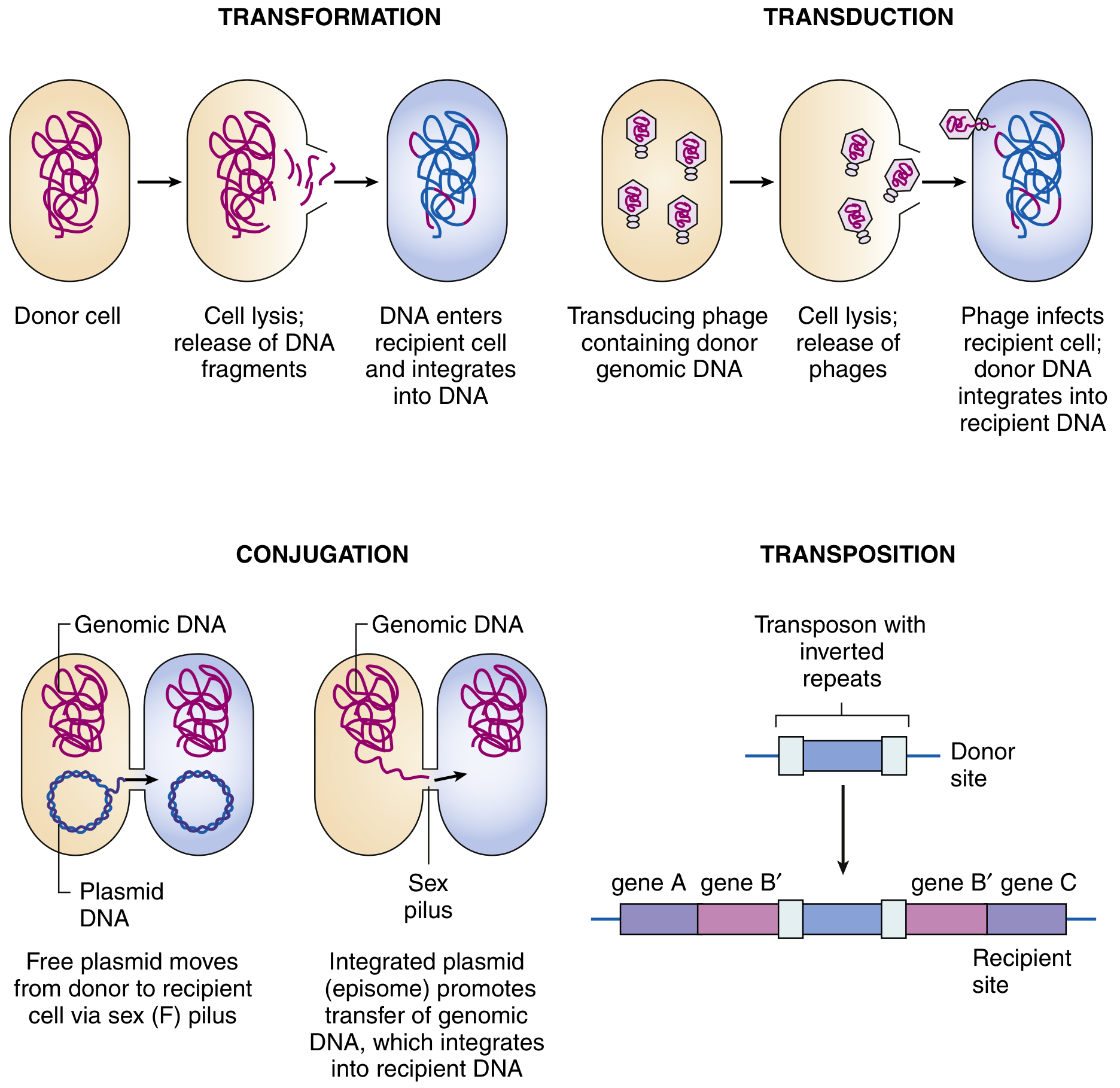
Types of Conjugation
1. F+ × F− Conjugation (Plasmid Transfer)
- F+ donor transfers a copy of the F plasmid to the F− recipient.
- Result: Recipient becomes F+; no chromosomal genes transferred in most cases.
- Very efficient and rapid transfer.
2. Hfr × F− Conjugation (High-Frequency Recombination)
- The F factor integrates into the bacterial chromosome at specific IS element sites, converting the F+ cell into an Hfr (High-Frequency Recombination) cell - this is the episome state.
- During conjugation, transfer begins from the origin of transfer (oriT) within the integrated F and proceeds linearly through the chromosome.
- Chromosomal genes are transferred at high frequency in the order they appear on the chromosome from the integration point.
- However, since the chromosome is very large (~100 minutes to fully transfer at 37°C), conjugation is usually interrupted before completion - so only genes near the integration point are frequently transferred.
- The F factor itself (its trailing end) is the last to be transferred and is usually not completely transferred to the F− recipient.
- Result: Recipient acquires chromosomal genes but remains F− (does not become F+).
- Used to construct genetic linkage maps of E. coli (distances measured in minutes).
3. F' (F-prime) × F− Conjugation (Sexduction)
- When the integrated F factor in an Hfr cell excises imprecisely from the chromosome, it may carry along a piece of flanking chromosomal DNA - forming an F' (F-prime) plasmid.
- This F' plasmid carries both F factor genes AND some bacterial chromosomal genes.
- On conjugating with F− cells, the chromosomal genes are transferred along with the F factor.
- Result: Recipient becomes F' - a partial diploid (merodiploid) carrying two copies of certain genes (one on chromosome, one on the F' plasmid). Used for complementation studies.
Summary Table of Conjugation Types
| Type | Donor | Recipient becomes | Chromosomal transfer | F factor transfer |
|---|---|---|---|---|
| F+ × F− | F+ cell | F+ | Very rare / no | Yes (complete) |
| Hfr × F− | Hfr cell | Remains F− | High frequency (partial) | Incomplete (usually no) |
| F' × F− | F' cell | F' (merodiploid) | Yes (F'-carried genes) | Yes (with chromosomal fragment) |
Significance of Conjugation in Medicine
-
Antibiotic resistance spread - The most clinically important consequence. The R factor (Resistance factor / R plasmid) is a conjugative plasmid with two components:
- RTF (Resistance Transfer Factor): carries conjugation machinery (tra genes)
- r determinants: carry genes encoding resistance enzymes (e.g., β-lactamases, aminoglycoside-modifying enzymes)
- R factors can transfer multiple drug resistance in a single conjugation event - the principal mechanism behind multidrug-resistant hospital infections.
-
Spread of virulence genes - Plasmids carrying toxin genes, invasin genes, and other virulence factors spread between bacteria via conjugation.
-
Inter-species transfer - Conjugation can occur between different bacterial species and even between bacteria and eukaryotic cells (plants, fungi), making it an important evolutionary mechanism.
-
Genetic mapping - Interrupted mating experiments with Hfr strains were used to construct the first complete genetic map of E. coli.
Key Points to Remember
- Conjugation requires direct cell contact (unlike transformation and transduction)
- Transferred DNA is single-stranded (rolling circle replication)
- DNase does not inhibit conjugation (unlike transformation which uses free naked DNA)
- The F pilus is a type IV secretion structure and a receptor for male-specific bacteriophages (MS2, f2, Qβ phages)
- Hfr conjugation produces F− recipients (not F+ ) because the trailing end of F is usually not transferred
- The R factor is responsible for transferable (infectious) drug resistance - the most medically relevant form of conjugation
toxic shock syndrome
toxic shock syndrome staphylococcal streptococcal diagnosis criteria management 2025
Toxic Shock Syndrome (TSS)
Definition
Types
| Feature | Staphylococcal TSS | Streptococcal TSS (STSS) |
|---|---|---|
| Causative toxin | TSST-1 (mainly); also enterotoxins B/C | Streptococcal pyrogenic exotoxins (SPE-A, B, C) |
| Blood cultures | Usually negative | Often positive (bacteremia common) |
| Skin findings | Diffuse erythroderma + desquamation | Rash less constant; often necrotizing fasciitis |
| Association | Tampons, wound infection, nasal packing | Wound infection, cellulitis, necrotizing fasciitis |
| Mortality | ~3-5% | 30-70% (much higher) |
Causative Organisms and Toxins
Staphylococcal TSS
- Organism: Staphylococcus aureus
- Primary toxin: TSST-1 (Toxic Shock Syndrome Toxin-1) - responsible for ~75% of menstrual TSS and ~50% of non-menstrual TSS
- Other SAgs implicated: enterotoxins B, C (especially in non-menstrual TSS)
- TSST-1 is uniquely able to be readily absorbed across mucosal surfaces (unlike other superantigens) - key to menstrual TSS
- (Sherris & Ryan's Medical Microbiology 8e; Goldman-Cecil Medicine)
Streptococcal TSS
- Organism: Streptococcus pyogenes (Group A Strep)
- Primary toxins: Streptococcal Pyrogenic Exotoxins A, B, C (SPE-A, SPE-B, SPE-C) - formerly called erythrogenic toxins
- Additional factor: M protein mediates neutrophil activation and vascular injury
- (Henry's Clinical Diagnosis and Management 23e)
Pathogenesis: The Superantigen Mechanism
- Antigen processing → peptide fragment loaded into MHC class II groove → presented to specific T cells (~1 in 10,000 T cells respond)
- From T cells: IL-2, IFN-γ, TNF-β (lymphotoxin)
- From APCs/macrophages: IL-1, TNF-α, IL-6
- Fever (IL-1, TNF-α acting on hypothalamus)
- Hypotension and shock (TNF-α, IL-1 causing vasodilation + capillary leak)
- Multi-organ failure (renal, hepatic, pulmonary, CNS)
- Rash (direct toxin effect + immune-mediated)
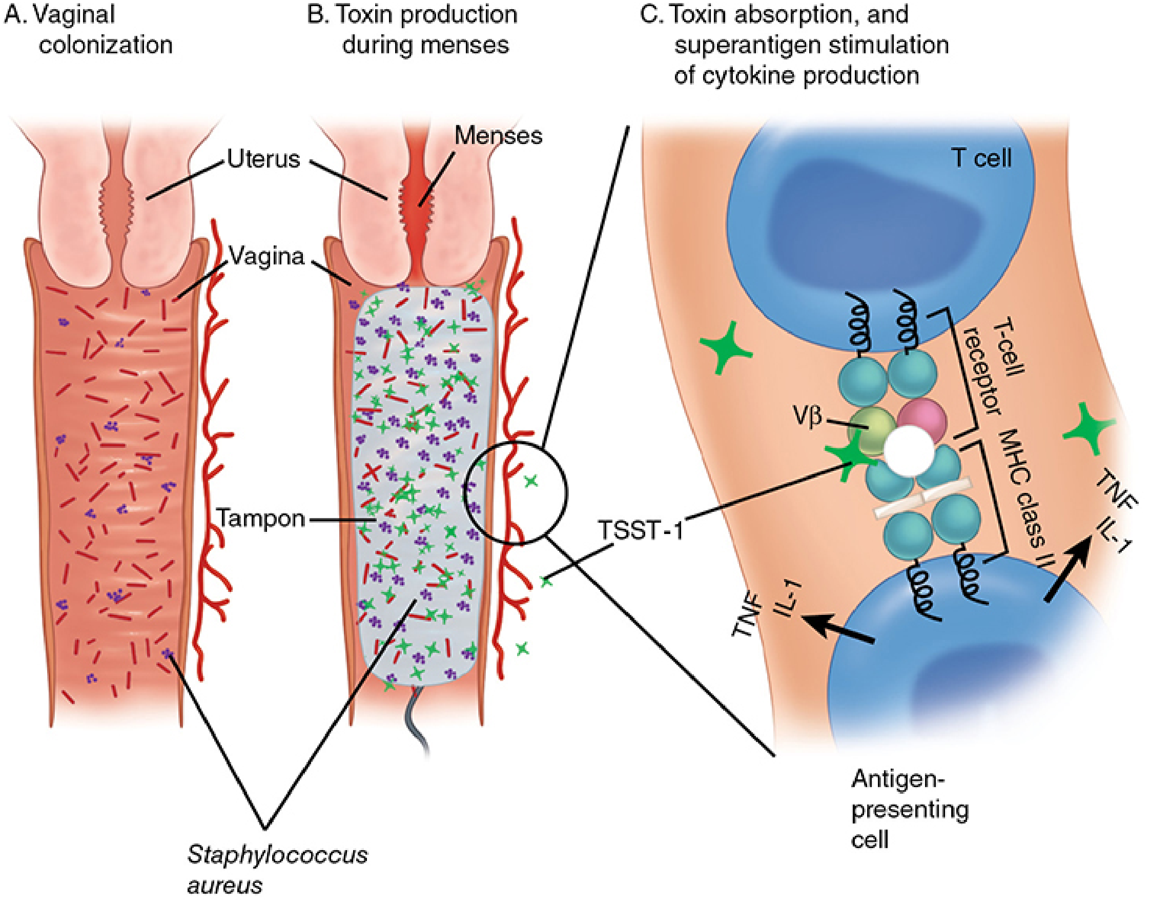
Predisposing Conditions
Staphylococcal TSS
- Menstrual (classic): Young women (15-25 years), high-absorbency tampon use
- Only <15% of women carry vaginal S. aureus; of these, <20% produce TSST-1
- Tampons create neutral pH (6.5-8), elevated pCO₂ and pO₂ - optimal conditions for TSST-1 production
- TSS occurs only in individuals with no pre-existing antibody to TSST-1
- Non-menstrual: Surgical wounds, nasal packing, postpartum, burns, sinusitis, influenza A
- Staphylococcal TSS is a localized infection (usually no bacteremia) with systemic toxin absorption
Streptococcal TSS
- Often follows skin/soft tissue infection: necrotizing fasciitis, myositis, cellulitis, bacteremia
- Can follow varicella infection (chickenpox) in children
- IV drug users, elderly, immunocompromised
Clinical Features
Onset
- Fever - high (≥38.9°C / ≥102°F)
- Myalgias, headache, malaise
- Vomiting and diarrhea (early)
- Hypotension - systolic BP <90 mmHg (adults); orthostatic drop ≥15 mmHg
The Rash (Staphylococcal TSS - Hallmark)
- Diffuse macular erythroderma - "sunburn-like" blanching rash
- Involves palms and soles (distinguishing feature)
- Desquamation of involved skin (full-thickness peeling) at 1-2 weeks, especially palms, soles, fingers, toes
Mucosal Involvement
- Strawberry tongue, pharyngeal hyperemia
- Conjunctival injection / conjunctivitis
- Vaginal mucosal ulceration (in menstrual TSS)
Multi-Organ Involvement (≥3 systems required for diagnosis)
| System | Manifestation |
|---|---|
| GI | Vomiting, diarrhea, abdominal pain |
| Musculoskeletal | Myalgia, CK >2x normal |
| Mucous membranes | Vaginal/oropharyngeal/conjunctival hyperemia |
| Renal | BUN/creatinine ≥2x normal, pyuria |
| Hepatic | Bilirubin/AST/ALT ≥2x normal |
| Hematologic | Platelets <100,000/mm³ |
| CNS | Disorientation, altered consciousness (without focal signs) |
| Cardiopulmonary | ARDS, heart failure |
CDC Diagnostic Criteria
Staphylococcal TSS (CDC)
- Fever ≥38.9°C
- Rash - diffuse macular erythroderma
- Desquamation (1-2 weeks after onset)
- Hypotension (SBP ≤90 mmHg)
- Multi-system involvement (≥3 organ systems)
- Confirmed: All 5 clinical + lab criteria met
- Probable: 4 of 5 clinical + lab criteria met
Streptococcal TSS (CDC/CSTE)
- A1: GAS from sterile site (blood, CSF, tissue) = Confirmed
- A2: GAS from non-sterile site (throat, wound) = Probable
- B1: Hypotension (SBP ≤90 mmHg)
- B2: ≥2 of: renal impairment, coagulopathy, liver impairment, ARDS, extensive tissue necrosis/necrotizing fasciitis, erythematous rash
Differences: Staphylococcal vs. Streptococcal TSS
| Feature | Staph TSS | Strep TSS |
|---|---|---|
| Blood culture | Usually negative | Often positive |
| Rash | Diffuse erythroderma + desquamation | Variable; less prominent |
| Necrotizing fasciitis | Rare | Common (defining feature) |
| Source of infection | Localized (tampon, wound) | Often invasive bacteremia |
| Age group | Young women (menstrual) | Any age |
| Recurrence | Yes (if no antibody developed) | Less common |
| Mortality | ~3-5% | 30-70% |
Management
1. Source Control
- Remove any foreign body immediately (tampon, nasal packing, wound packing)
- Surgical debridement of necrotizing fasciitis or deep tissue infection (critical in streptococcal TSS - do not delay)
2. Resuscitation
- Aggressive IV fluid resuscitation (capillary leak causes massive fluid loss)
- Vasopressors (norepinephrine) for refractory shock
- Note: patients with TSS may show early myocardial dysfunction - consider inotropes earlier than in standard septic shock
- Steroids for refractory shock per septic shock protocol
3. Antibiotics
- Cover both staphylococci and streptococci empirically, even if source appears minor
| Drug | Role |
|---|---|
| Beta-lactam (nafcillin, oxacillin, or cefazolin) | Bactericidal for MSSA |
| Vancomycin | Cover MRSA (especially nosocomial or uncertain) |
| Clindamycin | Toxin suppression - inhibits ribosomal protein synthesis, reducing toxin production; also provides post-antibiotic effect against GAS |
| Clindamycin + beta-lactam | Combination preferred for streptococcal TSS |
- Clindamycin is added specifically to suppress toxin synthesis - it acts even on non-replicating bacteria (unlike beta-lactams which only work on dividing bacteria)
4. Intravenous Immunoglobulin (IVIG)
- Contains pooled antibodies against TSST-1 and streptococcal toxins
- Neutralizes circulating superantigens and blocks T-cell activation
- Recommended for severe/refractory STSS and for cases not responding to antibiotics + source control
- Efficacy in staphylococcal TSS less well established
- (CDC Clinical Guidance 2024; Merck Manual Sept 2025)
5. Monitoring
- Continuous monitoring: renal, hepatic, bone marrow, cardiopulmonary function
- Blood, wound, vaginal, throat cultures (to confirm organism and guide antibiotic de-escalation)
Immunity and Recurrence
- TSS occurs in individuals who lack antibody to TSST-1
- After recovery, some patients fail to mount an adequate antibody response
- Recurrence of menstrual TSS is possible (up to 30% without preventive measures) - patients should avoid tampons after an episode
- (Sherris & Ryan 8e)
Key Points Summary
- TSS = superantigen (TSST-1 / SPE) → MHC II + TCR Vβ cross-linking → 5-25% T cells activated → cytokine storm
- Staphylococcal TSS: localized infection, negative blood culture, erythroderma + desquamation, lower mortality
- Streptococcal TSS: invasive disease, positive blood culture, necrotizing fasciitis, very high mortality
- Management: remove source + aggressive resuscitation + beta-lactam + clindamycin (toxin suppression) + IVIG in severe cases
- Diagnosis is clinical - do not wait for culture confirmation to start treatment