I now have comprehensive content from multiple authoritative textbooks plus excellent diagrams. Let me compile the full answer.
Edema: Investigations and Treatment
Definition and Overview
Edema is an accumulation of interstitial fluid within tissues. Fluid can also collect in body cavities - pleural (hydrothorax), pericardial (hydropericardium), or peritoneal (ascites/hydroperitoneum). Severe, generalized edema with profound subcutaneous swelling is called anasarca.
Pathophysiology (Understanding Before Treating)
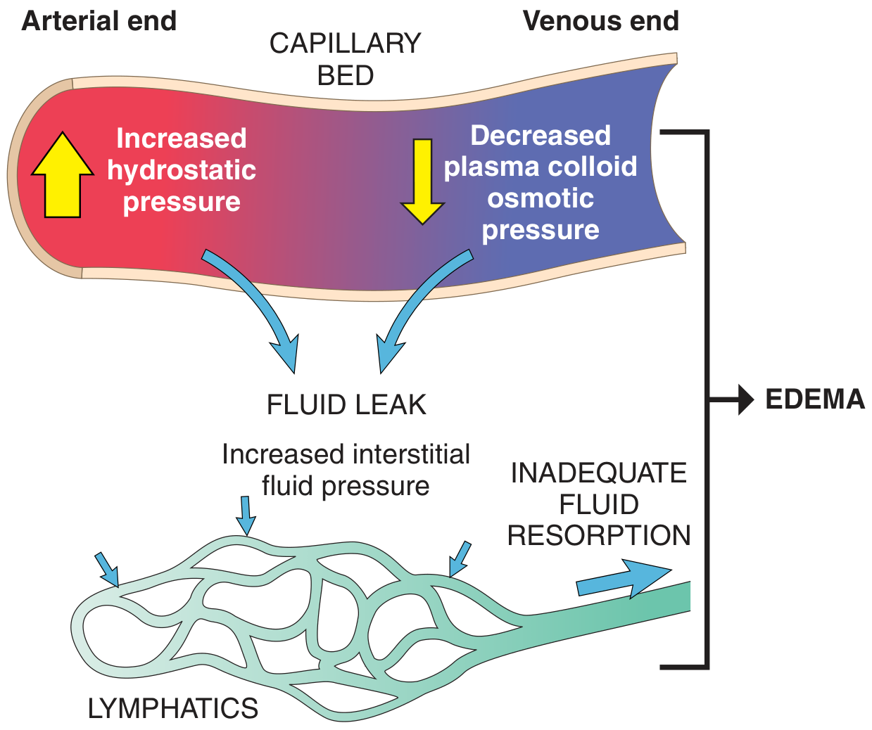
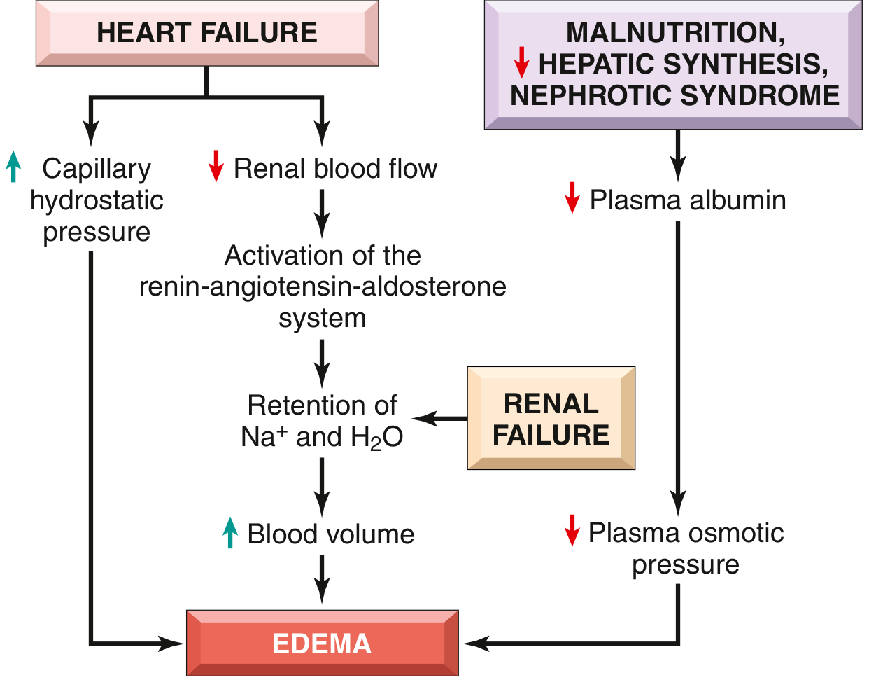
Fluid movement between the vascular and interstitial spaces is governed by two opposing forces: vascular hydrostatic pressure and plasma colloid osmotic pressure. Edema results when one or more of these four mechanisms are disrupted:
Causes of Edema
| Mechanism | Examples |
|---|
| Increased hydrostatic pressure | Congestive heart failure, DVT, venous obstruction, constrictive pericarditis, cirrhosis, prolonged limb dependency |
| Reduced plasma osmotic pressure | Nephrotic syndrome (protein loss), cirrhosis (decreased synthesis), malnutrition/kwashiorkor |
| Lymphatic obstruction | Filariasis (elephantiasis), malignancy, post-surgical (e.g., axillary node resection), radiation |
| Sodium and water retention | Renal failure, hyperaldosteronism |
| Increased capillary permeability | Inflammation, allergy, angioedema, burns, sepsis |
(Robbins & Kumar Basic Pathology, Table 3.1)
Investigations
The workup is directed by the clinical context - bilateral vs. unilateral, pitting vs. non-pitting, and associated symptoms.
Initial Blood Tests
| Test | What It Evaluates |
|---|
| Serum albumin | Hypoalbuminemia (nephrotic, cirrhosis, malnutrition) |
| Urea, creatinine, eGFR | Renal function - nephrotic/nephritic, CKD |
| LFTs (ALT, AST, bilirubin, ALP) | Hepatic synthesis failure, cirrhosis |
| Serum electrolytes (Na, K) | Hyponatremia (dilutional), hyperaldosteronism |
| CBC | Anemia, infection |
| BNP / NT-proBNP | Heart failure (very sensitive marker) |
| TFTs (TSH, T4) | Hypothyroidism (myxedema) |
| Coagulation studies (PT, APTT) | Hypercoagulability in nephrotic syndrome |
Urine Tests
| Test | What It Evaluates |
|---|
| Urine protein:creatinine ratio (PCR) | Nephrotic range proteinuria (PCR >2 in first morning void) |
| 24-hour urine protein | >3.5 g/day = nephrotic range |
| Urinalysis (dipstick + microscopy) | Hematuria, casts (nephritic vs. nephrotic) |
| Urine sodium | Differentiates prerenal from intrinsic renal causes |
Immunological/Serological Tests
(For suspected secondary nephrotic syndrome or systemic disease)
- ANA, dsDNA - systemic lupus erythematosus
- ANCA - vasculitis
- Serum complement (C3, C4) - membranoproliferative GN, SLE
- Hepatitis B, C serology - membranous nephropathy
- Serum immunoglobulins, SPEP - multiple myeloma, amyloidosis
- HIV serology
Imaging
| Modality | Indication |
|---|
| Chest X-ray | Pulmonary edema, cardiomegaly, pleural effusion |
| Echocardiogram | Ventricular function, EF assessment, pericardial disease |
| Doppler ultrasound (limbs) | DVT in unilateral lower limb edema |
| Renal ultrasound | Kidney size, echogenicity, obstruction |
| Abdominal USS | Ascites, portal hypertension, liver echogenicity |
| CT angiography | Suspected pulmonary embolism, renal vein thrombosis |
(Tintinalli's Emergency Medicine; Barash Clinical Anesthesia)
Renal Biopsy
- Indicated when: renal cause is suspected but undefined, steroid-resistant nephrotic syndrome, persistent elevated creatinine, hypocomplementemia, or gross hematuria - Tintinalli's Emergency Medicine
Treatment
1. General Principles
Treatment focuses on:
- Correction of the underlying cause (most important)
- Judicious diuretic use
- Sodium and fluid restriction
- Monitoring of renal function
- Anticoagulation for DVT prevention (especially in nephrotic syndrome - hypercoagulable state)
- Patient mobilization
(Goldman-Cecil Medicine)
2. Dietary and Non-Pharmacological Measures
- Low-sodium diet (2 g/day) - reduces sodium-driven fluid retention
- Fluid restriction - in hypervolemic states
- Leg elevation - for dependent edema
- Compression stockings - for lymphedema and venous insufficiency
- Increased protein intake - caution: in nephrotic syndrome, high protein paradoxically worsens proteinuria
3. Pharmacological Treatment
A. Diuretics (Cornerstone of Treatment)
| Drug Class | Example | Mechanism | Use |
|---|
| Loop diuretics | Furosemide, bumetanide, torsemide | Block Na-K-2Cl in thick ascending limb | First-line for acute pulmonary edema, heart failure, renal impairment. Rapid onset. |
| Thiazide diuretics | Hydrochlorothiazide, metolazone | Block NaCl in distal convoluted tubule | Hypertension with mild edema, synergistic with loop diuretics |
| Aldosterone antagonists | Spironolactone (25 mg/day), eplerenone | Block mineralocorticoid receptor | Heart failure (dominant mechanism), cirrhotic ascites, secondary hyperaldosteronism |
| Potassium-sparing | Amiloride, triamterene | Block ENaC in collecting duct | Avoid hypokalemia; note: ENaC activation is key in nephrotic sodium retention |
- Loop diuretics are drugs of choice for pulmonary edema and acute/chronic peripheral edema from heart failure or renal impairment - Lippincott Pharmacology
- In profound hypoalbuminemia (nephrotic syndrome), diuretics may be ineffective; albumin infusion (0.5-1.0 g/kg) followed by furosemide may be required, with ICU monitoring - Tintinalli's Emergency Medicine
- Oral spironolactone 25 mg once daily is useful in heart-failure-dominant edema; afterload reduction may also help - Goldman-Cecil Medicine
- Loop diuretics + hypertonic saline are useful for life-threatening hyponatremia with edema - Goodman & Gilman
B. Vasodilators (Acute Pulmonary Edema)
- IV nitrates (nitroglycerin/nitroprusside) - reduce preload and afterload, useful in acute cardiogenic pulmonary edema without hypotension
- Morphine (historical use) - reduces sympathetic tone; now used cautiously
- Inotropes (dobutamine, milrinone) - in cardiogenic shock with pulmonary edema
(Morgan & Mikhail's Clinical Anesthesiology)
C. RAAS Inhibitors
- ACE inhibitors / ARBs - reduce afterload, lower glomerular pressure (also reduce proteinuria in nephrotic syndrome), useful in heart failure
D. Corticosteroids (Nephrotic Syndrome)
- Prednisone 2 mg/kg/day (max 60 mg/day) in children with nephrotic syndrome
- Minimal change disease and mesangial proliferative nephritis: often steroid-responsive
- Membranous nephropathy: may respond
- FSGS: typically steroid-resistant - requires calcineurin inhibitors (cyclosporine, tacrolimus)
- Tintinalli's Emergency Medicine
E. Treatment of Specific Causes
| Cause | Targeted Treatment |
|---|
| Heart failure | Loop diuretics, ACEi/ARB, beta-blockers, aldosterone antagonists, device therapy |
| Nephrotic syndrome | Corticosteroids, immunosuppressants (cyclophosphamide, mycophenolate), low-salt diet, statin for hyperlipidemia |
| Cirrhosis/ascites | Spironolactone ± furosemide, paracentesis (large-volume), TIPS, treat underlying liver disease |
| DVT | Anticoagulation (heparin/LMWH, then warfarin or DOAC), compression |
| Lymphedema | Compression garments, manual lymphatic drainage, exercise, rarely surgery |
| Hypothyroidism | Thyroid hormone replacement (levothyroxine) |
| Malnutrition | Nutritional rehabilitation (high-quality protein, calories) |
| Angioedema (C1-INH deficiency) | C1-INH concentrate, icatibant, fresh frozen plasma |
4. Monitoring During Treatment
- Renal function (urea, creatinine) - diuretics can worsen renal perfusion
- Electrolytes - hypokalemia and hypomagnesemia with loop diuretics; hyperkalemia with spironolactone
- Daily weight - best measure of fluid balance (target 0.5-1 kg/day loss)
- Blood pressure - avoid hypotension especially with aggressive diuresis
- Urine output - oliguria may indicate over-diuresis or worsening renal failure
Summary Algorithm
EDEMA DETECTED
    |
    +-- Unilateral? --> Consider DVT (Doppler USS), lymphedema, trauma/infection
    |
    +-- Bilateral/generalized?
           |
           +-- Raised JVP, S3 gallop, orthopnea --> Heart Failure
           |       --> BNP, echo, CXR; loop diuretics, RAAS blockade
           |
           +-- Heavy proteinuria, hypoalbuminemia --> Nephrotic Syndrome
           |       --> Urine PCR, serum albumin, renal biopsy; steroids, diuretics
           |
           +-- Jaundice, spider naevi, ascites --> Liver Cirrhosis
           |       --> LFTs, USS abdomen; spironolactone + furosemide
           |
           +-- Raised creatinine, oliguria --> Renal Failure
           |       --> eGFR, urine studies; renal replacement if severe
           |
           +-- Low albumin, no protein loss --> Malnutrition/Protein deficiency
                   --> Nutritional support
Key Sources:
- Robbins & Kumar Basic Pathology - pathophysiology and causes
- Goldman-Cecil Medicine - clinical management principles
- Lippincott Illustrated Reviews: Pharmacology - diuretic pharmacology
- Goodman & Gilman's Pharmacological Basis - loop diuretic therapeutics
- Tintinalli's Emergency Medicine - nephrotic syndrome workup and ED management
- Comprehensive Clinical Nephrology, 7th Ed. - nephrotic mechanisms
- Morgan & Mikhail's Clinical Anesthesiology - acute pulmonary edema management