

My toung becomes very sensitive to spices from past 7days
glossitis tongue inflammation sore red tongue
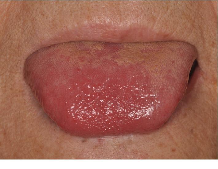
This clinical photograph shows a close-up, frontal view of a human tongue protruded from the oral cavity. The primary finding is focal erythema (redness) and inflammation at the apex (tip) and anterior dorsal surface of the tongue. The affected area appears moist, glossy, and more intensely red compared to the more posterior regions. There is evidence of filiform papillae atrophy in the erythematous zone, giving it a smoother appearance, while the posterior dorsal surface exhibits a yellowish-tan coating or slight discoloration. The surrounding perioral skin appears unremarkable. This visual evidence of glossitis and dysgeusia in a febrile patient with elevated inflammatory markers (CRP and WBC) is clinically significant in evaluating systemic infections or inflammatory conditions, such as infective endocarditis (IE) following dental procedures. The image serves as an educational example of oral manifestations of systemic disease and localized glossal inflammation.
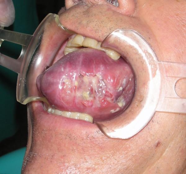
This clinical intraoral photograph demonstrates severe macroglossia (volumetric increase of the tongue) and glossitis. The tongue exhibits a diffuse red-violet or erythematous-purpuric discoloration suggesting inflammation or vascular congestion. The dorsal surface and left lateral margin show well-delimited necrotic-ulcerative lesions characterized by yellowish-white fibrinopurulent exudate and irregular tissue breakdown. Evidence of focal hemorrhage is visible, which may be secondary to local trauma or bite injuries against the teeth due to the significant swelling. The image is captured using a lip retractor to reveal the extent of the pathology in the oral cavity. This photograph is educationally relevant for oral medicine, pathology, and surgery, illustrating severe inflammatory or traumatic glossal manifestations and the clinical presentation of macroglossia.
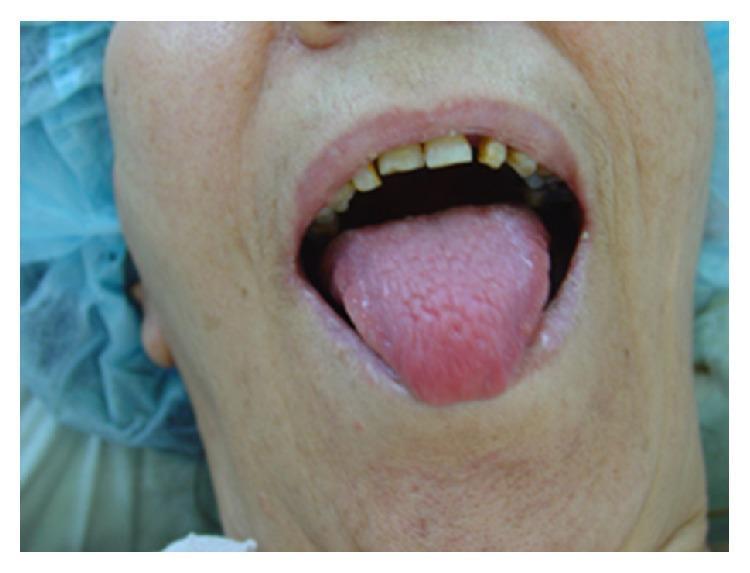
Clinical photograph of the oral cavity showcasing glossitis. The tongue is prominently displayed, appearing bright red (erythematous) and moderately enlarged. The dorsal surface shows signs of atrophy, with a smoother-than-normal texture and partial loss of the standard lingual papillae architecture. In the background, the maxillary teeth exhibit signs of dental attrition and discoloration, and the gingiva appears pale. This presentation is characteristic of nutritional deficiencies or systemic syndromes such as glucagonoma (associated with necrolytic migratory erythema). The image serves as a clinical reference for identifying atrophic glossitis during a physical examination of the head and neck. It is intended for medical students and clinicians to recognize oral manifestations of systemic disease.
Tongue Sensitivity to Spices - Assessment
What the Image Shows
Most Likely Causes
1. Nutritional Deficiency (Most Common Clinical Cause)
- Signs: burning/sensitive tongue, taste changes, angular cheilitis (cracks at corners of mouth)
- Common in vegetarians, people with poor diet, or those with gut absorption issues
- Andrews' Diseases of the Skin confirms: "The patches are painful and sensitive, so eating may be difficult and taste impaired" with atrophic glossitis from nutritional deficiency
2. Transient Lingual Papillitis ("Lie Bumps")
3. Oral Candidiasis (Fungal/Thrush) - Mild Form
- Check: Can the white coating be scraped off? If yes - more likely thrush.
4. Toothpaste/Mouthwash Irritation (Sodium Lauryl Sulfate)
5. Dry Mouth (Xerostomia)
6. Burning Mouth Syndrome / Glossodynia
What to Look Out For (Red Flags - See a Doctor If You Notice These)
- White patches that cannot be scraped off (could be leukoplakia)
- Ulcers that don't heal in 2-3 weeks
- Tongue becomes progressively smoother and glossier
- Tongue pain at rest, not just with spices
- Weight loss, fatigue, or pallor alongside the tongue symptoms (suggests systemic nutritional deficiency or anemia)
What You Can Do Now
| Action | Why |
|---|---|
| Get a blood test: CBC, Vitamin B12, folate, iron, ferritin | Rule out deficiency as the #1 cause |
| Switch to SLS-free toothpaste (e.g., Sensodyne, biotene) | Reduces mucosal stripping |
| Stay well hydrated | Helps saliva protect the tongue |
| Avoid very spicy, acidic, or hot foods temporarily | Allows mucosa to recover |
| Avoid alcohol-based mouthwashes | They dry out and irritate the tongue |
| If white coating worsens, see a doctor | May need antifungal (nystatin/fluconazole) |
When to See a Doctor
- A blood workup (Vitamin B12, iron studies, CBC)
- Examination for oral thrush or other causes
- If deficiency is found, appropriate supplementation resolves the sensitivity in most cases
Disclaimer: This is for informational purposes and not a substitute for a professional medical evaluation. Please see a doctor for a proper diagnosis.