
Use of valsalva maneuver clinically
I'll read the medical library skill first, then search for comprehensive information on the Valsalva maneuver.
Reading File
Now I'll search the medical library and PubMed in parallel for comprehensive coverage.
Finding Sources
Finding Sources
Searching PubMed
valsalva maneuver clinical applications
Excellent results. Now I'll read the key sections from the most authoritative sources in parallel.
Reading File
Reading File
Reading File
Reading File
Now let me fetch additional content on the cardiac murmur effects, Harriet Lane modified Valsalva, and the 4 phases in detail.
Reading File
Reading File
Finding Sources
Finding Sources
Let me get the four-phase description from Fuster & Hurst's and the table from Tintinalli's.
Reading File
Reading File
I now have comprehensive data from multiple authoritative sources. Let me also fetch the Valsalva 4-phase image for visual clarity.
Loading Image
Finding Sources
I now have all the information needed to write a comprehensive, well-organized answer. Let me compile it.
Valsalva Maneuver - Clinical Uses
The Valsalva maneuver is a forced expiration against a closed glottis (or bearing down as if to defecate), generating an intrathoracic pressure of approximately 30-40 mm Hg for at least 10 seconds. It was first described by Antonio Maria Valsalva (1666-1723) in the context of expelling pus from the middle ear.
Physiology: The Four Phases
The maneuver produces predictable, sequential hemodynamic changes that form the basis for all its clinical uses:
| Phase | What Happens | Hemodynamic Effect |
|---|---|---|
| I - Onset of straining | Increased intrathoracic pressure transmitted to aorta | Transient rise in systolic BP |
| II - Sustained strain (active phase) | Compressed veins → decreased venous return → decreased stroke volume → decreased pulse pressure | Reflex tachycardia; BP falls |
| III - Release of strain | Sudden drop in intrathoracic pressure | Further brief BP fall |
| IV - Post-strain overshoot | Venous return restored + peripheral vasoconstriction (from phase II) → BP rises above baseline | Reflex bradycardia via baroreceptors |
During phase II, the decrease in arterial pressure inhibits baroreceptors, producing tachycardia and increased peripheral resistance. When the glottis opens (phase IV), blood is pumped by a sympathetically activated heart into a constricted circulation, causing BP overshoot and vagally mediated bradycardia.
- Ganong's Review of Medical Physiology, 26e
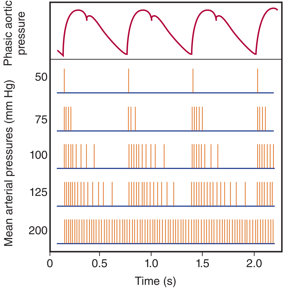
Clinical Uses
1. Termination of SVT (Supraventricular Tachycardia)
This is the most common acute use. Increased intrathoracic pressure reduces venous return, which reflexively increases vagal tone via baroreceptor activation, slowing SA and AV node conduction.
Technique:
- Patient supine with IV access and cardiac monitor
- Take a deep breath, close lips around thumb, attempt to exhale without releasing air
- Alternatively, bear down against the clinician's hand placed on the abdomen
- Strain sustained for 10-20 seconds
- Roberts and Hedges' Clinical Procedures in Emergency Medicine
Modified Valsalva (superior efficacy):
- Position patient at 45 degrees, apply standard Valsalva strain
- Immediately reposition supine with passive leg raise at 45 degrees for 15 seconds
- The leg raise augments venous return during phase IV, enhancing the vagal response
- Has a greater success rate at restoring sinus rhythm than standard Valsalva
- The Harriet Lane Handbook, 23rd ed.
In children: The diving reflex (ice-cold water applied to face for 15-30 seconds) is a practical alternative when the child cannot cooperate with the maneuver.
2. Differentiation of Cardiac Murmurs
This is the most important bedside use in cardiology. During phase II, most systolic murmurs decrease (due to reduced LV filling and stroke volume). The key exceptions are:
| Condition | Effect on Murmur During Strain Phase | Mechanism |
|---|---|---|
| HOCM (hypertrophic obstructive cardiomyopathy) | Increases | Smaller LV cavity → more LVOT obstruction |
| Mitral valve prolapse | Increases + click moves earlier (toward S1) | Smaller LV → leaflets prolapse earlier into atrium, longer murmur |
| Aortic stenosis | Decreases | Less flow across fixed stenosis |
| Mitral regurgitation | Decreases (softer) | Left-sided response to reduced preload |
| VSD | Decreases | Reduced LV-RV pressure gradient |
During Valsalva, the transient reduction in LV size increases the degree of LVOT obstruction in HOCM, increasing murmur intensity. In contrast, the aortic stenosis murmur decreases because of reduced blood flow across the stenotic valve.
- Rosen's Emergency Medicine
The examiner should document murmur change only after 6-8 beats during the strain phase, as this is the time needed for the change in blood volume to circulate through the pulmonary vasculature to the left heart.
- Fuster and Hurst's The Heart, 15th ed.
Note: If tachycardia is absent during phase II, there is autonomic dysfunction, beta-blockade, or elevated left-sided filling pressures - in these cases, the murmur response is unreliable. - Fuster and Hurst's The Heart, 15th ed.
Bedside maneuver comparison table (HOCM vs MVP):
| Intervention | HOCM | Mitral Valve Prolapse |
|---|---|---|
| Valsalva strain phase | Murmur increased | Click closer to S1, murmur increased |
| Standing after squatting | Murmur increased | Click closer to S1, murmur increased |
| Passive leg elevation | Murmur decreased | Click closer to S2, murmur decreased |
| Hand grip | Murmur decreased | Click closer to S1, murmur increased |
| Squatting | Murmur decreased | Click closer to S2, murmur decreased |
Tintinalli's Emergency Medicine
3. Assessment of Autonomic Nervous System Function
The Valsalva maneuver is a standard bedside test for autonomic integrity, particularly baroreceptor and sympathetic function.
Protocol: Patient blows into a closed mouthpiece to raise intrathoracic pressure to 40 mm Hg for 12 seconds, while BP and HR are continuously monitored.
Normal response: BP and HR follow the four phases as described above (including reflex tachycardia in phase II and bradycardia + BP overshoot in phase IV).
Abnormal responses:
- In sympathetic dysfunction: heart rate changes still occur (baroreceptors and vagus are intact), but the BP rise in phase IV is absent
- In complete autonomic insufficiency (e.g., MSA, diabetic autonomic neuropathy): all phases are abnormal - the BP just progressively falls without recovery, and HR changes are absent
- In primary hyperaldosteronism: HR changes and BP overshoot in phase IV are absent; returns to normal after tumor removal
- Ganong's Physiology; Braunwald's Heart Disease
4. Radiological Use - Hernias and Pelvic Floor Assessment
During fluoroscopy or imaging, a sustained Valsalva maneuver increases intra-abdominal pressure, which helps demonstrate:
- Small inguinal or femoral hernias
- Pelvic floor defects
- Rectocele, enterocele, or other masses
The radiologist should perform examinations with and without a prolonged Valsalva maneuver to demonstrate small defects.
- Sleisenger and Fordtran's Gastrointestinal and Liver Disease
5. Ear and Sinus Equalization (ENT)
The Valsalva maneuver is used to open the Eustachian tube by raising nasopharyngeal pressure, equalizing middle ear pressure. This is used:
- In divers / aviators to prevent barotrauma
- To relieve ear discomfort during ascent/descent
- As a test of Eustachian tube patency
The larynx closes tightly during the maneuver, stabilizing the thorax for muscular actions and also allowing increased intra-abdominal pressure for defecation, vomiting, and childbirth. - K.J. Lee's Essential Otolaryngology
6. Cough Headache and Exertional Headache Workup
Primary cough headaches are precipitated by the Valsalva maneuver (coughing, lifting, straining). Recognition is important because:
- They must be differentiated from secondary causes (subarachnoid hemorrhage, posterior fossa lesions)
- Symptom to Diagnosis: An Evidence-Based Guide, 4th ed.
7. Valsalva Retinopathy
Excessive or forceful Valsalva (heavy lifting, vomiting, prolonged coughing, strenuous bowel movement) causes a sudden rise in intraocular venous pressure, leading to:
- Pre-retinal (sub-ILM) hemorrhages at the macula - typically bilateral, bright red with dark center
- Usually self-limiting and resolves in weeks to months
- Clinically important to distinguish from diabetic retinopathy or other retinal hemorrhages
- Wills Eye Manual; Barash's Clinical Anesthesia
Patients with known proliferative retinopathy should be counseled to avoid excessive Valsalva maneuvers during labor.
- Rosen's Emergency Medicine
8. Anesthesia - Intubation and Emergence
- Vigorous Valsalva maneuvers (bucking on endotracheal tube during emergence, protracted vomiting) can precipitate Valsalva retinopathy in the perioperative setting
- Monitored during anesthesia as a test of autonomic integrity
- Barash's Clinical Anesthesia, 9e; Miller's Anesthesia, 10e
Performing the Maneuver - Practical Steps
- Patient supine (or semirecumbent for modified version)
- Cardiac monitor, IV access, resuscitation equipment ready (if for SVT)
- Patient takes a deep breath, places thumb in closed mouth, attempts to exhale
- Alternatively: "Bear down as if straining during a bowel movement"
- Maintain for 10-20 seconds
- Observe for SVT termination, murmur change, or hemodynamic response
Contraindications and Precautions
- Dyspnea or tachypnea (patient cannot cooperate or sustain the maneuver)
- Severe hypertension (initial BP surge may be dangerous)
- Known proliferative retinopathy (risk of vitreous/retinal hemorrhage)
- Recent myocardial infarction or acute aortic pathology (avoid BP surges)
- Recent eye surgery
Summary of Clinical Applications at a Glance
| Application | Basis |
|---|---|
| Terminate SVT | Vagal tone increase → AV node slowing |
| Differentiate HOCM vs AS murmur | HOCM increases; AS decreases |
| MVP diagnosis | Earlier click, longer murmur |
| Autonomic function testing | Absence of HR/BP phases = autonomic dysfunction |
| Hernia/pelvic floor imaging | Increased intra-abdominal pressure unmasks defects |
| Eustachian tube equalization | Opens ET by nasopharyngeal pressure |
| Cough headache evaluation | Trigger for primary cough headache |
| Valsalva retinopathy | Cause of sub-ILM macular hemorrhages |
Key sources: Ganong's Review of Medical Physiology, 26e | Fuster and Hurst's The Heart, 15th ed. | Roberts and Hedges' Clinical Procedures in Emergency Medicine | Braunwald's Heart Disease | Tintinalli's Emergency Medicine | Harriet Lane Handbook, 23rd ed. | Wills Eye Manual | Sleisenger and Fordtran's GI Disease
Anesthetic clinical uses of valsalva maneuver
Finding Sources
Finding Sources
Good leads. Now read the Miller's Anesthesia dedicated section and the Barash sections in parallel, plus the surgical uses.
Reading File
Reading File
Reading File
Finding Sources
Finding Sources
Finding Sources
Finding Sources
Reading File
Reading File
Finding Sources
Reading File
Reading File
Reading File
Finding Sources
Finding Sources
Your credits are exhausted. You can purchase a top-up or subscribe to a plan to continue using Orris. If you're already subscribed, your daily credits will reset tomorrow.
This is a shared conversation. Sign in to Orris to start your own chat.