
Interstitial Lung Disease (ILD): Classification and HRCT Patterns
HRCT patterns interstitial lung disease UIP NSIP
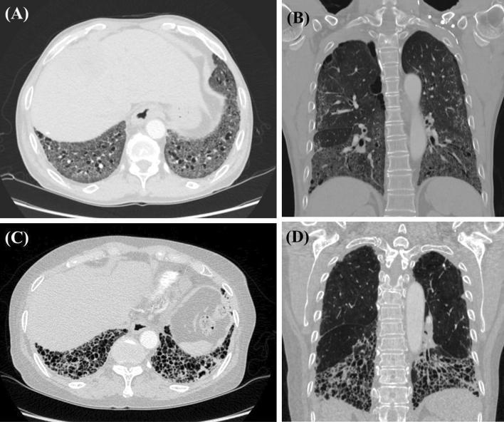
High-resolution computed tomography (HRCT) comparison of interstitial lung disease (ILD) patterns in systemic sclerosis (SSc). Figures A (axial) and B (coronal) display a Non-Specific Interstitial Pneumonia (NSIP) pattern characterized by bilateral, diffuse ground-glass opacities and mild traction bronchiectasis with a distinct basal predominance. Figures C (axial) and D (coronal) demonstrate a Usual Interstitial Pneumonia (UIP) pattern. This pattern is marked by extensive subpleural honeycombing (clusters of cystic airspaces), exuberant traction bronchiectasis, and severe architectural distortion predominantly involving the lower lung zones. These diagnostic images serve to illustrate the distinct radiologic morphology between NSIP and UIP patterns in patients with connective tissue disease-associated ILD, highlighting key features such as distribution, presence of honeycombing, and the degree of fibrotic changes essential for clinical classification and management.
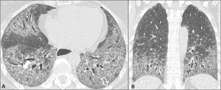
High-resolution computed tomography (HRCT) of the chest in a patient with scleroderma-associated nonspecific interstitial pneumonia (NSIP). Image A (axial view) demonstrates bilateral ground-glass attenuation interspersed with fine linear reticular opacities. Hallmark signs of fibrotic lung disease, including traction bronchiectasis and bronchiolectasis, are visible within the areas of lung parenchyma distortion. A characteristic feature of NSIP—subpleural preservation—is indicated by an open arrow, showing a narrow band of relatively spared lung tissue immediately adjacent to the pleura. Image B (coronal reformatted view) highlights the classic symmetric and predominantly basal distribution of the interstitial changes, with opacities increasing in severity toward the lung bases. These findings collectively represent the common imaging presentation of cellular and fibrotic NSIP within the context of systemic autoimmune disease. This diagnostic imaging serves as a clinical reference for identifying patterns of interstitial lung disease and differentiating NSIP from other patterns such as usual interstitial pneumonia (UIP).
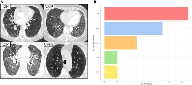
This composite educational resource consists of a diagnostic imaging panel (A) and a comparison bar chart (B) illustrating interstitial lung disease (ILD) patterns in patients with Sjögren's syndrome. Panel A displays axial High-Resolution Computed Tomography (HRCT) slices of four distinct ILD patterns: Usual Interstitial Pneumonia (UIP) characterized by peripheral reticulation and honeycombing; Non-Specific Interstitial Pneumonia (NSIP) showing more uniform ground-glass opacities; Desquamative Interstitial Pneumonia (DIP) with diffuse ground-glass attenuation; and Combined Pulmonary Fibrosis and Emphysema (CPFE) demonstrating upper-lobe emphysematous lucencies alongside fibrotic changes. Panel B is a horizontal bar chart quantifying the prevalence of these CT disease patterns among the study cohort. The chart indicates that UIP is the most frequent pattern, followed by NSIP, unspecific changes (Unspez), and lastly DIP and CPFE, which show equal, lower prevalence. This visual aid is intended for medical education regarding the radiologic classification and epidemiological distribution of pulmonary manifestations in systemic autoimmune diseases.
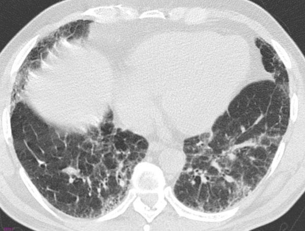
High-resolution computed tomography (HRCT) axial scan of the chest at the level of the lower lobes demonstrating parenchymal abnormalities. The image shows bilateral, predominantly peripheral, and basal patchy ground-glass opacities (GGO), appearing as areas of hazy increased lung attenuation that do not obscure the underlying pulmonary vasculature. Intermixed with the GGO is a fine reticular pattern, characterized by linear opacities representing interstitial thickening. While the biopsy confirmed Usual Interstitial Pneumonia (UIP), the HRCT lacks definitive features of a UIP pattern, such as honeycombing or significant traction bronchiectasis; instead, the diffuse, symmetric distribution and predominant GGO are more suggestive of a Nonspecific Interstitial Pneumonia (NSIP) pattern. This case serves as a clinical example of the discordance between radiologic patterns and histologic findings in interstitial lung disease (ILD), highlighting the importance of multidisciplinary diagnosis.
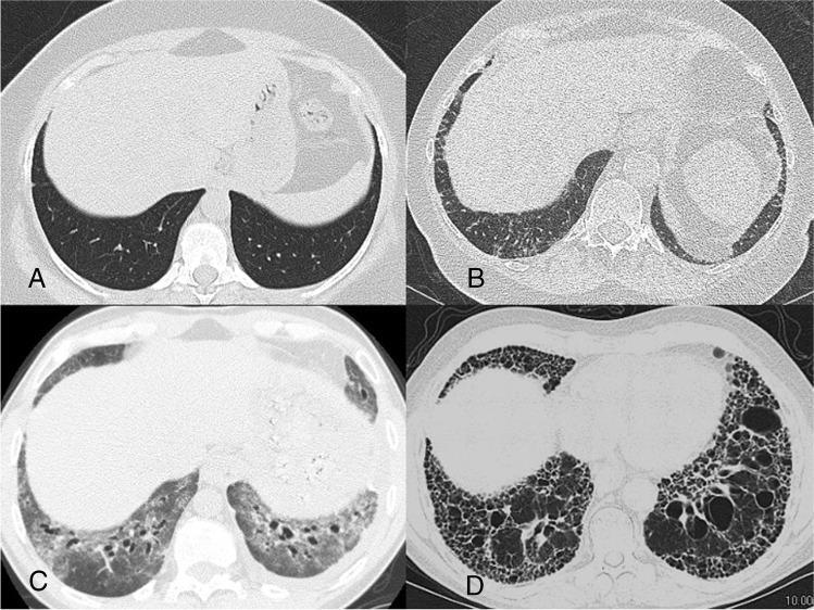
This composite diagnostic image features four axial High-Resolution Computed Tomography (HRCT) scans of the lung bases, illustrating various manifestations of Interstitial Lung Disease (ILD) commonly associated with Systemic Sclerosis (SSc).
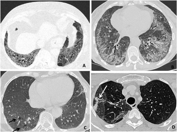
This composite of four axial high-resolution computed tomography (HRCT) scans illustrates various radiologic patterns of Rheumatoid Arthritis-associated Interstitial Lung Disease (RA-ILD). Panel A demonstrates a Usual Interstitial Pneumonia (UIP) pattern, characterized by bibasilar, subpleural honeycombing (clusters of cystic airspaces), reticular opacities, and traction bronchiectasis. Panel B displays a Nonspecific Interstitial Pneumonia (NSIP) pattern, showing diffuse, patchy ground-glass opacities and septal thickening with relative subpleural sparing. Panel C reveals a Lymphocytic Interstitial Pneumonia (LIP) pattern, highlighted by perivascular thin-walled lung cysts (indicated by black arrows). Panel D illustrates an Organizing Pneumonia (OP) pattern, featuring focal areas of parenchymal consolidation (indicated by a white arrow). These images serve as educational benchmarks for differentiating pulmonary manifestations in systemic autoimmune diseases. The radiological findings are critical for determining prognosis and guiding therapeutic strategies, such as distinguishing between predominantly fibrotic (UIP) versus inflammatory (NSIP/OP) processes.
UIP pattern HRCT honeycombing subpleural basal idiopathic pulmonary fibrosis
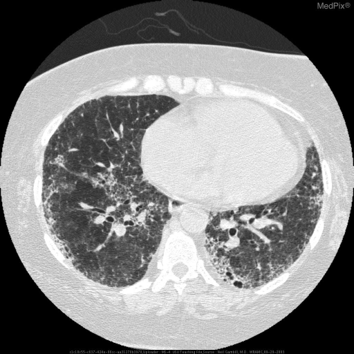
**Imaging Modality:** High-resolution computed tomography (HRCT) of the chest, axial section.
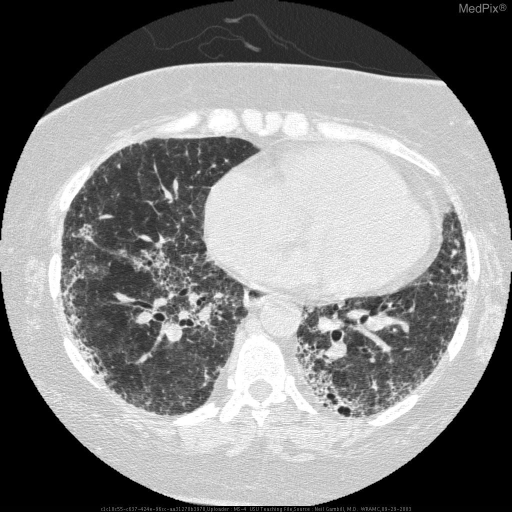
**Imaging Modality:** High-Resolution Computed Tomography (HRCT) of the chest.
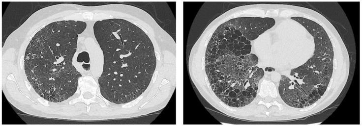
Two axial slices of high-resolution computed tomography (HRCT) of the chest demonstrating a classic Usual Interstitial Pneumonia (UIP) pattern. The images show significant bilateral lung parenchymal abnormalities with a subpleural and basal predominance. Key radiologic features include extensive honeycombing, characterized by clustered, thick-walled subpleural cystic airspaces, and prominent reticular opacities. Additionally, there is evidence of traction bronchiectasis, where the fibrotic destruction of the lung architecture has caused irregular dilation of the bronchi. These findings are consistent with end-stage pulmonary fibrosis, often seen in idiopathic pulmonary fibrosis or connective tissue disease-associated interstitial lung disease (such as systemic sclerosis). The images serve as an educational example of advanced architectural distortion and the characteristic distribution of UIP-pattern fibrosis, distinguishing it from other interstitial lung diseases by the presence of honeycombing and peripheral gradient.
Interstitial Lung Disease (ILD): Classification and HRCT Patterns
What Is ILD?
Section 1 - Clinical Classification of ILD
Known Causes
| Category | Examples |
|---|---|
| Connective tissue disease (CTD)-related | RA, SSc, SLE, dermatomyositis/polymyositis, Sjögren, MCTD |
| Environmental / occupational | Asbestosis, silicosis, coal worker's pneumoconiosis, berylliosis |
| Drug/radiation-induced | Amiodarone, methotrexate, nitrofurantoin, bleomycin, checkpoint inhibitors |
| Hypersensitivity pneumonitis | Farmer's lung, bird fancier's lung |
| Smoking-related | RB-ILD, DIP, combined pulmonary fibrosis and emphysema |
Unknown Causes (Idiopathic / IIP)
| Group | Entities |
|---|---|
| Chronic fibrosing IPs | IPF (UIP pattern), Idiopathic NSIP |
| Acute/subacute IPs | Cryptogenic organizing pneumonia (COP), Acute interstitial pneumonia (AIP) |
| Smoking-related IPs | RB-ILD, Desquamative interstitial pneumonia (DIP) |
| Rare IIPs | Idiopathic LIP, Idiopathic pleuroparenchymal fibroelastosis (PPFE) |
Section 2 - HRCT in ILD: Why It Matters
- Characterize the pattern of disease (fibrotic vs. inflammatory vs. combined)
- Define anatomical distribution (subpleural, peribronchial, upper vs. lower zone)
- Guide biopsy site selection
- Replace biopsy entirely when HRCT findings are "typical" (e.g., classic UIP pattern in the right clinical context)
Section 3 - Major HRCT Patterns and Their Signatures
3A. UIP Pattern (Usual Interstitial Pneumonia)
- Subpleural, basal-predominant distribution; often bilateral and heterogeneous
- Honeycombing - clustered cystic airspaces (3-10 mm), subpleural, basal
- Traction bronchiectasis/bronchiolectasis - irregular dilation of airways within fibrosis
- Reticular pattern - fine irregular lines
- Ground glass opacity: present but NOT predominant
- Upper lobe subpleural disease, when present, tends to be anterior
| Category | Features |
|---|---|
| Typical UIP | Subpleural + basal honeycombing ± traction bronchiectasis |
| Probable UIP | Subpleural + basal reticular pattern + peripheral traction bronchiectasis; no honeycombing |
| Indeterminate for UIP | Subtle reticulation ± mild GGO/distortion; heterogeneous/asymmetric distribution without features inconsistent with UIP |
| Alternative diagnosis | Peribronchovascular/perilymphatic/upper-mid lung distribution; cysts, marked mosaic attenuation, profuse micronodules, nodules, or consolidation |
3B. NSIP Pattern (Non-Specific Interstitial Pneumonia)
- Bilateral, symmetric, lower-lobe-predominant distribution (but without the obvious apical-basal gradient of UIP)
- Ground-glass opacity - present in ~50% of cases; often the dominant finding
- Reticular opacities with lower-zone predominance
- Traction bronchiectasis (fibrotic NSIP)
- Subpleural sparing - a narrow band of relatively preserved lung immediately adjacent to the pleura (key distinguishing feature from UIP)
- No honeycombing (or very limited, small cysts in late disease)
- Homogeneous involvement without obvious craniocaudal gradient
3C. OP Pattern (Organizing Pneumonia / Cryptogenic Organizing Pneumonia)
- Patchy air space consolidation - bilateral, often peripheral (subpleural) and/or peribronchovascular
- Lower-lung-zone predominance
- Small nodular opacities and bronchial wall thickening/dilation
- Reverse halo sign (atoll sign) - central GGO surrounded by denser crescentic consolidation; seen in ~20% of COP cases; highly suggestive
- CT findings are often far more extensive than the chest radiograph suggests
- Opacities tend to migrate and may decrease even without treatment
- Consolidation associated with resolution; reticular opacities with persistence
- Honeycombing: rare, late manifestation only
3D. AIP Pattern (Acute Interstitial Pneumonia / Diffuse Alveolar Damage)
- Bilateral diffuse GGO - often with consolidation
- Dependent consolidation and air bronchograms
- Rapid onset (days to weeks); often preceded by a prodrome
- Architectural distortion and traction bronchiectasis develop in surviving patients (organizing phase)
3E. DIP Pattern (Desquamative Interstitial Pneumonia)
- Diffuse ground-glass opacity - the dominant finding, often widespread
- Lower-lobe predominance
- Cystic spaces scattered within areas of GGO (variable, not subpleural clusters)
- Minimal or no honeycombing
- Irregular lines (reticular) in a minority
3F. RB-ILD (Respiratory Bronchiolitis-ILD)
- Diffuse patchy centrilobular ground-glass nodules (3-5 mm, poorly defined)
- Upper-lobe or diffuse distribution
- Mild air trapping
- No honeycombing
3G. LIP (Lymphoid Interstitial Pneumonia)
- Ground-glass opacity (bilateral, diffuse)
- Thin-walled perivascular cysts - a characteristic hallmark (arrows on HRCT)
- Lower-lobe predominance
- Reticular or nodular opacities
3H. Sarcoidosis
- Perilymphatic micronodules - along bronchovascular bundles, interlobar fissures, and costal pleura; this distribution is the key differentiating feature from other ILDs
- Upper and mid-lung predominance
- Bilateral hilar and mediastinal lymphadenopathy (very characteristic)
- Peribronchovascular consolidation (galaxy sign/confluent nodules)
- Ground-glass opacity less common
- Fibrosis in late-stage: upper-lobe retraction, traction bronchiectasis
3I. Hypersensitivity Pneumonitis (HP)
- Centrilobular nodules (ground-glass, ill-defined) - subacute/chronic
- Mosaic attenuation / air trapping (lobular) on expiratory images - a hallmark
- Upper and mid-lung predominance (unlike IPF/NSIP)
- In chronic fibrotic HP: UIP-like features may develop (honeycombing, traction bronchiectasis), making differentiation from IPF difficult
- Absence of subpleural sparing and presence of centrilobular nodules + air trapping help distinguish from UIP/IPF
3J. LAM and LCH
- Diffuse, bilateral, thin-walled cysts of uniform size distributed throughout both lungs (no zonal predominance)
- Normal intervening lung parenchyma
- Predominantly in women of child-bearing age
- Irregularly shaped cysts + nodules
- Upper and mid-lung predominance with costophrenic angle sparing
- Associated with smoking
Section 4 - Chest Radiograph Patterns as Diagnostic Clues
| CXR Pattern | Suggested ILD |
|---|---|
| Decreased lung volumes | IPF, NSIP, DIP, CTD-ILD, asbestosis |
| Increased/preserved volumes | LCH, LAM, RB-ILD, sarcoidosis, COP |
| Micronodules | HP, sarcoidosis, RB-ILD, infection |
| Honeycombing | IPF, fibrotic NSIP, asbestosis, chronic HP |
| Migratory/fleeting infiltrates | COP, HP, eosinophilic pneumonia |
| Pneumothorax | LCH, LAM, tuberous sclerosis, neurofibromatosis |
| Hilar/mediastinal adenopathy | Sarcoidosis, LIP, silicosis, berylliosis |
| Upper-mid lung zone | HP, sarcoidosis, LCH, PPFE, silicosis |
| Lower-lung zone | IPF, fibrotic NSIP, asbestosis, CTD-ILD |
| Peripheral predominance | IPF, fibrotic NSIP, COP, eosinophilic pneumonia |
Section 5 - Contrasting Histopathologic Features of Major IIPs
| Feature | IPF (UIP) | NSIP | COP | AIP (DAD) | DIP |
|---|---|---|---|---|---|
| Temporal pattern | Heterogeneous (mixed old + new fibrosis) | Homogeneous | Homogeneous | Homogeneous (phases) | Homogeneous |
| Fibrosis | Dense, subpleural | Uniform, often cellular | Absent/minimal | Organizing (early), fibrosis (late) | Minimal |
| Fibroblastic foci | Prominent | Sparse | Absent | Absent | Absent |
| Honeycombing | Yes | Rare | No | No | No |
| Inflammation | Mild | Moderate to marked | Patchy plugs of granulation tissue | Hyaline membranes, edema | Diffuse macrophage filling |
| Granulomas | No | No | No | No | No |
Section 6 - Comparative Clinical/HRCT Summary Table
| IPF | NSIP | RB-ILD | SSc-ILD | Sarcoidosis | |
|---|---|---|---|---|---|
| Demographics | Older male, smoker | Middle-aged female, never-smoker | Active smoker | Women (SSc) | Variable |
| HRCT pattern | Bilateral basal subpleural reticulation, honeycombing, traction bronchiectasis. Classic UIP = diagnostic | Peripheral subpleural GGO + reticular; traction bronchiectasis common, honeycombing rare | Diffuse patchy centrilobular GGO nodules | UIP or NSIP; dilated esophagus, vascular enlargement | Perilymphatic nodules, hilar adenopathy, peribronchovascular |
| Biopsy needed? | Not if typical UIP | Usually required | Often clinical | Context-dependent | Often biopsy-confirmed |
| Prognosis | 50% mortality at 3-5 yr | 18% mortality at 5 yr | 25% mortality at 7 yr | 20-30% at 10 yr | Generally good |
Section 7 - Diagnostic Approach
- History: Duration, tempo of progression, exposures (occupational, environmental, drugs, birds), smoking, family history, extrapulmonary symptoms (skin, joints, Raynaud's)
- Exam: Basal crackles, clubbing (common in IPF), extrapulmonary CTD signs
- Labs: CBC, ESR, ANA, RF, anti-CCP, anti-Scl-70, anti-Jo-1, ANCA as guided by clinical context
- HRCT chest: The primary imaging modality; defines pattern, distribution, and guides next steps
- PFTs: Typically show restrictive defect (reduced TLC, DLCO); obstructive pattern in some (HP, LAM)
- BAL: Useful in sarcoidosis (high lymphocytes, elevated CD4:CD8), HP (lymphocytosis), eosinophilic pneumonia
- Biopsy: Transbronchial lung biopsy, cryobiopsy, or surgical lung biopsy (VATS) when HRCT is non-diagnostic
- Multidisciplinary discussion (MDD): The gold standard for ILD diagnosis - improves diagnostic accuracy and confidence
Key Points to Remember
- Honeycombing = UIP/IPF until proven otherwise; subpleural and basal is the hallmark
- Subpleural sparing = NSIP; distinguishes it from UIP
- Consolidation migrating over time = COP; look for the reverse halo sign
- Perilymphatic nodules + hilar adenopathy = Sarcoidosis
- Bilateral diffuse thin-walled cysts = LAM (in young women); LCH has irregular cysts + nodules, upper zone
- Centrilobular GGO nodules + air trapping + upper zone = HP
- GGO + cysts + smoking history = DIP
- UIP pattern is not synonymous with IPF - it can occur in CTD-ILD, chronic HP, drug-induced ILD, and asbestosis