
Ewing sarcoma detailed note for clinical orthopaedics 15 marks bpt mgr university exam
Ewing sarcoma bone X-ray histology onion skin periosteal reaction
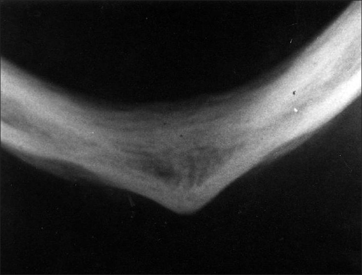
**Imaging Modality:** Intraoral occlusal radiograph (X-ray). **Anatomical Region:** Anterior mandible and symphysis menti, viewed from a submental-vertex/occlusal perspective. **Observed Pathology:** The image demonstrates a characteristic "onion-skin" appearance, indicating periosteal reaction. There is evidence of proliferative periostitis (Garre's osteomyelitis) characterized by multiple radiopaque laminations of new bone formed parallel to the cortical surface. **Characteristic Visual Features:** * **Periosteal New Bone Formation:** Distinct, concentric layers of radiopaque bone resembling onion layers. * **Cortical Expansion:** Bilateral expansion of the mandibular cortex, most prominent at the symphysis. * **Bone Density:** Mixed radiolucent and radiopaque appearance within the expanded cortical layers. * **Symmetry:** The periosteal thickening appears relatively symmetrical across the midline of the mandibular arch. **Diagnostic Differentiating Features:** The presence of thin, parallel, circumferential laminations of bone without significant cortical destruction or sunburst spiculations is highly suggestive of Garré’s chronic nonsuppurative sclerosing osteomyelitis, distinguishing it from aggressive malignancies like osteosarcoma or Ewing sarcoma.
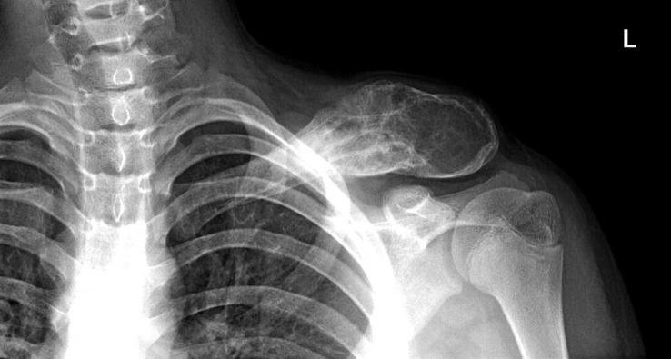
This diagnostic x-ray radiography displays an anteroposterior view of the left clavicle and shoulder region. The image demonstrates a large, aggressive osteolytic lesion primarily involving the distal half of the clavicle. Key radiological features include a 'moth-eaten' pattern of bone destruction and a lamellated (onion-skin) periosteal reaction, which are characteristic findings of highly aggressive primary bone tumors such as Ewing sarcoma. The lesion is expansile, causing significant enlargement and thinning of the cortical bone with focal disruption of the periosteum. Additionally, there is a prominent soft tissue mass or swelling overlying the affected bone segment, suggesting extraosseous extension of the pathology. The acromioclavicular and glenohumeral joints appear preserved. This clinical imaging is essential for medical students and radiology residents to recognize aggressive bone malignancies in pediatric or adolescent patients, emphasizing the importance of identifying specific periosteal reactions and bone destruction patterns in musculoskeletal oncology.
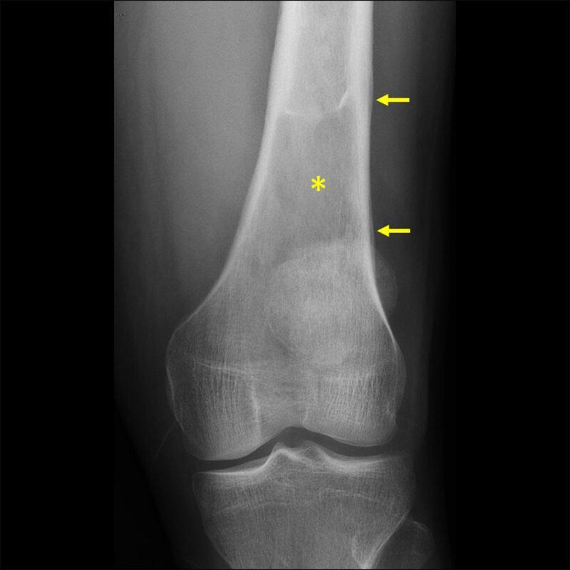
This diagnostic x-ray shows an anteroposterior (AP) view of the distal femur and knee joint, demonstrating a primary bone pathology. A prominent intramedullary lytic lesion is visible in the distal femoral shaft (indicated by a yellow asterisk), characterized by an area of decreased bone density and loss of normal trabecular pattern. Along the lateral aspect of the femoral diaphysis, there is a distinct lamellated (onion-skin) periosteal reaction, marked by yellow arrows. This aggressive periosteal pattern suggests a rapidly progressing process. Despite the intramedullary involvement and periosteal lifting, the cortical integrity appears largely preserved without gross pathological fracture. This radiographic presentation is clinically significant in the study of musculoskeletal oncology, commonly associated with conditions such as Ewing sarcoma or osteomyelitis. The image serves as an educational resource for identifying aggressive bone lesions, distinguishing between different patterns of periosteal reaction, and understanding the radiological evaluation of pediatric or adolescent bone tumors.
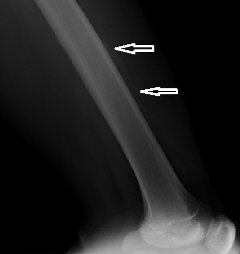
**Imaging Modality:** Conventional radiography (X-ray) **Anatomical Region:** Left femur, lateral (profile) view, including the mid-diaphysis and distal metaphysis. **Observed Pathology:** Multilamellar periosteal reaction (also known as "onion-skin" appearance) located along the mid-diaphyseal cortical surface. **Characteristic Visual Features:** * **Periosteal Apposition:** Linear, layered radiodense streaks parallel to the long axis of the femoral shaft, indicated by white arrows. * **Cortical Integrity:** The underlying cortex appears thickened but lacks overt evidence of gross destruction or aggressive osteolysis in the visualized segment. * **Soft Tissue:** Subtle increased radiopacity in the adjacent soft tissues, suggesting swelling or hematoma formation. **Clinical Context & Differentiation:** This aggressive-appearing periosteal reaction is consistent with a subperiosteal hematoma (often associated with hemophilia pseudotumor) or can mimic primary bone malignancies such as Ewing sarcoma. The distribution is localized to the mid-shaft of the long bone. **Key Diagnostic Features:** Multi-layered periosteal thickening; absence of Codman’s triangle or sunburst pattern; localized to the diaphysis of the femur.
Ewing sarcoma small round blue cells histology microscopy
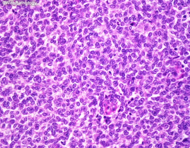
This is a light microscopy image of a bone tumor biopsy stained with Hematoxylin and Eosin (H&E). The image demonstrates the classic small round blue cell morphology of Ewing sarcoma. The tumor is composed of uniform, small, primitive-appearing cells with scant pale eosinophilic or clear cytoplasm and rounded to oval nuclei with evenly dispersed chromatin and small or inconspicuous nucleoli. Cytoplasmic borders are indistinct, producing a syncytial appearance. Mitotic activity is variable and may be increased. Intracytoplasmic glycogen is common and can be highlighted with periodic acid–Schiff (PAS) staining; this feature supports the diagnosis of this entity. The tumor cell nests may arrange around delicate vasculature to form pseudorosettes; true Homer-Wright rosettes may be present in some cases. The stroma is a delicate vascular network; necrosis is frequent, appearing as large geographic areas or smaller foci. Together these features produce a dense, sheets-and-nests pattern with a high nuclear-to-cytoplasmic ratio typical of small round blue cell tumors. The image is diagnostic when integrated with immunohistochemical and molecular studies (e.g., CD99 positivity, EWSR1 rearrangement) but the morphology shown is highly characteristic of Ewing sarcoma. This histology is pathognomonic in the appropriate clinical context and is essential for prognosis and treatment planning.
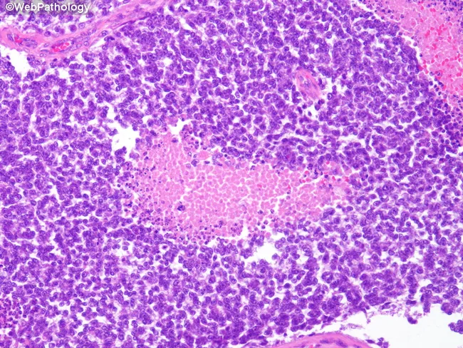
Imaging modality: Histopathology using light microscopy of formalin-fixed, paraffin-embedded bone tissue stained with Hematoxylin and Eosin (H&E). The slide displays a skeletally derived lesion with features classic for Ewing sarcoma. At high cellular density, there are sheets of small round blue cells with scant cytoplasm and round to oval nuclei showing hyperchromasia and brisk mitotic activity. The tumor cells are arranged in uniform clusters, separated by delicate fibrous strands, creating a highly cellular, often undulating landscape. Areas of geographic necrosis are frequently seen within the tumor, producing pink, acellular zones that disrupt cohesive cell architecture. Cytoplasmic scantiness and high nuclear-to-cytoplasmic ratio are typical; occasional rosette-like formations (Homer-Wright) may be present but are not required for diagnosis. The overall histologic pattern corresponds to a high-grade small round blue cell neoplasm in bone. Although not shown in this image, CD99 membrane positivity is commonly observed in Ewing sarcoma and, together with molecular testing for EWSR1-FLI1 fusion (t(11;22)) or related rearrangements, strengthens the diagnosis. Clinically, this histology correlates with aggressive bone lesions in children and adolescents and has important differential considerations including lymphoma, rhabdomyosarcoma, neuroblastoma, and osteosarcoma. Correlation with radiology and immunohistochemistry is essential for definitive diagnosis and prognostic assessment in clinical practice overall.
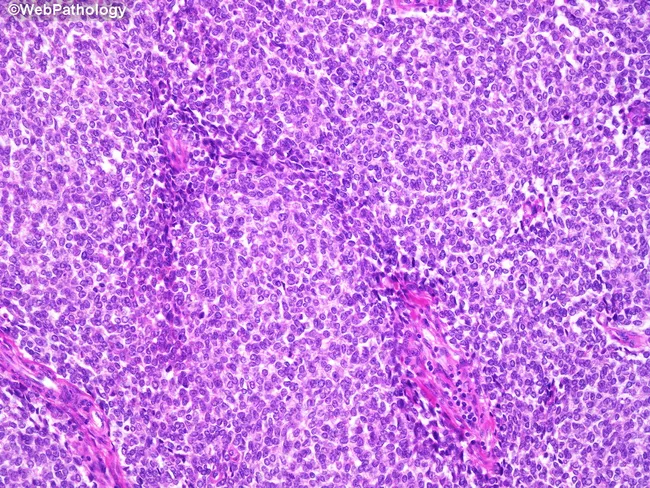
This section presents a high-magnification light microscopy image of a small round blue cell tumor, consistent with Ewing sarcoma, stained with Hematoxylin and Eosin. The specimen is a bone/soft tissue biopsy section examined under bright-field microscopy. The tumor displays sheets of uniform, small, primitive-appearing cells with round to oval nuclei, finely dispersed chromatin, and small or inconspicuous nucleoli. Cytoplasm is scant, pale eosinophilic or clear, producing a high nuclear-cytoplasmic ratio and indistinct cytoplasmic borders that contribute to a syncytial-appearing cellular nest. Mitoses are variable, sometimes brisk. Intracytoplasmic glycogen is a key feature that can be highlighted by Periodic acid–Schiff (PAS) staining (PAS-positive granules). Tumor cells frequently organize around delicate vasculature, forming pseudorosettes; true Homer-Wright rosettes may be present in some fields. The stroma is scant but vascular, and extensive necrosis can be seen as geographic areas or scattered foci. Immunophenotype is typically CD99 (MIC2) positive and molecular testing often reveals EWSR1-FLI1 or related translocations; these ancillary tests are essential for confirmation. Distinguishing from other small round blue cell tumors is critical because prognosis and therapy differ. This histologic pattern supports a diagnosis of Ewing sarcoma in the appropriate clinical context and guides multimodal treatment planning.
EWING SARCOMA
Detailed Note for Clinical Orthopaedics | 15 Marks | BPT MGR University
DEFINITION
EPIDEMIOLOGY / INCIDENCE
- Third most common nonhematologic primary bone malignancy overall
- Second most common (after osteosarcoma) bone sarcoma in patients under 30 years
- Most common bone sarcoma in children under 10 years
- Incidence: fewer than 1 per 1 million per year; approximately 200 cases/year in the USA
- Age: 5-25 years (peak in second decade); range from infants to elderly
- Sex: Slight male predominance
- Race: Exceedingly rare in individuals of African descent; most common in Caucasians
- No known predisposing factors
PATHOGENESIS / MOLECULAR BIOLOGY
- Chromosomal translocation t(11;22)(q24;q12) is present in >90% of cases - this creates an EWS-FLI1 fusion gene (chimeric oncogene)
- Other translocations: t(21;22)(q22;q12) and t(7;22)(p22;q12) in remaining cases
- The EWS-FLI1 fusion protein binds chromatin and dysregulates transcription, leading to uncontrolled growth and abnormal differentiation
- Cell of origin: Likely mesenchymal stem cells or primitive neuroectodermal cells (uncertain)
SITES OF INVOLVEMENT
| Location | Features |
|---|---|
| Metaphysis of long bones (most common) | Femur, tibia, humerus - often extends into the diaphysis |
| Flat bones | Pelvis, scapula, ribs (shoulder and pelvic girdles) |
| Diaphysis | Classic teaching description |
| Spine | Rare (3-15%); sacrum most common spinal site |
| Small bones | Hands and feet - rare |
| Extraosseous | ~20% of cases are extraskeletal (extraosseous Ewing sarcoma) |
- Large portions of the entire bone may be involved (unlike most other bone tumors)
- "Skip" metastases are NOT reported in Ewing sarcoma (contrast with osteosarcoma)
CLINICAL PRESENTATION
- Pain - almost universal; insidious onset; initially mild and intermittent
- May mimic musculoskeletal strain; average diagnosis delay = 34 weeks
- Average patient delay: 15 weeks (symptoms to first visit)
- Average physician delay: 19 weeks (first visit to diagnosis)
- Swelling over the affected area
- Local warmth and erythema - may simulate osteomyelitis or cellulitis
- Fever (systemic)
- Tender, palpable soft-tissue mass (often very large)
Ewing sarcoma closely mimics osteomyelitis - fever, erythema, raised WBC, elevated ESR, and CRP are common. A needle aspirate may grossly resemble pus! Always send specimens for both culture AND pathology.
INVESTIGATIONS
Laboratory Findings
| Test | Finding |
|---|---|
| WBC count | Elevated (leukocytosis) |
| ESR | Elevated |
| CRP | Elevated |
| LDH (Lactate Dehydrogenase) | Elevated - poor prognostic indicator |
| Hemoglobin | May be low (anemia) |
Imaging
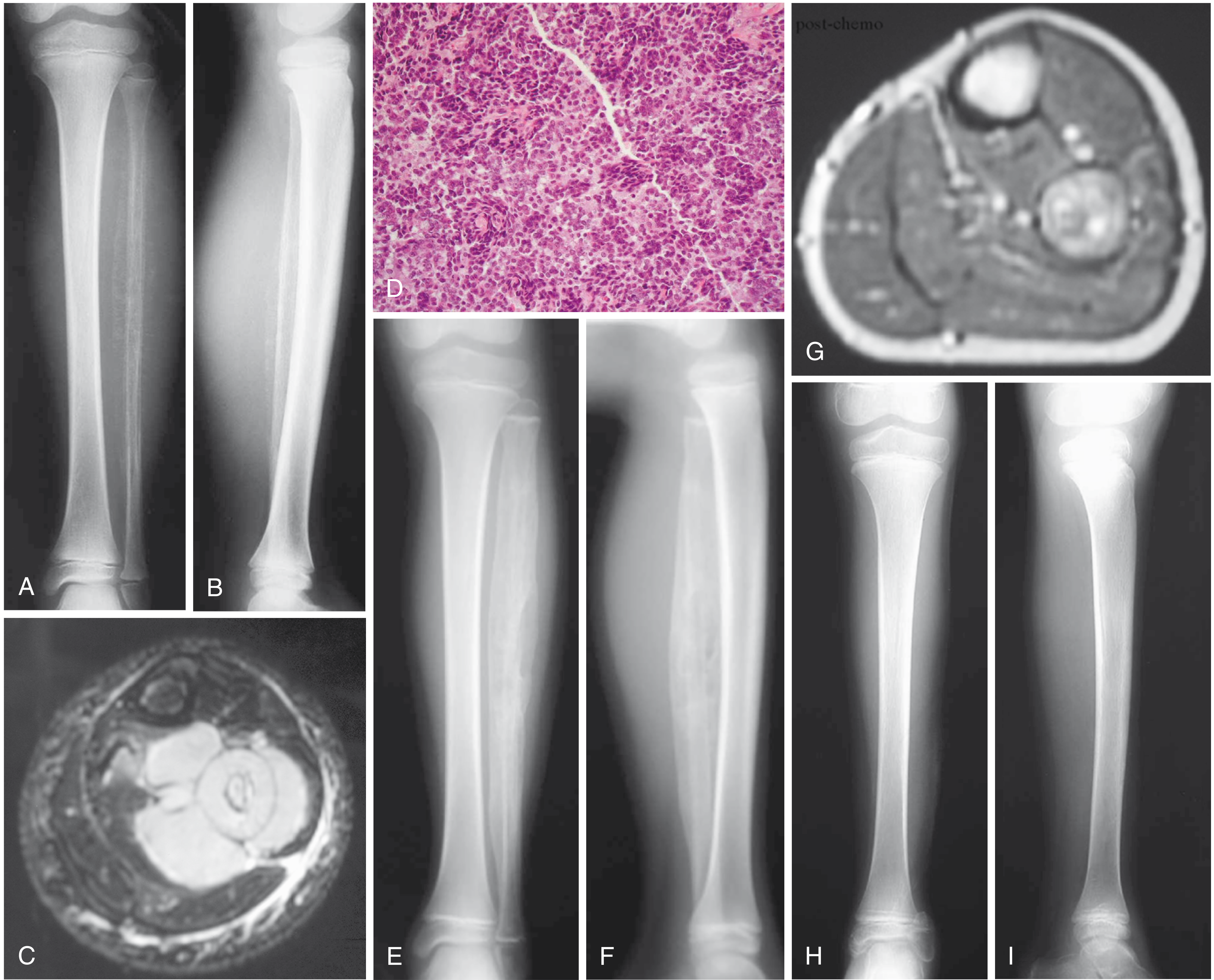
- Permeative/moth-eaten pattern of bone destruction
- "Onion skin" periosteal reaction - pathognomonic - due to alternating layers of tumor and reactive periosteal bone
- Soft tissue shadow often visible
- Diaphyseal involvement classic; metaphyseal more common in reality
- In flat bones: nonspecific destructive lesion
- Shows full extent of tumor - always image entire bone
- Soft-tissue mass extent (often disproportionately large)
- Isointense on T1; slightly hyperintense on T2
- Intense gadolinium enhancement (hypercellular tumor)
- Mandatory for surgical planning
- Baseline and staging - lungs are the most common site of metastases
- Bone is the second most common site of metastases; bone scan mandatory
- New standard for initial staging, recurrence detection, and metastatic disease detection
- High sensitivity and accuracy; can replace blind bone marrow biopsy in some centers
- Performed at some centers to rule out diffuse systemic disease
HISTOPATHOLOGY
Gross Appearance
- Tumor is soft, tan-white in color
- Frequently shows areas of hemorrhage and necrosis
- Arises in medullary cavity, invades cortex, periosteum, and soft tissue
Microscopic Appearance (H&E)
- Sheets of uniform small round blue cells - classic "small round blue cell tumor"
- Cells slightly larger than lymphocytes; scant cytoplasm
- Clear cytoplasm due to glycogen content (PAS-positive, diastase-sensitive)
- High nuclear-to-cytoplasmic ratio
- Homer-Wright rosettes (circular groupings with central fibrillary core) may be present
- No bone or cartilage production by tumor cells
- Very little intercellular matrix
Special Stains and Markers
| Stain/Marker | Result |
|---|---|
| PAS (Periodic Acid-Schiff) | Positive (glycogen in cytoplasm) |
| MIC-2 (CD99) | Positive - specific immunohistochemical marker |
| Cytokeratin | Negative (rules out metastatic carcinoma) |
| NSE (Neuron-specific enolase) | May be positive |
DIFFERENTIAL DIAGNOSIS
- Osteomyelitis - most important clinical DD; can mimic Ewing sarcoma
- Osteosarcoma (small cell variant) - produces osteoid matrix
- Lymphoma of bone (Non-Hodgkin Lymphoma)
- Neuroblastoma (in children, usually metastatic to bone)
- Rhabdomyosarcoma
- Leukemia (acute lymphoblastic leukemia)
- PNET (Primitive Neuroectodermal Tumor)
- Metastatic carcinoma (in older adults)
Cytogenetic or immunohistochemical studies are required to differentiate Ewing sarcoma from other small blue cell tumors.
STAGING
- All Ewing sarcomas are considered high grade (Stage IIB or III)
- Histologic grade has no prognostic significance within Ewing sarcoma (all are high grade)
- ~25% of patients have detectable metastases
- Lungs (most common), followed by bone
TREATMENT
1. Chemotherapy (Systemic)
- Mandatory - both neoadjuvant (before local treatment) and adjuvant (after)
- Before chemotherapy era: survival <10%; now 60-75% long-term survival
- Standard regimen (VAC/IE alternating):
- Vincristine + Doxorubicin (Adriamycin) + Cyclophosphamide (VAC)
- Alternating with Ifosfamide + Etoposide (IE)
- Neoadjuvant chemotherapy goals: treat micrometastases, reduce tumor size (facilitates surgery), assess chemosensitivity
2. Local Treatment (Surgery vs. Radiation)
| Factor | Surgery | Radiation |
|---|---|---|
| Local recurrence rate | <10% (wide resection) | Slightly higher |
| Preferred when | Limb accessible, acceptable functional deficit | Unresectable, central, pelvis, spine |
| Complications | Surgical morbidity | Growth disturbance, secondary malignancy, radiation necrosis |
- Neoadjuvant chemotherapy given first
- Repeat staging (radiograph, MRI) after chemotherapy
- If wide resection is feasible with acceptable functional deficit → Surgery preferred
- If wide margin difficult OR unacceptable functional deficit → Radiation is acceptable alternative
- Radiation can also be used adjuvantly after marginal/contaminated resection
- Treatment plan individualized after discussion with patient/family
3. High-Dose Chemotherapy + Stem Cell Rescue
- For metastatic disease and recurrence
PROGNOSIS
Good Prognostic Factors
- Localized disease (no metastases)
- Distal extremity location
- Small tumor size
- Early diagnosis
- Good histologic response to neoadjuvant chemotherapy (>90% necrosis = good response)
Poor Prognostic Factors
| Factor | 5-Year Survival |
|---|---|
| Distant metastases at presentation | 20-30% (worst) |
| Local recurrence | ~20% |
| Distant relapse | ~10% |
| Large tumor size | Poor |
| Pelvic/axial location | Poor |
| Older age at presentation | Poor |
| Fever, anemia, elevated LDH, WBC, ESR | Worse prognosis |
- Overall 5-year survival with localized disease: 60-75%
- Histologic grade has no prognostic significance (all Ewing sarcomas are high grade)
- Relapse within 1 year of primary treatment = worse prognosis than late relapse
RECURRENCE
- Poor prognosis despite aggressive salvage treatment
- Salvage options: further surgery, radiation, and/or high-dose chemotherapy
- Local recurrence: ~20% 5-year survival
- Distant relapse: ~10% 5-year survival
- Time to relapse has prognostic significance
SUMMARY TABLE: EWING SARCOMA vs. OSTEOSARCOMA
| Feature | Ewing Sarcoma | Osteosarcoma |
|---|---|---|
| Age | 5-25 yrs (peak 2nd decade) | 10-20 yrs (peak 2nd decade) |
| Location | Diaphysis/metaphysis; flat bones | Metaphysis of long bones |
| Genetics | t(11;22) EWSR1-FLI1 | p53, Rb mutations |
| Histology | Small round blue cells; no matrix | Pleomorphic cells; osteoid production |
| X-ray | "Onion skin" periosteal reaction | Sunburst periosteal reaction + Codman triangle |
| Radiosensitivity | Radiosensitive | Radioresistant |
| Treatment | Chemo + surgery/radiation | Chemo + surgery (radical) |
| Skip lesions | Not reported | Reported |
| Survival (localized) | 60-75% | ~65-70% |
- Campbell's Operative Orthopaedics 15th Ed 2026 - Primary orthopaedic reference
- Robbins & Kumar Basic Pathology - Molecular pathology and histology
- Grainger & Allison's Diagnostic Radiology - Imaging features
- Goldman-Cecil Medicine - Epidemiology and clinical features