
Diabetic foot ulcer detailed notes for surgery mbbs
diabetic foot ulcer Wagner classification diagram
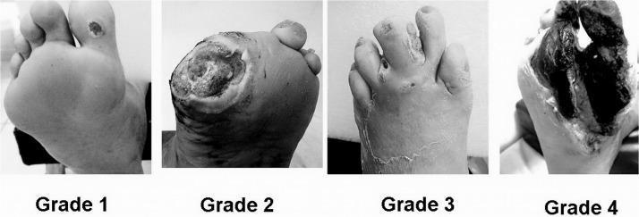
A comparative clinical photograph set illustrating the Wagner Classification System for diabetic foot ulcers across four stages (Grade 1 to Grade 4). Grade 1 (Superficial Ulcer) shows a small, localized skin lesion on the plantar surface near the base of the toes with no deep tissue involvement. Grade 2 (Deep Ulcer) depicts a larger, deeper wound penetrating to the tendon or joint capsule without abscess or osteomyelitis. Grade 3 (Osteitis/Infection) demonstrates a deep ulcer associated with significant inflammation, purulent discharge, and clinical signs of underlying bone infection or deep tissue abscess. Grade 4 (Forefoot Gangrene) shows extensive necrosis and blackening of the tissue localized to the forefoot or toes, indicating severe vascular compromise and tissue death. This series serves as an educational tool for medical professionals to differentiate between stages of diabetic foot pathology, guiding clinical management and prognosis assessment.
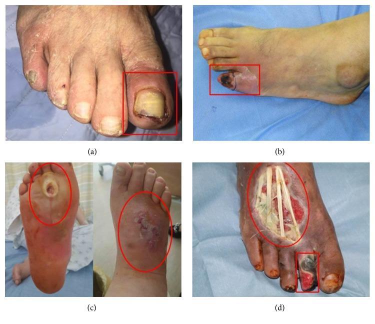
A series of four clinical photographs (a-d) demonstrating the progression of diabetic foot ulcers categorized by the Wagner classification system. (a) Wagner Grade 1: A superficial ulcer on the left hallux (big toe) featuring erythema, periungual inflammation, and a thickened, discolored yellowish toenail, indicative of an ischemic etiology. (b) Wagner Grade 2: A deep ulcer on the fifth digit (pinky toe) showing more extensive tissue involvement and localized cyanosis. (c) Wagner Grade 3: A neuropathic perforating ulcer extending from the plantar surface to the dorsum of the foot, associated with deep infection, abscess, and clinical signs of osteomyelitis. (d) Wagner Grade 4: Advanced diabetic foot pathology exhibiting local gangrene of the second toe and a large, deep dorsal foot ulcer. The necrotic tissue is blackened, and the wound bed is exposed, revealing underlying anatomical structures such as tendons. This set illustrates the clinical manifestations of ischemic, neuropathic, and mixed-type diabetic foot disease for educational use in wound care and endocrinology.
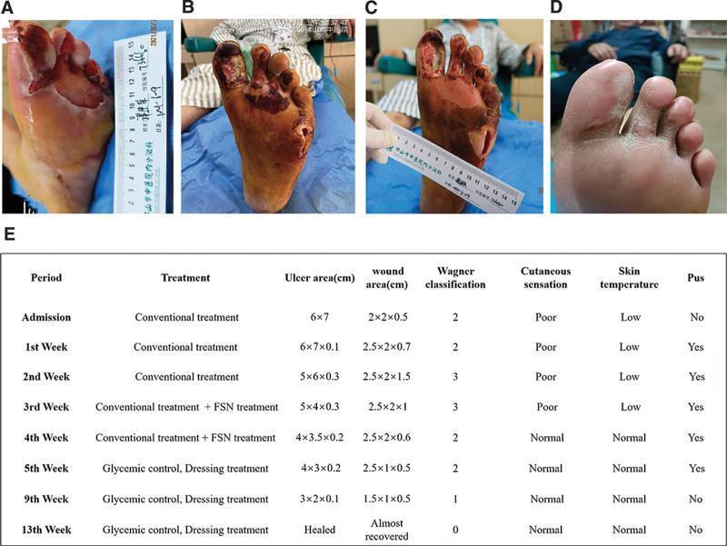
This composite figure presents a clinical progression timeline of a complex diabetic foot ulcer (DFU) on the left foot, categorized under the Wagner classification system. (A) Initial presentation shows a large, 6x7 cm ulcer on the plantar forefoot with surrounding maceration and a lateral abscess. (B) Following initial debridement, the wound exhibits deep tissue involvement (Wagner Grade 3), with necrotic tissue extending to the tendons and musculature of the first through third toes, accompanied by a deep 2x2x0.7 cm abscess cavity. (C) Mid-treatment status demonstrates significant clinical improvement; the wound bed appears ruddy with healthy granulation tissue, reduced exudate, and diminishing necrotic debris. (D) Final follow-up at three months shows complete secondary intention healing with full epithelialization and stable scar tissue formation. (E) An accompanying summary table provides longitudinal data on treatment phases (including Fu's subcutaneous needling), ulcer/wound dimensions, Wagner grade transition (from Grade 3 to 0), and clinical markers such as cutaneous sensation, skin temperature, and presence of pus.
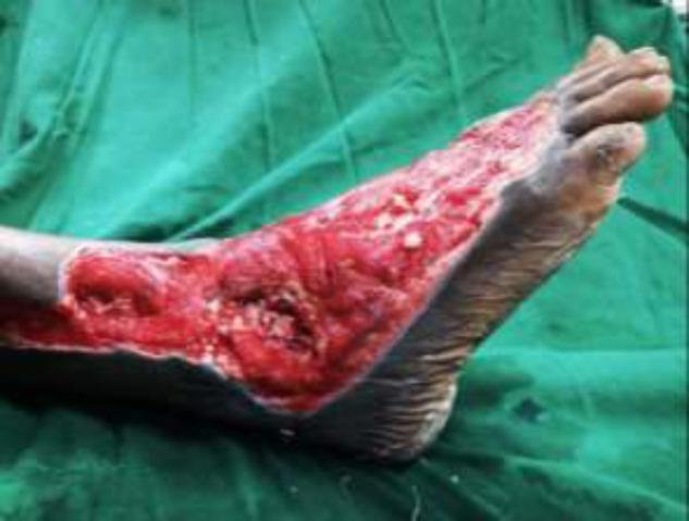
Clinical photograph of a right foot demonstrating a severe, extensive Wagner Grade III or IV diabetic foot ulcer. The wound spans the lateral and dorsal aspects of the foot, extending from the distal lower leg across the ankle joint to the midfoot region. The ulcer bed is characterized by beefy red granulation tissue and deep cavitation, with visible exposure of underlying musculoskeletal structures including the ankle joint. There are focal areas of yellow/white slough and fibrin, indicative of potential infection or chronic inflammatory processes. The wound margins are irregular but well-demarcated against the surrounding hyperpigmented, intact skin. This image serves as a clinical example of complex diabetic wound progression requiring advanced surgical intervention, such as debridement and microvascular flap reconstruction. Key educational concepts include diabetic foot classification, wound bed assessment, and the management of deep tissue infections with joint involvement.
Charcot foot neuropathic diabetic foot deformity
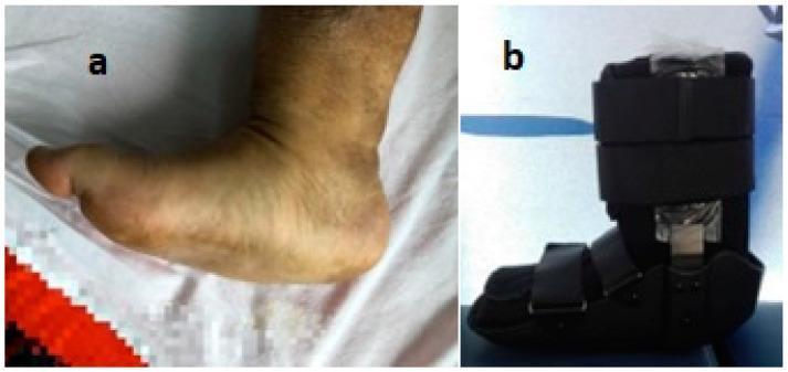
This composite image illustrates the clinical presentation and management of a neuropathic diabetic foot. Panel (a) is a clinical photograph showing a Charcot foot deformity in a side profile. Key visual features include a significant collapse of the longitudinal arch (rocker-bottom foot), localized swelling, and medial bulging consistent with midfoot joint destruction and bone displacement. The skin exhibits mild hyperpigmentation and texture changes associated with chronic inflammation. Panel (b) shows a diagnostic and therapeutic device: a personalized neuropathic diabetic foot orthosis. This black, boot-like orthopedic device is designed for off-loading therapy and immobilization to prevent further structural damage or ulceration. It features multiple wide, adjustable velcro straps for secure fixation, a rigid exterior shell for stabilization, and integrated metallic structural supports. The images collectively demonstrate the progression of diabetic neuroarthropathy and the specialized orthopedic intervention required for pressure redistribution and limb preservation.
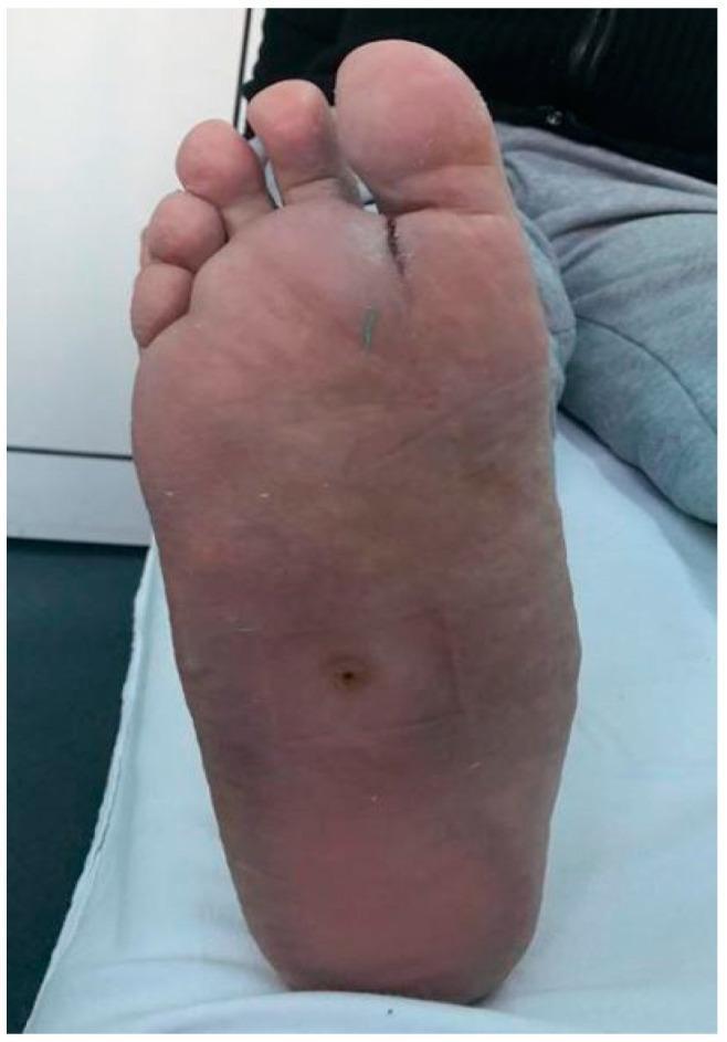
This clinical photograph displays the plantar surface of a human foot, illustrating complications of diabetic foot disease and neuropathic osteoarthropathy (Charcot foot). The image reveals two distinct areas of concern: a small, circular, well-circumscribed neuropathic ulcer located in the central midfoot region, and a linear skin break or fissure proximal to the second and third toes featuring a greenish exudate, suggestive of localized infection or drainage. The foot demonstrates a characteristic 'rocker-bottom' deformity associated with Charcot osteoarthropathy, characterized by a flattened or protruding longitudinal arch. The surrounding skin shows areas of hyperkeratosis (callus formation) and mild desquamation. This visual material is used in medical education to demonstrate the clinical presentation of the high-risk diabetic foot, emphasizing the relationship between sensory neuropathy, structural deformity, and the development of pressure-related ulcerations.
Diabetic Foot Ulcer (DFU) - Detailed Notes for Surgery MBBS
1. DEFINITION AND EPIDEMIOLOGY
- Lifetime risk of DFU in a diabetic patient: 15%
- Annual incidence: 1-2%
- 15-20% of DFUs lead to lower extremity amputation (LEA)
- ~85% of all lower limb amputations are preceded by a DFU
- Foot infections are the leading cause of hospital admissions in diabetic patients
- 5-year mortality of DFU complications rivals that of many cancers (~30.5%)
2. PATHOPHYSIOLOGY - THE TRIAD
A. Peripheral Neuropathy (Most Important)
- Sustained hyperglycemia → glucose converted to sorbitol and fructose (polyol pathway) → impairs ATPase pumps → structural breakdown + impaired axonal transport
- Excess glucose interacts with intracellular proteins/nucleotides → normal cellular function impaired
- Mitochondria increase free radical production → oxidative stress
- Basement membranes of vasa nervorum become glycosylated and thickened → chronic ischemia of nerves
- Sensory neuropathy: Loss of light touch, temperature first → progresses proximally ("glove and stocking" distribution) → painless injuries go unnoticed
- Motor neuropathy: Intrinsic foot muscle wasting → claw toes, hammer toes, foot drop → altered plantar pressure → ulceration (especially 1st and 5th metatarsal heads, great toe)
- Autonomic neuropathy: Decreased sweating → dry, cracked, fissured skin → portal of entry for infection; altered blood distribution to skin
- 10-g Semmes-Weinstein monofilament (primary screen)
- Plus at least one of: pinprick, temperature, vibration, or proprioception testing
- Atrophy of extensor digitorum brevis = early motor neuropathy sign
B. Peripheral Vascular Disease (Ischemia)
- Hyperglycemia → glycosylation of endothelial basement membranes → poor O2 diffusion
- Arteriosclerosis + medial calcinosis of arterial walls → narrowed lumen → poor perfusion
- Diabetic PAD primarily affects tibial and femoral-popliteal distribution
- ABI (Ankle-Brachial Index) is unreliable in diabetics due to medial calcinosis causing non-compressible arteries (falsely elevated ABI)
- More reliable tests: Toe-Brachial Index (TBI), toe pressure (normal ≥60 mmHg), transcutaneous oxygen (TcPO2), waveform analysis
C. Impaired Wound Healing
- Hyperglycemia → decreased function of neutrophils, macrophages, and fibroblasts
- Epigenetic modification of macrophages → persistent pro-inflammatory state → fails to transition to proliferation/remodeling phase
- Bacterial colonization + biofilm formation further inhibits healing
- Wound infection is usually polymicrobial: gram-positive aerobes (Staphylococci most common), gram-negative bacilli, and obligate anaerobes
3. SITES OF PREDILECTION
- Plantar surface of 1st metatarsal head (greatest pressure in push-off)
- Plantar surface of 5th metatarsal head
- Great toe tip / dorsum (ischemic)
- Heel (pressure ulcers in bedridden patients)
- 5th toe and between toes
4. CLINICAL FEATURES
Neuropathic Ulcer vs. Ischemic Ulcer
| Feature | Neuropathic | Ischemic |
|---|---|---|
| Site | Plantar, pressure points | Tips of toes, lateral foot, heel |
| Pain | Painless | Painful (rest pain, claudication) |
| Surrounding skin | Warm, well-perfused, dry | Cold, pale/mottled/cyanotic |
| Ulcer base | Pink, granulating | Pale, sloughy |
| Callus | Present (surrounding) | Absent |
| Pulses | Present | Absent/diminished |
| Neuropathy | Prominent | May be co-existent |
| Edema | May be present (Charcot) | Usually absent |
Inspection of the Foot (Systematic Exam)
- Inspect shoes: size, seams, abnormal wear patterns (pressure points)
- Calluses/corns = precursors to ulcer
- Claw/hammer toes = motor neuropathy
- Erythema, warmth, swelling = infection
- Wound size, depth, probing to bone
- Lymphangitis, spreading cellulitis (ascending infection)
5. CLASSIFICATION
A. Wagner-Meggitt Classification (Most Used - Must Know!)
| Grade | Description | Treatment |
|---|---|---|
| 0 | Skin intact; bony deformity - "at risk" foot | Extra-depth shoes, pressure-relief insoles, regular review |
| 1 | Superficial ulcer; no tendon/bone involvement | In-office debridement + Total Contact Cast (TCC) |
| 2 | Deep ulcer with exposed tendon or joint capsule | Formal operative debridement + TCC once wound bed healthy |
| 3 | Extensive ulcer with exposed bone / osteomyelitis / abscess | Surgical debridement of bone/osteomyelitis, antibiotics |
| 4 | Localized forefoot gangrene | Revascularization assessment + partial foot amputation |
| 5 | Extensive whole foot gangrene | Revascularization + below-knee or above-knee amputation |
B. Brodsky Depth-Ischemia Classification (Modification of Wagner-Meggitt)
- Grade A: Normal vascularity
- Grade B: Ischemia without gangrene - noninvasive vascular studies + revascularization if indicated
- Grade C: Partial (forefoot) gangrene - vascular studies + revascularization + partial foot amputation
- Grade D: Complete foot gangrene - BKA or AKA
C. WIfI Classification (Society for Vascular Surgery - Modern)
- Better predicts risk of major amputation at 1 year and benefit of revascularization
- Correlates with wound healing time and 1-year amputation-free survival
6. INVESTIGATIONS
Bedside
- Probe-to-bone test: Metal probe inserted into ulcer; if bone is felt = highly specific for osteomyelitis (PPV ~89%)
- Monofilament test: 10-g Semmes-Weinstein for neuropathy assessment
Blood Tests
- CBC (leukocytosis in infection, but often normal in DFU)
- ESR, CRP (may be only mildly elevated - unreliable)
- HbA1c (glycemic control; target <7%; >7% = independent risk factor for DFU)
- Blood cultures if systemically unwell
- Serum albumin, total protein (nutritional status for surgery planning - albumin <2.5 g/dL = surgery should be delayed)
Imaging
-
X-ray foot (3 views): First-line; assess for:
- Osteomyelitis (bony destruction, periosteal reaction) - low sensitivity early
- Gas in soft tissues (necrotizing infection/gas gangrene)
- Charcot deformity, prior amputations, structural deformity
- Cortical erosion, "sucked candy" appearance of bone
-
MRI: Most sensitive and specific for osteomyelitis; detects early bone marrow edema; also shows extent of soft tissue infection - investigation of choice
-
Bone scan (Tc-99m): When MRI unavailable; sensitive but not specific
-
Tagged WBC scan (In-111): Better specificity than bone scan for osteomyelitis
-
CT scan: Shows gas in soft tissues, bony destruction (useful in necrotizing fasciitis)
Vascular Studies
- ABI (unreliable in diabetics due to non-compressible calcified arteries)
- Toe-Brachial Index (TBI): Preferred; normal >0.7; <0.4 = severe ischemia
- Toe pressures: Normal ≥60 mmHg
- TcPO2 (transcutaneous O2): >40 mmHg = adequate healing potential
- Doppler waveforms: Triphasic > biphasic > monophasic (worst)
- CT/MR angiography or conventional angiography: For revascularization planning
Microbiology
- Superficial wound swabs are NOT RELIABLE for determining deep infection organisms
- Tissue biopsy > deep wound cultures > superficial swabs
- Bone biopsy = gold standard for osteomyelitis (gives culture + sensitivities)
7. OSTEOMYELITIS IN DFU
- Most important risk factor = presence of a foot ulcer
- Organisms: S. aureus, β-haemolytic streptococci, aerobic gram-negative bacilli, Pseudomonas (over-represented); anaerobes present in abscesses/devitalized tissue
- Diagnosis confirmed by combination of:
- Probe-to-bone test positive +
- Elevated inflammatory markers (ESR/CRP) +
- Abnormal plain radiograph
- MRI = most clinically useful diagnostic tool
- Gold standard = bone biopsy and culture
8. MICROBIOLOGY OF DFU INFECTION
| Severity | Organisms |
|---|---|
| Mild (superficial) | S. aureus, Streptococcus (Group A, B) |
| Moderate-Severe | Polymicrobial: Staph, Strep + gram-negative bacilli (E. coli, Klebsiella, Proteus) |
| Severe (deep, necrotic) | Above + Pseudomonas aeruginosa + anaerobes (Bacteroides, Clostridia) |
- MRSA increasingly prevalent in hospital-acquired infections
- Empirical antibiotic principles:
- Mild-moderate: Cover gram-positive cocci (S. aureus including MRSA, Streptococci)
- Severe: Add cover for Pseudomonas + consider metronidazole for anaerobes
9. PEDAL SEPSIS (Surgical Emergency)
- Features: Fever, tachycardia, hypotension, altered mental status, acidosis, multi-organ failure
- Exam: Spreading erythema with cellulitis tracking proximally up the calf, lymphangitis, purulent drainage, tissue necrosis
- Imaging: Plain X-ray/CT showing gas in soft tissues
- Management: Immediate surgical consultation; source control is URGENT
- Do not delay surgery for imaging when clinical picture is clear
10. MANAGEMENT
A. Non-Surgical / Conservative
- Target HbA1c <7%
- Tight glucose control halts neuropathy progression; may be partially reversed
| Device | Notes |
|---|---|
| Total Contact Cast (TCC) | Gold standard; reduces plantar pressure 60%; non-removable; weekly cast changes; requires uninfected wound |
| Fixed ankle walking boot | Good alternative; less effective than TCC |
| Removable walker boot | Allows dressing changes; patient compliance issue |
| Wedge shoes / heel-out shoes | For specific locations |
| NOT below-ankle prosthetics | Insufficient offloading |
- Healthy wound → collagen dressing
- Fibrinous tissue → collagenase (Santyl) or MediHoney
- Deep healthy wound → VAC (Negative Pressure Wound Therapy/NPWT)
- Locally infected wound → Iodosorb or mupirocin (Bactroban)
- Highly exudative/macerated → alginate with silver, absorptive foams
- Simple option: moist saline- or Vaseline-impregnated gauze (daily or twice-daily changes)
- Multilayer compression (venous/lymphedema component)
- Hyperbaric Oxygen Therapy (HBOT): Increases tissue oxygenation, promotes angiogenesis; requires good perfusion (palpable pulses, normal TBI); several weeks of treatment
- Topical PDGF (Platelet-Derived Growth Factor - Becaplermin): For wounds failing conventional therapy
- Maggot (larval) therapy: Selective debridement of necrotic tissue; limited by patient/provider acceptability
- BP control (ACE inhibitor preferred)
- Statins for hyperlipidemia
- Antiplatelet therapy (aspirin) in PAD
- Tobacco cessation
- Regular foot inspection + podiatry care
- Professional nail trimming
- Moisturize; avoid walking barefoot; properly fitted shoes
B. Surgical Management
1. Debridement (Core Surgical Principle)
- Sharp debridement (scalpel/scissors) - most common; in-office or OR
- Hydrosurgical debridement (high-pressure irrigation; useful for superficial wounds)
- Mechanical debridement (scrubbing under anesthesia)
- Maggot therapy (selective biological debridement)
- Callus debridement essential as overriding callus hides the wound
- Primary closure: Rarely ideal (bacterial colonization, risk of deep infection, tension)
- Delayed primary closure: When wound is clean
- Secondary intention: Most common; wet-to-dry dressings
- NPWT (VAC therapy): Accelerates healing, reduces dressing changes
2. Management by Wagner Grade
| Grade | Surgical Management |
|---|---|
| 0 | Preventive; no surgery needed unless deformity correction required |
| 1 | In-office debridement; callus removal; TCC |
| 2 | Formal operative debridement of exposed tendon and non-viable tissue; TCC after clean wound bed |
| 3 | Surgical debridement of bone/osteomyelitis; bone biopsy; culture-directed antibiotics; consider bone resection |
| 4 | Revascularization assessment; partial foot amputation (ray, transmetatarsal); vascular bypass or angioplasty |
| 5 | Major amputation (BKA or AKA); revascularization if possible |
3. Surgical Offloading Procedures
- Metatarsal head resection: Corrects deformity, reduces pressure point; prevents recurrence
- Achilles tendon lengthening (TAL - Tendo-Achilles Lengthening):
- Equinus contracture is common; TAL offloads midfoot/forefoot
- Indications: Recurrent forefoot/midfoot ulceration + ulceration with equinus deformity
- Liquid silicone injections at pressure points
- Flexor tenotomies: For dorsal toe ulcers
- Joint resection for toe deformities
- Midfoot osteotomy or fusion: For midfoot collapse (Charcot); fusion if instability present
4. Vascular Intervention
- Toe pressure <60 mmHg (or 60-80 mmHg with non-healing wound at 4-6 weeks)
- Grade 1 ischemia and above (WiFi) - book for angiography
- Do not revascularize through an actively infected field - debride first, then revascularize
- Endovascular (PTA, stenting): First-line for short segment disease; decreased patency below the knee
- Open bypass (femoro-popliteal, femoro-tibial, pedal bypass): For extensive tissue destruction or tibial disease; better for large wounds needing robust flow
- Proximal angioplasty/bypass before infection surgery improves outcomes
5. Amputations
- "Amputation is not an easy option" in DFU - wound healing is problematic
- Excision must be adequate to remove all infected material
- Excess bone resected to allow tension-free skin closure
- If extensive peripheral neuropathy, BKA (with better sensation) preferred over more distal amputation
- Digital/Toe amputation - single digit for localized gangrene
- Ray amputation - toe + corresponding metatarsal shaft
- Transmetatarsal amputation (TMA) - all 5 metatarsals
- Lisfranc amputation - at tarsometatarsal joints
- Chopart amputation - at midtarsal joint (calcaneocuboid + talonavicular)
- Syme's amputation - ankle disarticulation with heel flap
- Below-knee amputation (BKA/transtibial) - most common major amputation; good rehab potential
- Above-knee amputation (AKA/transfemoral) - when BKA not feasible
11. CHARCOT NEUROARTHROPATHY
- Destructive joint disease due to severe sensory neuropathy
- Acute phase: Red, hot, swollen foot (may mimic cellulitis/DVT/osteomyelitis)
- Chronic phase: Collapse of midfoot → "rocker-bottom" deformity → creates new pressure points → ulceration
- Urgent management: Offloading in plaster cast (total contact cast); strict non-weight-bearing
- Unstable Charcot: May require midfoot fusion/osteotomy
- Any unexplained swelling, heat, ulcer, or deformity in a diabetic patient should be treated as Charcot until proven otherwise
12. MULTIDISCIPLINARY TEAM (MDT)
- Vascular surgeon
- Surgical podiatry
- Wound care nurse/specialist
- Endocrinologist
- Infectious disease specialist (complex infections)
- Plastic surgeon (reconstruction)
- Orthopedic foot & ankle surgeon (deformity)
- Dietitian (nutritional optimization)
13. ANTIBIOTICS IN DFU INFECTION
| Severity | Empirical Regimen |
|---|---|
| Mild | Oral: Flucloxacillin / Amoxicillin-clavulanate (cover Staph/Strep) |
| Moderate | IV: Amoxicillin-clavulanate or Co-amoxiclav; add MRSA cover (vancomycin/linezolid) if risk factors |
| Severe | Broad-spectrum IV: Piperacillin-tazobactam ± vancomycin ± metronidazole (Pseudomonas + anaerobe cover) |
| Osteomyelitis | Prolonged course (6 weeks); guided by bone biopsy culture |
- Superficial swabs are NOT reliable; deep tissue/bone cultures guide therapy
- Consider MRSA cover in severe/hospital-acquired infections
14. PREVENTION
- Annual foot examination (minimum) for all diabetics; more frequent if prior ulcer/deformity
- Patient education: Daily self-foot inspection, proper footwear, moisturizing, professional nail care
- No walking barefoot
- Well-fitted, cushioned shoes; custom orthotics after healed ulcer
- Treat calluses early (podiatry)
- HbA1c target <7%; treat hypertension, hyperlipidemia, and tobacco use
15. PROGNOSIS
- DFU with infection + ischemia = 90x higher amputation risk
- Infected ulcer alone = 40-55% chance of some form of amputation
- 5-year mortality of DFU complications (30.5%) comparable to many cancers
- Ulcer <3 cm² → better chance of healing without amputation
- Recurrence rate remains high at 3 years even with MDT care
HIGH-YIELD EXAM POINTS
- Triad: Neuropathy + Ischemia + Infection
- Most important risk factor for osteomyelitis: Presence of a foot ulcer
- Probe-to-bone test = bedside diagnosis of osteomyelitis
- Gold standard for osteomyelitis: Bone biopsy
- MRI: Most sensitive/specific imaging for osteomyelitis
- Best offloading device: Total Contact Cast (TCC)
- Wagner Grade 0 = at-risk foot (skin intact); Grade 5 = whole foot gangrene
- ABI unreliable in diabetics (calcified vessels) - use toe pressure/TBI instead
- Superficial swabs are NOT reliable - get deep/bone cultures
- Pedal sepsis = surgical emergency - immediate source control
- Achilles tendon lengthening for recurrent forefoot ulcers with equinus deformity
- Charcot foot presents as hot, swollen, red foot - urgent offloading in plaster
- Revascularize BEFORE debridement in subacute/chronic infection; DEBRIDE FIRST in acute infection
- HBOT - used when perfusion is good but wound fails to heal
- HbA1c target <7% for wound healing and prevention