
Uterus
Reading File
The query "Uterus" is quite broad. Let me clarify what you're looking for before diving into research.
Asking for Preferences
What aspect of the uterus would you like to explore?
All thing to study for 10 marks
Finding Sources
Searching the Web
uterus anatomy physiology pathology 10 marks exam topics MBBS
Now let me pull the actual textbook content from the most relevant sources - anatomy, histology, and pathology simultaneously.
Reading File
Reading File
Reading File
Now let me pull more pathology (fibroids, endometrial carcinoma) and the histology of the menstrual cycle.
Reading File
Reading File
Now let me fetch the key anatomical diagram and the histology diagram of endometrium layers.
Loading Image
Loading Image
Loading Image
Loading Image
Excellent content gathered. Now here is your complete, exam-ready 10-mark study note on the Uterus.
The Uterus - Complete 10-Mark Study Notes
1. GROSS ANATOMY
The uterus is a thick-walled, hollow, pear-shaped muscular organ located in the midline of the pelvis, posterior to the bladder and anterior to the rectum.
Size: ~8 cm long, 5 cm wide, 4 cm thick (non-pregnant)
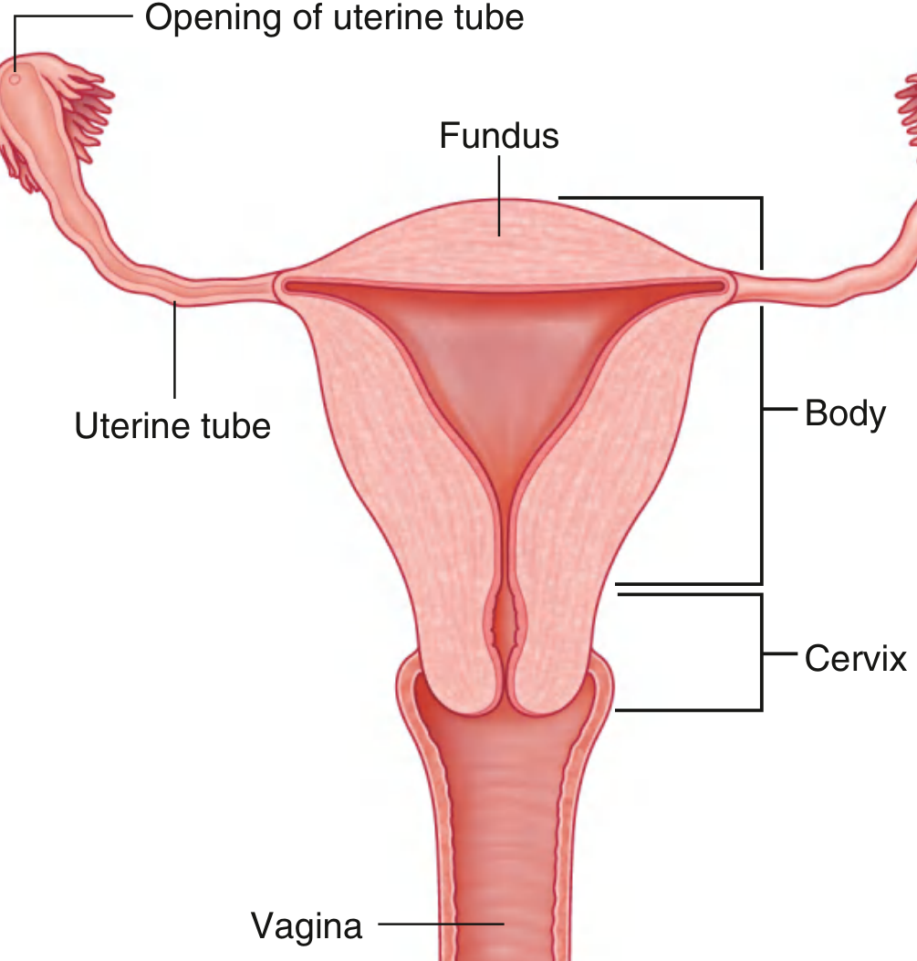
Parts of the Uterus
| Part | Description |
|---|---|
| Fundus | Rounded superior portion, above the opening of uterine tubes |
| Body (Corpus) | Main central portion; flattened anteroposteriorly; cavity is an inverted triangle |
| Isthmus | Narrow zone between body and cervix |
| Cervix | Lower cylindrical part; has internal os (opens to isthmus) and external os (opens to vagina) |
Position
- Normal position: Anteverted (long axis tilted forward relative to vagina) and Anteflexed (body bent forward at isthmus relative to cervix)
Ligaments (Support)
| Ligament | Function |
|---|---|
| Round ligament | Maintains anteversion |
| Broad ligament | Lateral peritoneal fold, contains uterine vessels |
| Uterosacral ligament | Main support; prevents backward displacement |
| Cardinal (Mackenrodt's) ligament | Most important support; prevents uterine prolapse |
| Pubocervical ligament | Anterior support |
- Primary support = pelvic floor muscles (levator ani), supplemented by ligaments.
Blood Supply
- Uterine artery (branch of internal iliac artery) - main supply
- At the isthmus it crosses the ureter ("water under the bridge" - clinically important in hysterectomy)
- Ascending branch anastomoses with the ovarian artery
- Venous drainage: uterine venous plexus → internal iliac vein
Lymphatic Drainage
- Upper body/fundus → para-aortic nodes
- Lower body/cervix → iliac (external & internal) nodes, obturator nodes
2. HISTOLOGY (Wall Layers)
The uterine wall has 3 layers (from outside in):
a) Perimetrium (outer)
- Peritoneal serosa (mesothelium) covering most of uterus
- Adventitia on anterior-inferior surface
b) Myometrium (middle - thickest)
- Bundles of smooth muscle in interwoven layers
- Rich in venous plexuses and lymphatics
- During pregnancy: undergoes hyperplasia + hypertrophy + increased collagen to withstand parturition
- After delivery: smooth muscle cells shrink, many undergo apoptosis
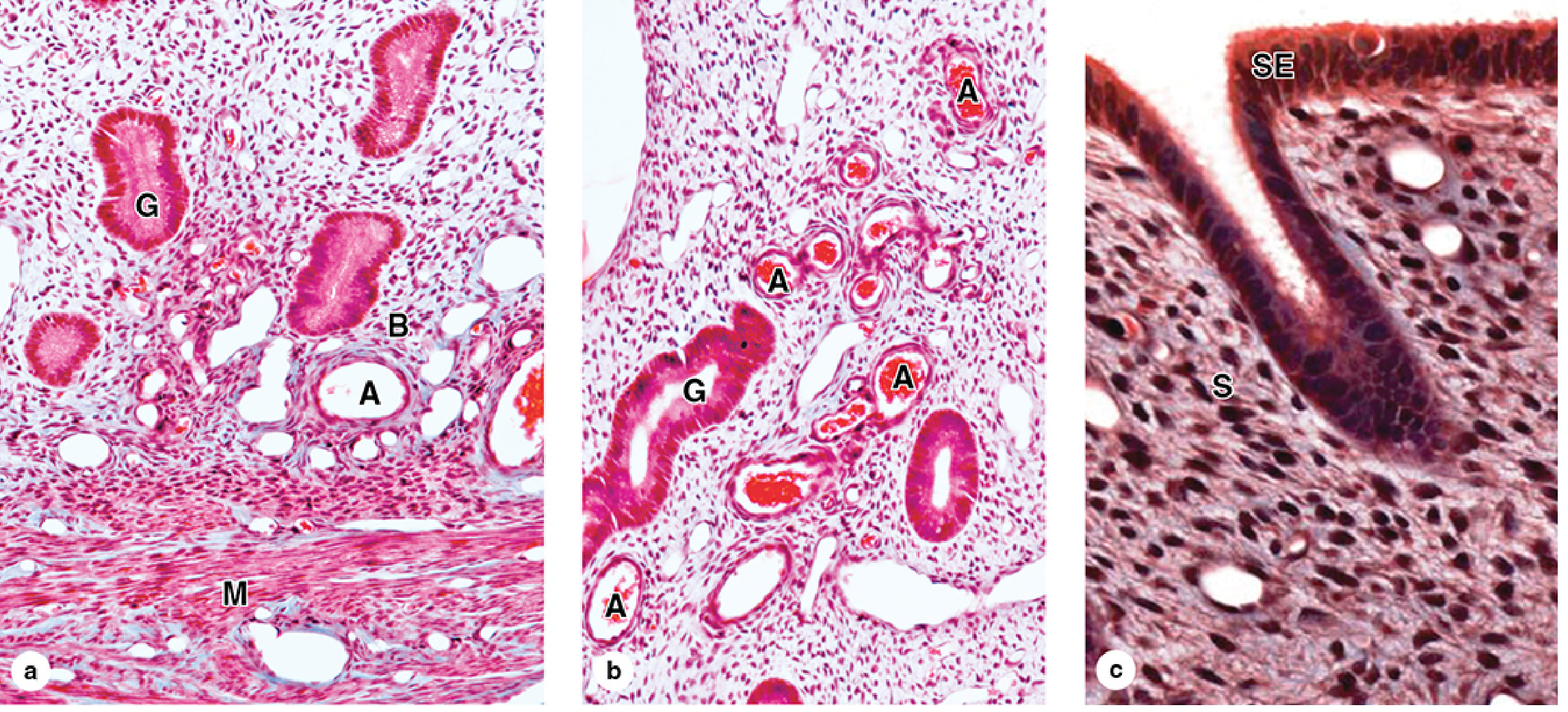
c) Endometrium (inner mucosa)
- Lined by simple columnar epithelium (ciliated + secretory cells)
- Stroma: type III collagen, abundant fibroblasts
- Contains tubular uterine glands penetrating full thickness
The endometrium has 2 concentric zones:
| Zone | Location | Sensitivity | Function |
|---|---|---|---|
| Basal layer | Adjacent to myometrium | Insensitive to hormones | Regenerates functional layer after menses |
| Functional layer | Superficial | Sensitive to estrogen/progesterone | Undergoes cyclic changes; shed during menses |
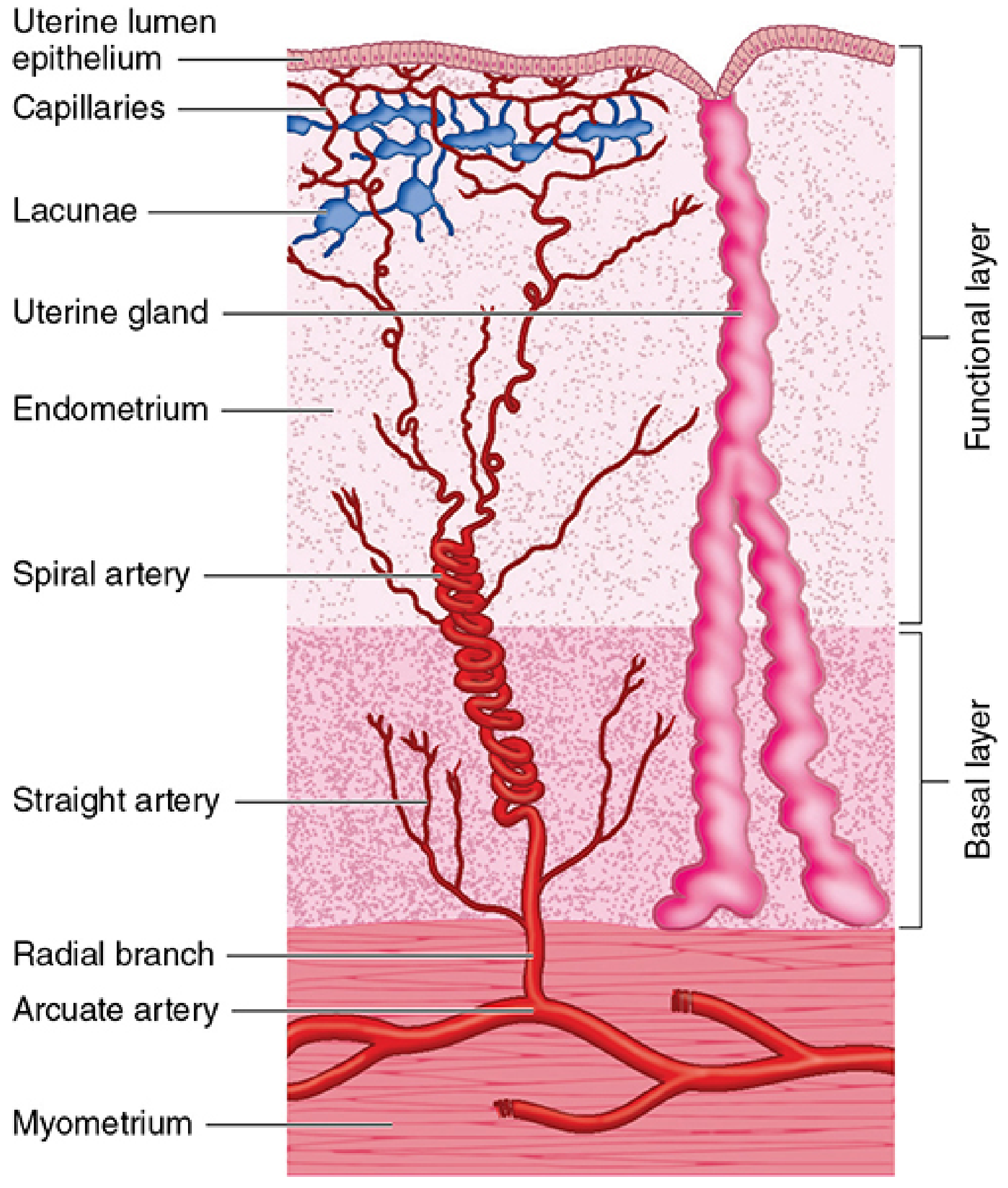
Blood Vessels of Endometrium (Critical for Menstruation)
Arcuate arteries (in myometrium) → radial branches → two types:
- Straight arteries - supply basal layer (NOT sensitive to progesterone withdrawal - why basal layer is retained)
- Spiral arteries - supply functional layer (sensitive to progesterone withdrawal → vasospasm → ischemia → menstrual shedding)
3. PHYSIOLOGY - MENSTRUAL / UTERINE CYCLE
The uterine cycle mirrors the ovarian cycle and lasts ~28 days.
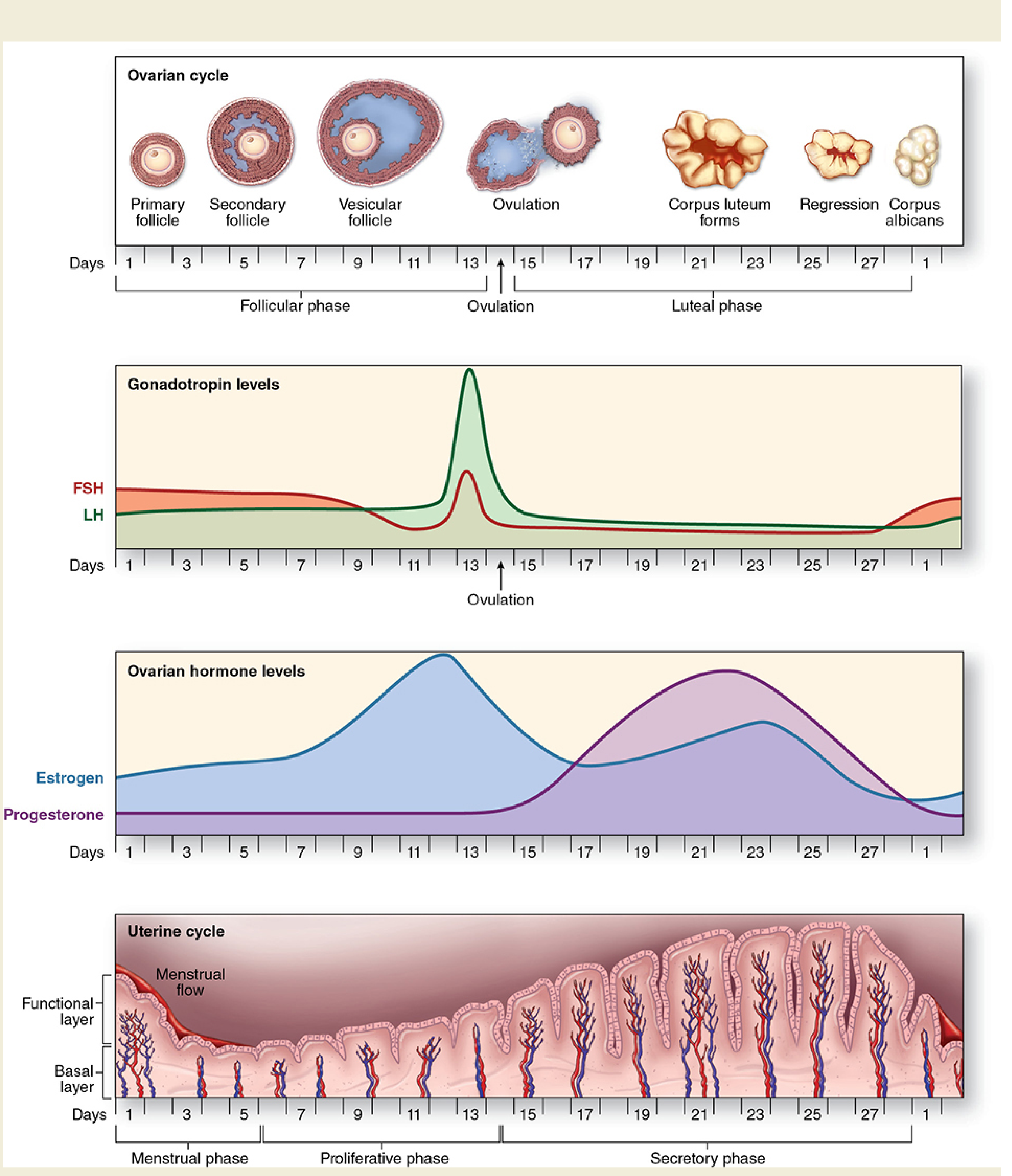
Phases of the Uterine Cycle
| Phase | Days | Hormone | Endometrial Changes |
|---|---|---|---|
| Menstrual phase | 1-5 | Estrogen + progesterone fall | Functional layer shed; spiral arteries spasm → ischemia |
| Proliferative phase | 6-14 | Rising estrogen (follicular phase) | Glands grow, straight & narrow; stroma thickens; epithelium regenerates |
| Secretory phase | 15-28 | Progesterone (corpus luteum) | Glands tortuous, sacculated, glycogen-rich secretions; stroma edematous; prepares for implantation |
- Day 14: LH surge triggers ovulation
- If no implantation: corpus luteum regresses → progesterone falls → spiral artery spasm → menstruation
- Implantation of blastocyst occurs in the body of the uterus, normally on day 20-23
4. PATHOLOGY (Diseases of the Uterus)
A. Endometritis
- Acute: bacterial, post-delivery/miscarriage; neutrophilic infiltrate
- Chronic: requires plasma cells for diagnosis (lymphocytes alone are normal); causes include PID (N. gonorrhoeae, C. trachomatis), tuberculosis (granulomatous endometritis)
- Features: fever, abdominal pain, menstrual irregularities
B. Adenomyosis
- Endometrial tissue (glands + stroma) present within the myometrium
- Induces reactive myometrial hypertrophy → enlarged, globular uterus
- Causes: menorrhagia, dysmenorrhea, pelvic pain (worse premenstrually)
- Can coexist with endometriosis
C. Endometriosis
- Endometrial glands + stroma outside the uterus
- Affects ~10% of women in reproductive years; ~50% with infertility
- Common sites: ovaries, pouch of Douglas, uterine ligaments, fallopian tubes
- Types: superficial peritoneal, ovarian (chocolate cysts), deep infiltrating
- Malignant transformation risk: mainly deep infiltrating type
- Pathogenesis theories:
- Regurgitation (retrograde menstruation) - most widely accepted
- Benign metastasis - via blood/lymphatics
- Metaplastic - from coelomic epithelium
- Stem/progenitor cell theory - bone marrow derived
D. Dysfunctional Uterine Bleeding (DUB)
- Abnormal uterine bleeding without organic cause
- Most common cause: anovulatory cycles (excess unopposed estrogen)
- Other causes: pituitary tumors (prolactin excess → reduced GnRH/LH/FSH), polycystic ovarian syndrome, obesity, luteal phase defect
Causes of AUB by Age:
| Age Group | Common Causes |
|---|---|
| Prepuberty | Precocious puberty |
| Adolescence | Anovulatory cycles, coagulation disorders |
| Reproductive age | Pregnancy complications, leiomyoma, adenomyosis, polyps, carcinoma |
| Perimenopause | Anovulatory cycles, hyperplasia |
| Postmenopause | Carcinoma, hyperplasia, polyps, atrophy |
E. Endometrial Hyperplasia
- Excess estrogen relative to progestin → exaggerated proliferation
- Causes: obesity (adipose converts precursors to estrogen), PCOS, granulosa-theca cell tumors, exogenous estrogen
| Type | Features | Risk of Carcinoma |
|---|---|---|
| Without atypia | Increased gland:stroma ratio | 1-3% |
| With atypia (EIN) | Complex glands + nuclear atypia + PTEN mutations | Significant; considered precursor to endometrial carcinoma |
- Treatment with atypia: hysterectomy (or high-dose progestins if fertility desired)
F. Endometrial Carcinoma
- Most common gynecologic malignancy
- Presents with postmenopausal bleeding (most common symptom)
- Two main types:
- Type I (endometrioid): Estrogen-related, arises from hyperplasia/EIN, PTEN mutations, better prognosis
- Type II (serous/clear cell): Not estrogen-related, TP53 mutations, worse prognosis
G. Leiomyoma (Fibroid)
- Most common uterine tumor; benign smooth muscle tumor of myometrium
- Estrogen-dependent; regress after menopause
- Presents with: menorrhagia, pelvic pain, pressure symptoms, infertility
- Malignant transformation to leiomyosarcoma is rare
5. EMBRYOLOGY (Development)
- Uterus develops from fusion of the paramesonephric (Mullerian) ducts
- In females: absence of Y chromosome, testosterone, and anti-Mullerian hormone → Mullerian ducts become dominant
- Incomplete fusion or failure of resorption leads to uterine anomalies (bicornuate, septate, unicornuate, didelphys uterus)
6. CLINICAL POINTS (For Exam MCQs/Short Notes)
| Key Fact | Detail |
|---|---|
| Most dangerous ligament relationship | Uterine artery crosses over the ureter at the isthmus - risk in hysterectomy |
| Most important support | Cardinal (Mackenrodt's) ligament |
| Most common uterine tumor | Leiomyoma |
| Most common gynecologic malignancy | Endometrial carcinoma |
| Spiral arteries role | Vasospasm during progesterone withdrawal → menstruation |
| Basal layer role | Regeneration of endometrium (never shed) |
| Chocolate cyst | Endometrioma (ovarian endometriosis) |
| Hysterectomy complication | Ureteric injury (water under the bridge) |
Sources: Gray's Anatomy for Students, p. 555 | Junqueira's Basic Histology (17e), p. 1129-1134 | Robbins & Kumar Basic Pathology, p. 690
Uterus : • Features • Blood supply • Nerve supply • Support ( Most asked ) • Lymphatic drainage ( Most asked ) Applied
Finding Sources
Finding Sources
Finding Sources
Finding Sources
Reading File
Reading File
Now let me get the specific sections on blood supply, nerve supply, and lymphatics from Berek & Novak's Gynecology and the broader pelvic anatomy section.
Finding Sources
Finding Sources
Reading File
Finding Sources
Reading File
Loading Image
Loading Image
All the key content is now in hand. Here is your focused, exam-oriented 10-mark answer on the Uterus.
UTERUS - Complete Exam Notes (10 Marks)
1. FEATURES
The uterus is a thick-walled, hollow, pear-shaped muscular organ situated in the midline of the pelvis, posterosuperior to the bladder and anterior to the rectum.
Size & Weight
- Length: ~8 cm | Width: ~5 cm | Thickness: ~4 cm
- Weight: ~60-80 g (nulliparous), ~100 g (parous)
Parts
| Part | Key Features |
|---|---|
| Fundus | Rounded, above uterine tube openings |
| Body (Corpus) | Flattened A-P; cavity = inverted triangle; uterine tubes open at upper corners |
| Isthmus | Narrow zone ~1 cm; becomes lower uterine segment in pregnancy |
| Cervix | Cylindrical; internal os (opens to isthmus), external os (opens to vagina) |
Normal Position
- Anteverted - long axis of uterus tilted forward relative to vagina (most common)
- Anteflexed - body bent forward at the isthmus relative to cervix
Wall Layers (3)
- Perimetrium - outer serosal/peritoneal covering
- Myometrium - thick smooth muscle (thickest layer); undergoes hyperplasia + hypertrophy in pregnancy
- Endometrium - inner mucosa; simple columnar epithelium; has functional layer (shed in menstruation) and basal layer (retained, regenerates)
Relations
- Anterior: Vesicouterine pouch + bladder
- Posterior: Rectouterine pouch (pouch of Douglas) + rectum
- Lateral: Broad ligament, uterine vessels, ureter
- Inferior: Vagina
2. BLOOD SUPPLY
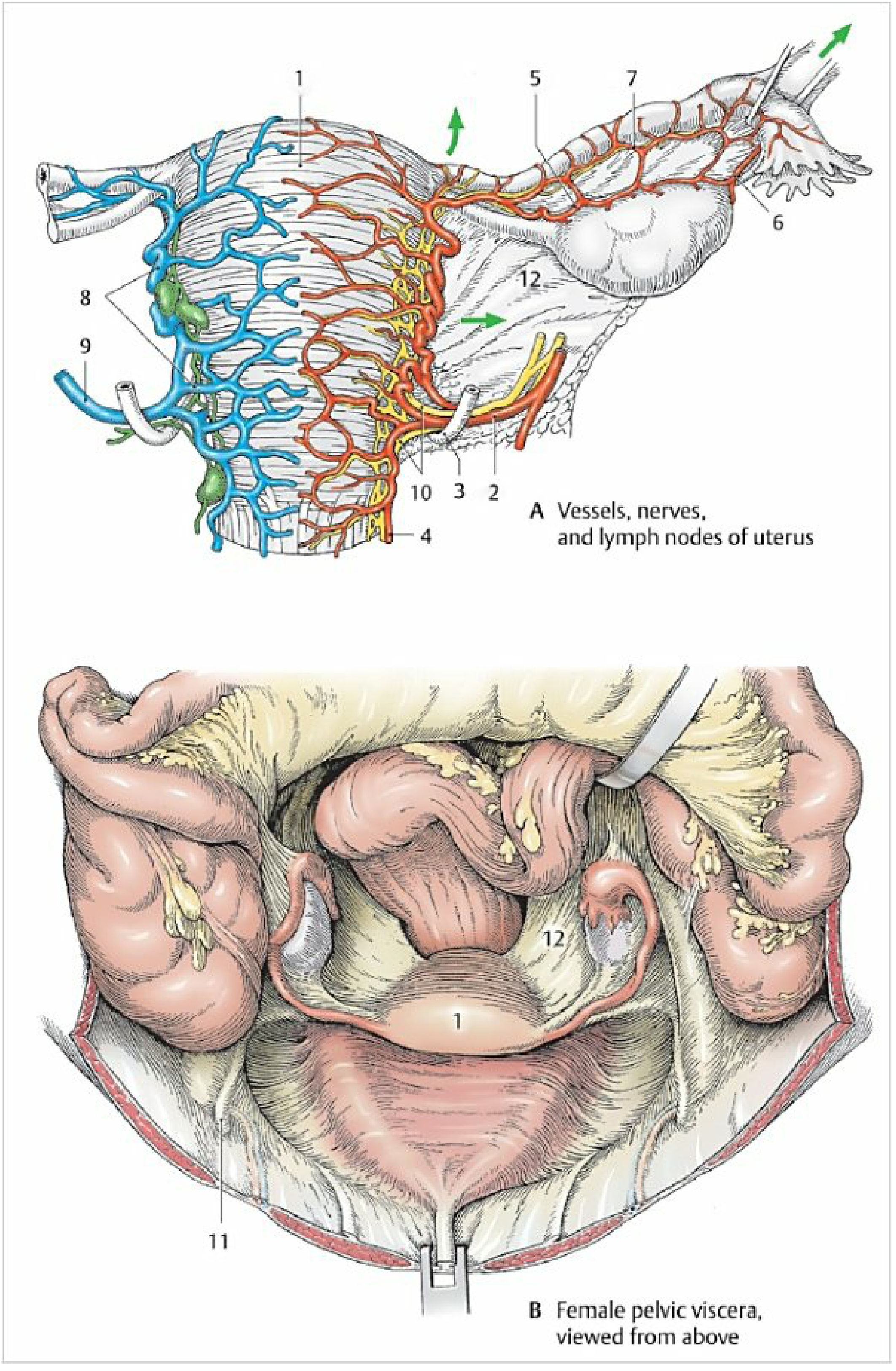
Arterial Supply
Main supply: Uterine Artery (branch of anterior division of internal iliac artery)
Course of the uterine artery:
- Runs in subperitoneal connective tissue
- Passes over the ureter at the base of the broad ligament (~1.5-2 cm lateral to the cervix)
- Reaches the lateral uterine wall near the cervix
- Divides into:
- Ascending branch - runs tortuously along the lateral wall → anastomoses with ovarian artery at the fundus; gives a tubal branch
- Descending branch (vaginal artery) - supplies cervix and upper vagina
Additional supply:
- Ovarian artery (branch of abdominal aorta) - anastomoses with ascending uterine artery at fundus
Venous Drainage
- Uterine venous plexus (valveless network around body and cervix, located in parametrium) → uterine veins → internal iliac veins
3. NERVE SUPPLY
Autonomic innervation only (uterus has no somatic sensation - pain is referred)
Source
- Inferior hypogastric plexus (pelvic plexus) + pelvic splanchnic nerves (S2-S4)
- These form the uterovaginal plexus (Frankenhäuser ganglion) - large ganglion cells located lateral to the cervix
Sympathetic (T10-L1)
- Via inferior hypogastric plexus
- Causes uterine contraction and vasoconstriction
Parasympathetic (S2-S4)
- Via pelvic splanchnic nerves
- Causes uterine relaxation and vasodilation
Pain Referral
- Uterine body pain - referred to T10-L1 dermatomes (umbilicus and groin)
- Cervical pain - referred to S2-S4 dermatomes (perineum, inner thigh)
4. SUPPORTS OF THE UTERUS ⭐ (Most Asked)
Primary support = pelvic floor muscles (levator ani) - the most important support
The ligaments provide secondary/accessory support:
A. MAIN LIGAMENTS
| Ligament | Attachment | Function |
|---|---|---|
| Cardinal (Mackenrodt's) ligament | Cervix → lateral pelvic wall (transverse cervical ligament) | Most important ligament - prevents uterine prolapse; chief support of cervix |
| Uterosacral ligament | Cervix/upper vagina → sacrum | Maintains anteversion; prevents retroversion; carries parasympathetic S2-S4 fibers |
| Round ligament | Uterine horn → labium majus (via inguinal canal) | Maintains anteversion; has smooth muscle + fibrous tissue |
| Pubocervical ligament | Cervix → pubic symphysis | Anterior support |
| Broad ligament | Lateral uterus → lateral pelvic wall | Peritoneal fold; not a true support ligament; contains uterine vessels, ureter, uterine tubes |
B. SECONDARY SUPPORT (condensations of endopelvic fascia)
- Parametrium (tissue around uterus/cervix)
- Perineal body
- Urogenital diaphragm
KEY EXAM POINT:
The cardinal (Mackenrodt's) ligament is the most important ligament support of the uterus. Damage to it (during childbirth) leads to uterine prolapse. The uterosacral ligament maintains anteversion and its damage causes retroversion.
5. LYMPHATIC DRAINAGE ⭐ (Most Asked)
Lymph from the uterus drains via three main routes:
| Region | Lymphatic Route | Nodes |
|---|---|---|
| Body + Fundus (upper) | Along suspensory ligament of ovary / ovarian vessels | Para-aortic (lumbar) nodes |
| Body + Fundus (middle) | Along broad ligament → external iliac nodes | External iliac nodes → common iliac |
| Fundus (accessory) | Along round ligament through inguinal canal | Superficial inguinal nodes |
| Cervix | Lateral (parametrium) | External iliac, internal iliac, obturator nodes |
| Cervix | Posteriorly | Sacral nodes |
| Cervix | Via broad ligament | Common iliac nodes |
Summary Diagram (Memory Aid)
FUNDUS ──────────────────────────────► Para-aortic nodes (main)
FUNDUS (via round lig.) ─────────────► Superficial inguinal nodes
BODY (via broad lig.) ───────────────► External/Common iliac nodes
CERVIX ──────────────────────────────► Internal iliac + Obturator + Sacral nodes
KEY EXAM POINT:
Para-aortic nodes are the primary nodes for the uterine fundus/body. The cervix drains to iliac nodes (internal + external + obturator + sacral). Sentinel node for cervical cancer = obturator node
6. APPLIED ANATOMY (Clinical Significance)
1. Uterine Artery - Ureter Relationship ("Water Under the Bridge")
- The uterine artery passes over the ureter at the base of the broad ligament (~1.5-2 cm lateral to the cervix)
- Mnemonic: "Water (ureter) under the bridge (uterine artery)"
- Clinical risk: During hysterectomy or ligation of uterine artery, the ureter can be accidentally cut or ligated
- Always identify the ureter before clamping the uterine artery
2. Uterine Prolapse
- Caused by damage to cardinal ligament + levator ani (most common after difficult childbirth)
- Types: 1st degree (cervix in vagina), 2nd degree (cervix at introitus), 3rd degree / procidentia (uterus outside vulva)
- Treatment: Pelvic floor exercises (mild), ring pessary, surgical repair (Manchester repair / hysterectomy)
3. Hysterectomy
- Surgical removal of uterus
- Main risk: Ureteric injury at the uterine artery ligation point
- Total abdominal hysterectomy (TAH) = body + fundus + cervix removed
- Radical hysterectomy (Wertheim's) = TAH + parametrium + upper vagina + pelvic nodes (for cervical cancer)
4. Carcinoma of Uterus / Cervix - Spread
- Spreads via lymphatics → internal and common iliac nodes, sacral nodes
- Can also spread via veins (lung metastases) and directly to bladder/rectum
5. Ectopic Pregnancy (Tubal)
- Blastocyst implants outside the uterus (most common: fallopian tube)
- Normal implantation: body of uterus (posterior wall near fundus, day 20-23)
- Uterine artery ligation may be used to control postpartum hemorrhage (PPH)
6. Lower Uterine Segment (LUS)
- Formed by the isthmus during pregnancy
- Site of lower segment cesarean section (LSCS) incision - less vascular, heals better
- Location of placenta previa if placenta implants here
7. Frankenhäuser's Ganglion Block
- Block of uterovaginal plexus (lateral to cervix) used in paracervical block for pain relief during labor or gynecological procedures
Sources: Color Atlas of Human Anatomy Vol.2 - Internal Organs, pp. 459-461 | Gray's Anatomy for Students, p. 555 | Campbell-Walsh Urology | Robbins Basic Pathology
This is a shared conversation. Sign in to Orris to start your own chat.