
orbital cellulitis opthalmology
orbital cellulitis proptosis eye
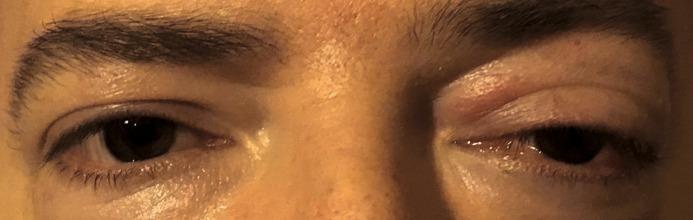
This clinical photograph provides a comparative view of the periocular region, highlighting unilateral pathology of the left eye. The right eye serves as a healthy control, showing normal orbital position, eyelid margin, and scleral appearance. In contrast, the left eye demonstrates significant proptosis (anterior displacement of the globe). Associated adnexal findings include pronounced edema and erythema of the upper eyelid, resulting in mechanical ptosis. The visible conjunctiva exhibits mild hyperemia (increased vascularity) and chemosis (conjunctival edema). These findings are characteristic of orbital inflammatory processes, such as orbital cellulitis, thyroid eye disease, or orbital pseudotumor. The image serves as an educational tool for identifying clinical signs of space-occupying or inflammatory lesions within the orbit, emphasizing the distinction between globe displacement and eyelid-specific pathology in ophthalmology and emergency medicine contexts.
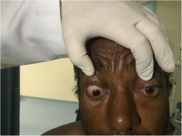
This clinical photograph shows a close-up frontal view of a patient's face during an ophthalmic examination, focusing on bilateral ocular pathology. A gloved hand is visible retracting the upper eyelids to reveal significant abnormalities. The left eye exhibits prominent proptosis (axial bulging) and severe chemosis (conjunctival edema), with marked ciliary injection and diffuse conjunctival erythema. There is evidence of matted eyelashes and purulent discharge along the lid margins. The right eye also demonstrates eyelid edema and conjunctival injection, though to a lesser degree than the left. The patient shows signs of ophthalmoplegia, as evidenced by a fixed, downward-turned gaze in the right eye despite attempted examination. These visual findings are highly characteristic of orbital cellulitis, potentially secondary to paranasal sinusitis. The image serves as an educational tool for identifying clinical red flags of orbital inflammation, including proptosis, chemosis, and impaired extraocular motility.

This clinical photograph shows a pediatric patient presenting with severe, unilateral periorbital inflammation of the right eye, consistent with orbital cellulitis. Key features include significant erythematous edema and distension of the upper and lower eyelids, resulting in complete mechanical ptosis and obscuration of the globe. There is visible epiphora (tearing) and evidence of serous discharge along the eyelid margin. The surrounding facial tissues show mild generalized swelling, while the contralateral eye appears unaffected. This image illustrates the typical clinical presentation of acute orbital infection in children, emphasizing the critical need for diagnostic differentiation from preseptal cellulitis to prevent sight-threatening complications such as abscess formation or intracranial spread. The case highlights the importance of monitoring for systemic signs, restricted extraocular motility, and proptosis in pediatric ophthalmology and emergency medicine contexts.
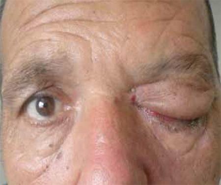
This clinical photograph displays a frontal view of a patient’s mid-face, highlighting a significant asymmetry between the orbits. The patient’s right eye appears unremarkable with normal eyelid position and globe alignment. In stark contrast, the left eye demonstrates severe periorbital pathology characterized by marked proptosis (exophthalmos) and intense inflammatory edema. The left upper and lower eyelids are severely swollen and erythematous, resulting in complete mechanical ptosis that nearly obscures the globe. There is visible crusting along the left lower lid margin and a focal area of hemorrhage or hyperemic tissue at the medial canthus. These clinical findings are consistent with orbital cellulitis or invasive rhino-orbital-cerebral mucormycosis, a diagnosis supported by the associated clinical history of diabetes and ophthalmoplegia. The image serves as an educational example of orbital apex syndrome or cavernous sinus involvement, where inflammatory mass effect leads to proptosis and paralysis of the extraocular muscles and eyelid levators.
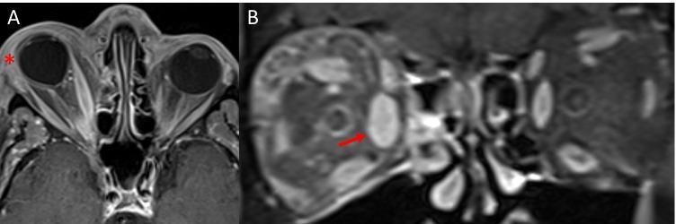
This diagnostic image consists of two panels (A and B) featuring fat-suppressed, contrast-enhanced T1-weighted MRI scans of the orbits, illustrating findings characteristic of orbital cellulitis. Panel A is an axial view showing significant preseptal soft tissue swelling (marked by a red asterisk), visible as hyperintense thickening anterior to the right globe. The right medial rectus muscle is notably enlarged and exhibits increased enhancement, accompanied by right axial proptosis (anterior displacement of the globe). Panel B is a coronal view highlighting the significant fusiform enlargement of the right medial rectus muscle (indicated by a red arrow). There is also evidence of intraconal fat stranding, characterized by streaky, hyperintense signal alterations within the normally suppressed orbital fat, signifying active inflammation and edema. These findings collectively demonstrate the hallmark radiological features used to differentiate orbital cellulitis from preseptal cellulitis and other inflammatory orbitopathies like thyroid eye disease.
"orbital cellulitis" AND management
Orbital Cellulitis (Ophthalmology Overview)
Etiology
- Most common route: direct extension from paranasal sinusitis, especially ethmoiditis (thin lamina papyracea is the weak point).
- Other sources: preseptal cellulitis, dacryocystitis/dacryoadenitis, hordeolum, dental infection, orbital/ocular trauma or surgery, hematogenous seeding, septic cavernous sinus thrombosis extension.
- Organisms: Streptococcus pneumoniae, S. aureus, S. pyogenes, Haemophilus influenzae (children, more so unvaccinated); post-traumatic cases favor Gram-negative rods; dental sources give mixed aerobes/anaerobes; immunocompromised/diabetic patients are at risk for fungal (Mucor, Aspergillus) invasive disease - a true emergency.
Clinical Features
- Rapid-onset pain worsened by eye movement, lid swelling, malaise, often reduced vision and diplopia, usually with recent sinonasal/respiratory symptoms.
- Signs: fever (often marked); tender, warm, erythematous, tense eyelids; chemosis and conjunctival injection; proptosis (may be masked by lid edema, or non-axial if a subperiosteal abscess is present); painful, restricted ophthalmoplegia; decreased visual acuity and color vision, relative afferent pupillary defect (suggests optic nerve compromise - a red flag); disc swelling/choroidal folds on fundoscopy.
- Key distinguishing point from preseptal cellulitis: preseptal disease has normal vision, pupils, motility, and no proptosis.
Complications
Workup
- History: trauma/surgery, ENT/dental infection, neck stiffness/mental status change, diabetes/immunosuppression.
- Full ophthalmic exam - APD, motility/pain, proptosis, retropulsion resistance, IOP, color vision, fundus.
- Vitals, mental status, neck flexibility, lymph nodes.
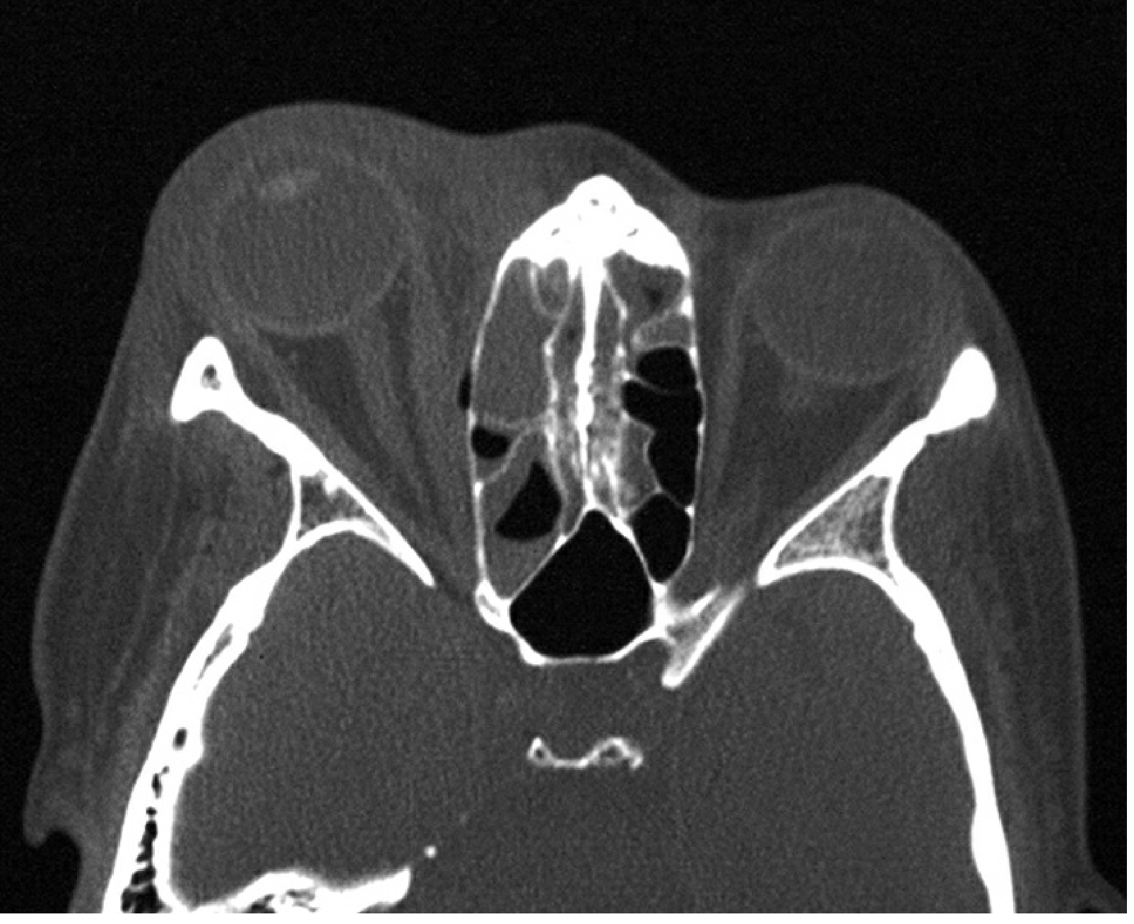
- Contrast CT of orbits, sinuses, and brain (axial/coronal) - confirms diagnosis, looks for subperiosteal/orbital abscess, sinus disease, cavernous sinus thrombosis, intracranial spread. MRI sometimes used.
- Labs: CBC with differential, blood cultures, nasal discharge culture; wound cultures if trauma-related.
- Lumbar puncture if meningeal/cerebral signs.
Treatment
- Hospital admission is mandatory with urgent ENT assessment, frequent ophthalmic review (VA, color vision, pupils checked at least every 4 hours initially), and low threshold for infectious disease/pediatric consultation.
- IV broad-spectrum antibiotics covering Gram-positive, Gram-negative, and anaerobic organisms (e.g., ceftazidime plus metronidazole per the Kanski regimen) for roughly 48-72 hours or until afebrile for 4 days, then oral therapy for 1-3 weeks total.
- Surgery: drainage of orbital/subperiosteal abscess considered early; sinus drainage if poor antibiotic response or severe sinus disease; emergency canthotomy/cantholysis if severe optic nerve compression; biopsy if the picture is atypical (to exclude fungal or neoplastic disease).
- Deteriorating optic nerve function on serial exams should prompt urgent surgical intervention.
Sources
- Kanski's Clinical Ophthalmology, A Systematic Approach, 10th ed., p. 139-140
- The Wills Eye Manual, 7th ed., section 7.3.1, p. 437-441
- Tintinalli's Emergency Medicine, "Postseptal or Orbital Cellulitis"
Recent evidence check
types
Types of Orbital Cellulitis / Orbital Complications (Chandler-Hubert Classification)
| Stage | Type | Description |
|---|---|---|
| I | Preseptal cellulitis | Infection confined to eyelid/periorbital soft tissue anterior to the orbital septum. Vision, pupils, and motility are normal; no proptosis. Less severe, more common. |
| II | Postseptal (orbital) cellulitis | Diffuse infection of the soft tissues posterior to the septum, within the orbit itself, without a discrete abscess. Produces proptosis, chemosis, painful ophthalmoplegia, possible visual/color vision impairment. |
| III | Subperiosteal abscess | Pus collects between the periosteum and the bony orbital wall (commonly medial wall, adjacent to the ethmoid sinus), displacing the globe (often non-axial proptosis/dystopia). |
| IV | Orbital abscess | Discrete collection of pus within the orbital soft tissue/fat itself (not just subperiosteal) - most severe local stage, high risk of marked proptosis, ophthalmoplegia, and optic nerve compromise. |
| V | Cavernous sinus thrombosis | Infection spreads posteriorly via the ophthalmic veins into the cavernous sinus. Presents with bilateral eye involvement, rapidly progressive chemosis/proptosis, multiple cranial nerve (III, IV, V1/V2, VI) palsies, fever, and can be fatal - a true emergency requiring neurosurgical/ID involvement. |
Additional ways orbital cellulitis is subdivided in the literature
- By anatomic compartment relative to the septum: preseptal vs. postseptal (orbital) - the fundamental first-line distinction, since preseptal disease lacks proptosis, ophthalmoplegia, and visual/pupillary compromise.
- By causative organism/etiology:
- Bacterial (most common): S. pneumoniae, S. aureus, S. pyogenes, H. influenzae (children); Gram-negative rods post-trauma; mixed aerobes/anaerobes from dental sources.
- Fungal (invasive rhino-orbital-cerebral mucormycosis/aspergillosis): seen in diabetics (especially with ketoacidosis) and other immunocompromised patients; rapidly progressive, life-threatening, and requires urgent surgical debridement plus antifungals rather than standard antibiotic therapy alone.
- By source: sinogenic (most common, especially ethmoiditis), odontogenic (dental), post-traumatic/post-surgical, and hematogenous.
Sources
- Kanski's Clinical Ophthalmology, A Systematic Approach, 10th ed., p. 138-140 (Chandler-Hubert classification, Types I-V)
- Scott-Brown's Otorhinolaryngology Head & Neck Surgery, Vol. 1 and Vol. 2, "Orbital Complications" sections
- Bailey and Love's Short Practice of Surgery, 28th ed., "Chandler classification of orbital complications"
- K.J. Lee's Essential Otolaryngology, "Ophthalmologic" chapter