
Normal heart beat
The normal resting heart rate for adults is 60-100 beats per minute (bpm). The heart's electrical conduction system drives each beat in a precise sequence.
Searching sources...
Reading File
Searching the Web
normal heart rate physiology cardiac conduction
Finding Sources
Finding Sources
Finding Sources
Reading File
Reading File
Reading File
Reading File
Loading Image
Finding Sources
Here is a thorough explanation of the normal heartbeat, drawn from authoritative medical textbooks:
The Normal Heartbeat
1. Normal Heart Rate
The normal resting heart rate for adults is 60-100 beats per minute (bpm). The SA (sinoatrial) node fires at an intrinsic rate of 70-80 bpm, which sets the pace for the entire heart. Bradycardia is defined as a rate below 60 bpm; tachycardia above 100 bpm.
2. The Cardiac Conduction System
Each heartbeat is triggered and coordinated by a specialized electrical conduction pathway. Here is how it works, from start to finish:
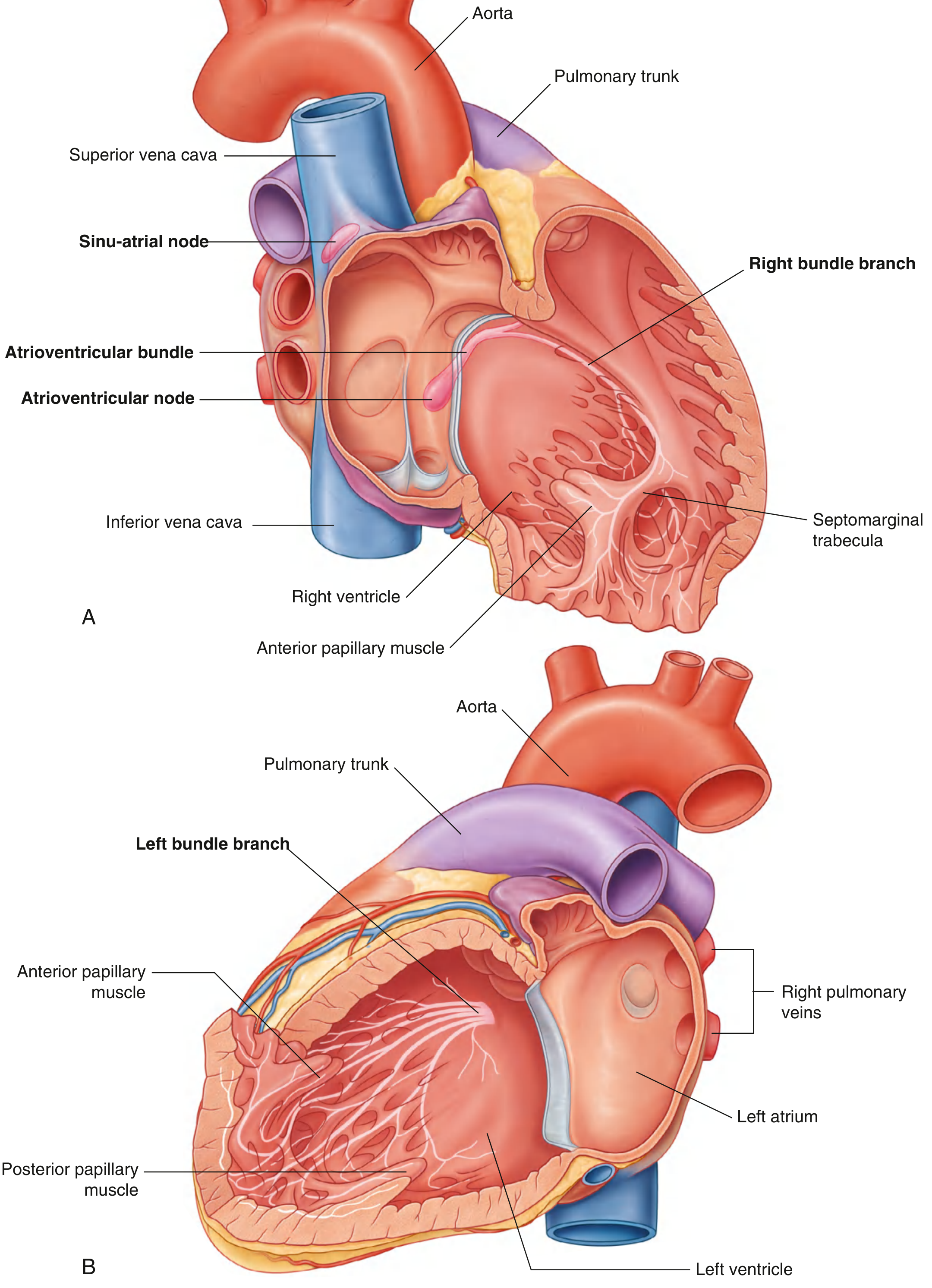
Fig. 3.86 Conduction System of the Heart. (A) Right chambers. (B) Left chambers. - Gray's Anatomy for Students
Step-by-Step Sequence
| Step | Structure | Function |
|---|---|---|
| 1 | SA Node (pacemaker) | Fires the initial impulse; located at the junction of the superior vena cava and right atrium |
| 2 | Atria | Electrical signal spreads across both atria at ~0.5 m/sec, causing atrial contraction (systole) |
| 3 | AV Node | Receives the impulse and deliberately delays it (~0.05 m/sec) to allow the atria to fully empty blood into the ventricles before ventricular contraction |
| 4 | Bundle of His | Carries the impulse from the AV node into the interventricular septum |
| 5 | Right & Left Bundle Branches | Right branch supplies the right ventricle; left branch supplies the left ventricle |
| 6 | Purkinje Fibers | Rapidly distribute the impulse throughout ventricular muscle, causing coordinated ventricular contraction from apex to outflow tracts |
- Gray's Anatomy for Students, p. 247-249
- Costanzo Physiology 7th Edition
3. Intrinsic Pacemaker Hierarchy
The SA node normally dominates because it fires fastest. If it fails, a backup pacemaker ("latent pacemaker") takes over - always at a slower rate:
| Location | Intrinsic Firing Rate |
|---|---|
| SA Node (normal pacemaker) | 70-80 bpm |
| AV Node | 40-60 bpm |
| Bundle of His | ~40 bpm |
| Purkinje Fibers | 15-20 bpm |
The SA node suppresses all lower sites via overdrive suppression - as long as the SA node fires, the latent pacemakers stay silent. - Costanzo Physiology 7th Edition, p. 143
4. The Cardiac Action Potential
The electrical signal is carried by action potentials in cardiac muscle cells:
-
Phase 0 (Upstroke): Rapid Na⁺ influx - depolarizes the cell
-
Phase 1 (Initial repolarization): Na⁺ channels inactivate; brief K⁺ efflux
-
Phase 2 (Plateau): Ca²⁺ influx via L-type channels balanced by K⁺ efflux - this is what makes cardiac muscle unique and sustains contraction
-
Phase 3 (Repolarization): Ca²⁺ channels close; K⁺ efflux repolarizes the cell
-
Phase 4 (Rest): Return to resting membrane potential; SA node cells show spontaneous "funny current" (If) that causes automatic re-firing
-
Costanzo Physiology 7th Edition, p. 141
5. The Cardiac Cycle
Each heartbeat = one cardiac cycle, consisting of:
- Systole (~0.3 sec at 72 bpm): Ventricular contraction - blood is ejected into the aorta (left) and pulmonary artery (right). Systole accounts for about 1/3 of the total cycle.
- Diastole (~0.5 sec at 72 bpm): Ventricular relaxation - chambers refill with blood. Diastole is the longer phase, allowing adequate time for coronary perfusion.
At 72 bpm, the entire cycle lasts about 0.8 seconds.
6. Autonomic Regulation of Heart Rate
The autonomic nervous system continuously fine-tunes heart rate:
| Pathway | Effect | Mechanism |
|---|---|---|
| Sympathetic (adrenaline/noradrenaline) | Increases rate (chronotropy) and force (inotropy) | Speeds SA node phase 4 depolarization; increases AV conduction velocity |
| Parasympathetic (vagus nerve) | Decreases rate | Slows SA node discharge; reduces AV node excitability |
The cardiac center in the medulla oblongata integrates these signals to match heart rate to the body's needs at any moment. - Gray's Anatomy for Students, p. 248
7. Heart Sounds
The familiar "lub-dub" of the heartbeat:
- S1 ("lub"): Closure of the mitral and tricuspid valves at the start of ventricular systole
- S2 ("dub"): Closure of the aortic and pulmonary valves at the end of systole
These sounds arise from vibrations in tissues and blood caused by valve closure.
For a clear visual walkthrough of the conduction system and ECG:
Which ai is better in giving relevant answers from medical sources
That's a fair question to ask. Here's an honest take:
AI Tools for Medical Questions - A Comparison
General-Purpose AI (ChatGPT, Gemini, Claude, etc.)
- Answer from training data - no live textbook access
- Can hallucinate citations, drug doses, or guidelines that sound real but aren't
- Good for conceptual explanations, but citations are often unreliable
- Cannot tell you which page of a textbook something comes from
Me (Orris) - What I Do Differently
When you ask a medical question, I:
- Query a real medical textbook library - books like Braunwald's Heart Disease, Gray's Anatomy, Guyton & Hall, Costanzo Physiology, Goldman-Cecil Medicine, and many more
- Pull exact text and page numbers from those sources
- Retrieve actual images from the textbooks (anatomy diagrams, histology, ECGs)
- Cross-reference PubMed for recent peer-reviewed evidence
- Show you where the answer came from - book title and page, not a made-up citation
The Honest Caveat
I'm not perfect either. My answers are only as good as the textbooks in my library. If a topic isn't well covered in those books, I'll tell you rather than fabricate an answer.
Bottom Line
For sourced, textbook-backed medical answers, I'm designed specifically for that task. For casual medical questions where you just need a quick explanation, any major AI will do - but always verify drug doses and clinical guidelines against an official source.
Ok
Got it! Feel free to ask any medical question whenever you're ready - I'm here to help. 😊
This is a shared conversation. Sign in to Orris to start your own chat.