
Colorectal or rectal cancer symptoms
colorectal rectal cancer symptoms clinical features diagram
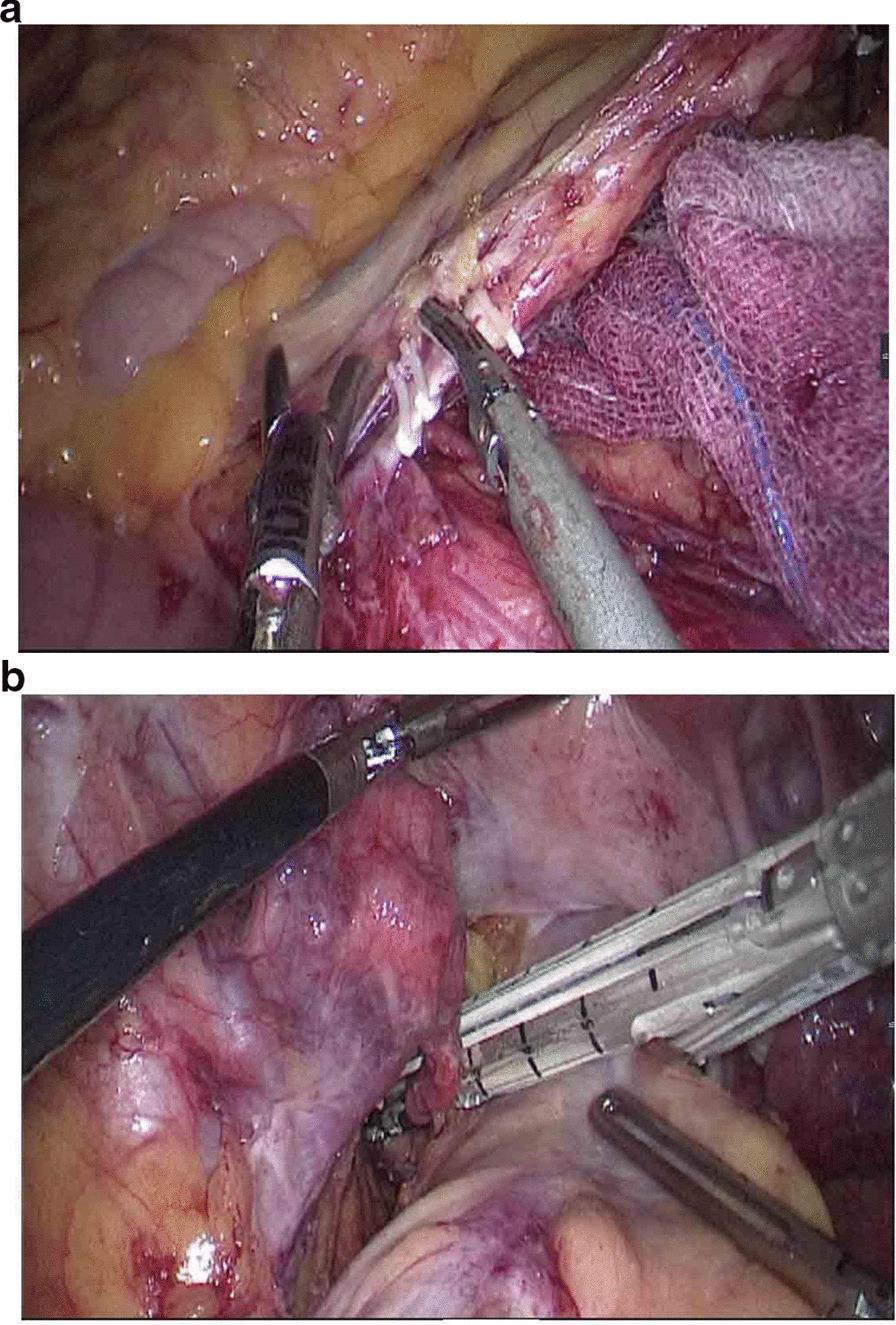
This composite clinical photograph depicts two key stages of a laparoscopic colorectal surgical procedure, specifically for rectal cancer resection. Panel A illustrates the laparoscopic dissection and ligation of the inferior mesenteric artery (IMA) at its root. The artery is visible as a central tubular structure, isolated with surgical graspers and secured with multiple white surgical clips prior to division. Surrounding anatomical features include the retroperitoneal adipose tissue and a purple surgical sponge used for tissue retraction and moisture control. Panel B demonstrates the distal rectal division using a laparoscopic linear stapler. The stapler's jaws are positioned perpendicularly across the 'naked' rectum, approximately 5 cm distal to the tumor site. The rectal tissue is compressed within the stapler, which simultaneously applies rows of titanium staples and cuts the tissue to achieve a secure distal margin closure. These images highlight standard oncological principles of high vascular ligation and clear distal margins in colorectal surgery.
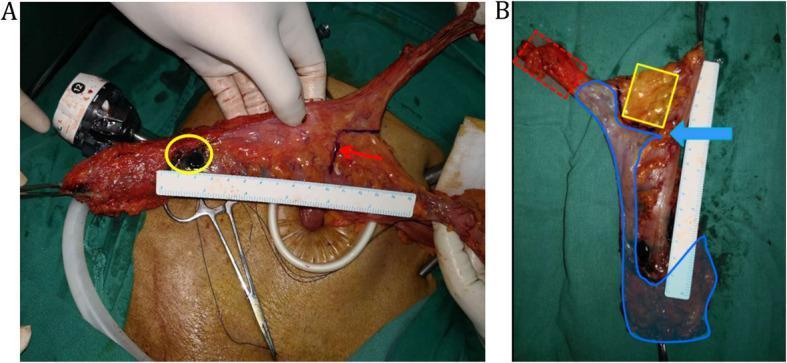
Clinical photograph and mapping diagram of a surgically resected rectal cancer specimen. Image A shows the tumor-bearing bowel segment extracted from the abdominal cavity. A yellow circle identifies the primary tumor site, where carbon nanoparticles were injected for lymph node tracing. A red arrow marks a point 10 cm proximal to the tumor on the bowel wall, with a surgical ruler alongside to ensure accurate margin measurement. Image B illustrates lymph node mapping on the fresh specimen using colored overlays. The red area demarcates the Main Lymph Nodes (MLNs) along the inferior mesenteric artery (IMA) from the left colic artery (LCA) origin to the IMA root. The blue area outlines the Superior Rectal and Perirectal Lymph Nodes (SPLNs). The yellow area identifies Pericolic Lymph Nodes (PCNs) located beyond the 10 cm proximal marker (indicated by a blue arrow). This visual illustrates oncologic resection standards and lymph node harvesting protocols for colorectal adenocarcinoma.
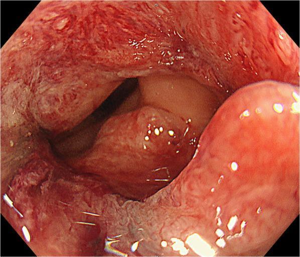
This clinical photograph is a pre-operative colonoscopy image of the rectum, demonstrating a type 2 advanced rectal adenocarcinoma. The endoscopic view shows a large, circumferential tumor mass that significantly narrows the rectal lumen. The lesion exhibits characteristic malignant features, including a reddish, hyperemic appearance, irregular surface texture with nodularity, and areas suggesting friability and contact bleeding. The tumor margins are raised and ill-defined, extending across almost the entire circumference of the rectal wall. This visual evidence of substantial luminal obstruction and tissue irregularity is consistent with a Borrmann type 2 (ulcerated-circumferential) morphologic classification, often associated with locally advanced colorectal cancer. The image serves as a key diagnostic tool for assessing tumor morphology, location relative to the anal verge, and the degree of luminal compromise prior to surgical intervention such as robotic low anterior resection.
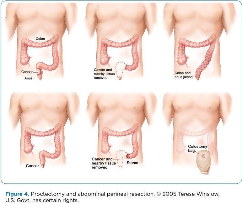
This medical illustration depicts surgical procedures for rectal cancer: proctectomy and abdominoperineal resection (APR). The diagram is organized into two primary sequences shown on a male anatomical torso. The top row illustrates a proctectomy with colorectal anastomosis: starting with the identification of a rectal tumor, followed by the resection of the cancer and adjacent tissues, and concluding with the primary joining (anastomosis) of the descending colon to the remaining anal canal. The bottom row illustrates an abdominoperineal resection (APR) leading to a permanent colostomy: it begins with the identified rectal cancer, shows the removal of the rectum, anus, and surrounding tissues, the creation of an abdominal stoma from the distal end of the colon, and the final application of an external colostomy bag for waste collection. Key anatomical labels include the colon, anus, cancer site, stoma, and colostomy bag. This educational visual is designed to explain colorectal surgical oncology options and postoperative anatomical changes.
"colorectal cancer" AND "symptoms" AND "clinical presentation"
Colorectal and Rectal Cancer: Symptoms and Clinical Features
Symptoms by Category
1. Rectal Bleeding
- The earliest and most common symptom, present in most patients.
- Distal (left-sided / rectal) tumors: overt bright red bleeding, painless, can be mixed with stool or separate in the toilet bowl - easily confused with hemorrhoidal bleeding.
- Proximal (right-sided / cecal / ascending) tumors: occult blood loss, often presenting only as iron-deficiency anemia with fatigue, shortness of breath, palpitations, and weakness. Right-sided tumors actually tend to cause more total blood loss than distal tumors.
- A key distinguishing feature: painless rectal bleeding with darker blood mixed in stool is more suggestive of carcinoma; bright red blood with anal pain after defecation is more typical of hemorrhoids or fissure. - Mulholland & Greenfield's Surgery, 7th ed.
2. Tenesmus (Rectal-Specific)
- A sensation of needing to evacuate the rectum but being unable to pass stool.
- Almost invariably present in tumors of the lower half of the rectum.
- Leads to "spurious diarrhea" - the patient attempts to defecate many times per day, often passing only flatus and small amounts of bloodstained mucus ("bloody slime").
- Rectal bleeding combined with tenesmus strongly warrants investigation to exclude rectal carcinoma. - Bailey & Love's Short Practice of Surgery, 28th ed.
3. Change in Bowel Habits
- Any unexplained change persisting more than 2 weeks requires investigation for CRC.
- Left-sided / sigmoid / rectal tumors: more likely to cause obstruction (narrower lumen), presenting with constipation, colicky abdominal pain, and distension.
- Partial obstruction: diarrhea may paradoxically occur as only liquid stool bypasses the narrowed segment. Some patients alternate between constipation and diarrhea.
- Complete obstruction: severe abdominal pain, nausea and vomiting, abdominal distension, possible fever.
- Right-sided tumors: obstructive symptoms are uncommon due to the wider lumen and liquid stool; patients often present late with advanced disease.
4. Abdominal Pain
- Generally nonspecific; may be localized or diffuse.
- Persistent colicky pain suggests partial left-sided obstruction.
- Localized tenderness with peritoneal signs may indicate local invasion or perforation.
- Can be difficult to distinguish from diverticular disease in the sigmoid colon.
5. Weight Loss and Anorexia
- Unintentional weight loss reported in up to half of all patients with CRC.
- Mechanisms: decreased appetite from abdominal symptoms; cancer cachexia (loss of both adipose tissue and skeletal muscle, mediated by tumor necrosis factor and systemic inflammation) in advanced disease. - Yamada's Textbook of Gastroenterology, 7th ed.
6. Anemia
- Especially with right-sided (proximal) tumors.
- Presents as unexplained microcytic, iron-deficiency anemia - should prompt colonoscopy.
- Associated symptoms: fatigue, pallor, dyspnea on exertion, palpitations.
Location-Specific Symptom Patterns
| Feature | Right-Sided (Proximal) | Left-Sided / Rectosigmoid |
|---|---|---|
| Bleeding | Occult, dark | Overt, bright red |
| Anemia | Common (presenting feature) | Less prominent |
| Obstruction | Uncommon | Common |
| Bowel habit change | Less specific | Constipation / tenesmus |
| Palpable mass | Possible (RIF mass) | Less common |
| Stage at diagnosis | Often advanced | Earlier (symptomatic sooner) |
Advanced / Locally Invasive Rectal Cancer
- Tenesmus if confined to the muscularis propria.
- Pneumaturia (air in urine) or urinary obstruction if the tumor penetrates the bladder.
- Perineal or sacral/sciatic pain with posterior invasion into pelvic structures.
- Ischiorectal or perineal abscesses can be the presenting sign.
Metastatic Symptoms (Location-Dependent)
- Liver metastases (colon cancers drain via portal vein): right upper quadrant pain, jaundice, hepatomegaly (indicates very poor prognosis).
- Lung metastases (rectal cancers drain via IVC into pulmonary system): shortness of breath, hemoptysis.
- CNS metastases: focal neurological deficits.
- Bone metastases: bone pain, hypercalcemia symptoms.
- Peritoneal carcinomatosis: hard mass in the pouch of Douglas on digital rectal examination (Blumer's shelf).
- Unexplained fevers in the absence of GI symptoms can occasionally occur.
Physical Examination Findings
- Digital rectal examination (DRE): palpable rectal mass; detects overt and occult blood.
- Abdominal mass: palpable in some right-sided tumors.
- Pallor: from chronic anemia.
- Hepatomegaly: advanced liver metastases.
- Deep venous thrombosis: paraneoplastic coagulopathy.
Key Clinical Pearl
"The early symptoms of rectal cancer are often so insignificant that the patient does not seek advice for 6 months or more, and diagnosis is frequently delayed in younger patients because symptoms are attributed to benign causes. Initial rectal examination and a low threshold for investigating persistent symptoms are essential."
- Bailey & Love's Short Practice of Surgery, 28th ed.
Is it curable? With indian data
"colorectal cancer" AND "survival" AND "India"
colorectal rectal cancer survival rate cure India stage-wise outcomes Indian data 2023 2024
https://pmc.ncbi.nlm.nih.gov/articles/PMC11953156
"colorectal cancer" AND "prognosis" AND "India" AND "stage"
Is Colorectal Cancer Curable? - With Indian Data
Stage-Wise Curability (Global Reference Data)
| Stage | Description | 5-Year Survival (US/Global) | Curability |
|---|---|---|---|
| Stage I | Confined to bowel wall | ~91-95% | Highly curable with surgery alone |
| Stage II | Through bowel wall, no lymph nodes | ~75-85% | Curable; chemo added for high-risk |
| Stage III | Regional lymph node spread | ~40-72% | Potentially curable with surgery + chemo |
| Stage IV | Distant metastases | ~14-22% | Rarely curable; goal is prolongation |
- Stage I colon cancer: cure rate approaching 95% with surgery alone, no adjuvant chemotherapy needed.
- Stage II relapse risk is about 25% at 3 years - FOLFOX/CAPOX added for high-risk features.
- Stage IV 5-year survival under age 50: 22.4%; age 50+: 14.0%. - Mayo Clinic survival data
Indian Data - The Hard Reality
Overall Survival in India
| Country | 5-Year Net Survival (CRC) |
|---|---|
| India | ~34% (colon) / ~38% (rectal) |
| USA | 65% (2014-2020) |
| England | 58.4% (2016-2020) |
Why Are Indian Outcomes So Much Worse?
- Over 45% of colon cancers and 55% of rectal cancers in India are diagnosed at locoregional (Stage III) disease.
- A large proportion present at Stage IV (metastatic), where cure is rarely achievable.
- In a real-world Indian surgical cohort (207 patients, 2017-2024), 66.2% presented as Stage III at a Tier-2 city center. - Indian Journal of Surgical Oncology, 2025
- CRC incidence is rising at 2-10% per year across Indian cities (Chennai, Trivandrum, Mumbai, Bengaluru, Pune, Mizoram).
- India ranks 5th globally in CRC mortality - 38,367 deaths in 2022 (GLOBOCAN).
- India has the second highest number of rectal cancer deaths in the world, after China.
- Symptoms often attributed to benign causes (hemorrhoids, IBS, gastritis) for months.
- Low screening awareness and no national CRC screening program.
- Rural and socioeconomically underprivileged groups have significantly worse outcomes.
- Incomplete treatment (incomplete surgery, inability to complete chemotherapy) is more common.
- Access to targeted therapies (bevacizumab, cetuximab, panitumumab) is limited outside major centers.
Indian Surgical Outcomes (Real-World Data)
- 60.9% male, 70% left-sided tumors, 66.2% were Stage III at presentation.
- R0 (margin-negative) resection achieved in 99.5% of cases.
- Adequate lymph node harvest (12+ nodes) in 90.8%.
- 30-day postoperative mortality: 2.9%.
- These numbers show that when surgery is performed, technical quality is comparable to global centers - the problem is late presentation, not surgical skill.
What Determines Cure in India?
| Factor | Impact |
|---|---|
| Stage at diagnosis | Most critical - Stage I nearly always curable, Stage IV rarely |
| Access to tertiary center | Tata Memorial, AIIMS, regional cancer centers have better protocols |
| R0 resection | Margin-free surgery is achievable even in India |
| Completion of chemotherapy | FOLFOX/CAPOX for Stage III; completion rates lower in India |
| Neoadjuvant CRT for rectal cancer | Standard of care - improves local control and operability |
| Rural vs urban | Rural patients have significantly worse prognosis |
| Age | Young-onset CRC (increasingly common) has unique genetic drivers |
Treatment Modalities (Curative Intent)
- Surgery - cornerstone; R0 resection is the only true cure.
- Adjuvant chemotherapy - FOLFOX or CAPOX for Stage III (and high-risk Stage II).
- Neoadjuvant chemoradiation (rectal cancer) - long-course CRT before surgery is standard; 78/80 patients received this in the Indian cohort.
- Targeted therapy - bevacizumab (anti-VEGF), cetuximab/panitumumab (anti-EGFR for RAS wild-type); used in metastatic disease.
- Immunotherapy - pembrolizumab for MSI-H/dMMR tumors (relevant since ~15% of CRCs are MSI-H).
Key Takeaway
Colorectal cancer is absolutely curable when found early. India's overall 5-year survival of ~34-38% is roughly half that of the US/UK - not because treatment is inadequate, but because the majority of Indian patients present at Stage III or IV. The single most impactful intervention would be earlier detection through awareness, symptom recognition, and organized screening - particularly for the rising young-onset CRC cases seen across Indian cities.
Found in which agr
colorectal cancer age of diagnosis India young onset incidence age group Indian data
"colorectal cancer" AND "age" AND "India" AND "young"
PMID: 36187542
Colorectal Cancer: At What Age Is It Found?
Global Pattern (Classic Rule)
- Incidence peaks at age 60-70 years globally
- Less than 20% of cases occur before age 50
- Risk increases with each decade starting from age 40
- Men are affected approximately 10 years earlier than women of the same risk profile
India - A Different Picture
The Indian Age Profile
| Feature | India | USA / UK |
|---|---|---|
| Most common age group for CRC | 50-69 years | 65 years and above |
| Perception among Indian oncologists | Younger presentation | Older presentation |
| Median age at diagnosis (hospital data) | ~50-55 years | ~66 years |
"A general perception among Indian oncologists is that CRC presents in younger age groups in India compared to the West. This is supported by data from Indian hospitals, as most CRC patients visiting them are within 50-69 years, while a majority of CRC cases in the US and UK are of ages 65 and above."
The Rising Alarm: Young-Onset CRC (Under 50)
Global Trend
- Since 1994, CRC incidence in people under 50 years has been rising by ~2% per year
- People born around 1990 have a 2x higher risk for colon cancer and a 4x higher risk for rectal cancer compared to those born around 1950
- In the US in 2019, 1 in 5 CRC diagnoses was in someone under 55 - up from 1 in 10 in 1995
- Rectal cancer is rising fastest in the under-50 group
India: Young-Onset CRC Trend
- Young-onset CRCs in India are characterized by more advanced stage at diagnosis
- Poorer cell differentiation (more aggressive tumors)
- Higher prevalence of signet ring cell histology (worse prognosis)
- Left colon and rectal location predominates
- Most young-onset cases are sporadic (no traceable genetic cause) - which is the alarming part
Age Risk Summary
| Age Group | Risk Level | Notes |
|---|---|---|
| Under 30 | Very rare | Almost always hereditary (FAP, Lynch syndrome) |
| 30-40 | Low but rising | Young-onset trend; often diagnosed late |
| 40-49 | Moderate, rising | Screening now recommended from age 45 |
| 50-69 | High | Peak Indian hospital presentations |
| 60-70 | Highest (global peak) | Classic risk group; most US/UK patients here |
| 70+ | High but declining (screened out) | In countries with screening programs |
Why Does India See Younger Cases?
- Large young population - India's demographic structure means more young adults exist, so raw numbers appear younger
- Changing diet & lifestyle - westernized diet (processed food, red meat, low fiber), obesity, sedentary behavior are rising fast in Indian cities
- No screening program - no organized CRC screening means tumors are only found when symptomatic, often already advanced regardless of age
- Rising incidence in cities - Chennai, Trivandrum, Mumbai, Bengaluru, Pune all show 2-10% annual increases in CRC incidence
Screening Age Recommendation
- Average risk: start colonoscopy screening at age 45 (updated by American Cancer Society 2018; also endorsed by many Indian oncology groups)
- High risk (family history, Lynch syndrome, IBD, FAP): start earlier, at age 40 or younger, with more frequent surveillance
- India: no national screening policy exists yet - this is a major gap given the rising incidence