
So the ha-- gastrohepatic... ... or the hepato-gastric ligament that separate the lesser and the greater sac on the right, while the gastroesophageal ligament separate the greater and the lesser sac on the left. Can you show me 3D picture of this and explain me how does it occur?
lesser sac greater sac omental bursa gastrohepatic ligament 3D anatomy diagram
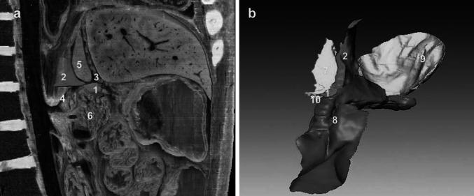
This educational image displays the complex anatomical relationships of the omental bursa (lesser sac) using a combination of a sagittal computerized tomography (CT) scan (Figure a) and a 3D anatomical reconstruction (Figure b). Figure a shows a sagittal plane identifying the foramen bursae omenti majoris (1), gastric bare area (2), hepatogastric ligament (3), left gastric artery (4), caudate lobe of the liver (5), and pancreas (6). The superior recess (7) is demonstrated with the caudate lobe serving as its right border and the hepatogastric ligament positioned anteriorly. Figure b provides an anterior 3D visualization of the communicative pathways between the superior and inferior recesses. It highlights the spatial configuration of the superior recess (7), inferior recess (8), splenic recess (9), and the vestibulum bursae omentalis (10). This visual resource is designed for advanced medical education to assist in understanding potential pathways for pancreatic fluid collections and surgical approaches to the lesser sac.
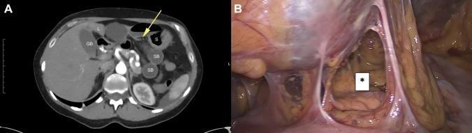
This figure presents diagnostic and intraoperative views of an isolated spontaneous lesser omental hernia. Panel A is an axial contrast-enhanced CT scan of the upper abdomen showing a 'closed-loop' internal hernia. Abnormally positioned, dilated small-bowel loops (SB) are incarcerated within the lesser omental sac (omental bursa), situated between the liver (L), stomach (S), and pancreas (P). A yellow arrow identifies the gastrohepatic ligament, indicating the site of herniation into the lesser sac. Panel B is a corresponding intraoperative laparoscopic photograph following the reduction of the herniated bowel. An asterisk (*) marks the defect in the lesser omentum, characterized by a clear opening with defined edges formed by thin, translucent mesenteric membranes. This illustrates a rare form of internal hernia where abdominal contents prolapse through the lesser omentum, often leading to acute small bowel obstruction (ileus). The images demonstrate the correlation between preoperative cross-sectional imaging and surgical findings in mechanical bowel obstruction.
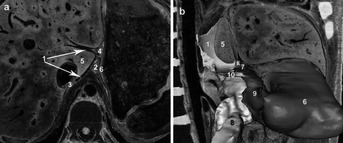
This figure provides a detailed anatomical visualization of the superior recess of the omental bursa (lesser sac) using cross-sectional imaging and 3D reconstruction. Figure (a) is a high-resolution axial cross-section viewed from the inferior aspect, demonstrating the superior recess (1) as it wraps around the caudate lobe of the liver (5). Key anatomical landmarks include the hepatogastric ligament (4) forming the anterior boundary, the hepatic bare area (3) positioned posteriorly, and the gastric bare area (2) located to the left, near the gastric wall (6). Figure (b) presents a 3D sagittal-oblique reconstruction from the right side, illustrating the longitudinal relationship between the superior recess (1), the vestibulum bursae omentalis (10), and the inferior recess (9). The foramen bursae omenti majoris (7) is highlighted as a critical transition point between these spaces. Other visualized structures include the pancreas (8) and the gastric wall (6). These images serve as educational resources for understanding the complex peritoneal folds and potential spaces for fluid collection in the upper abdomen, relevant for surgical planning and radiological interpretation of conditions like acute pancreatitis.
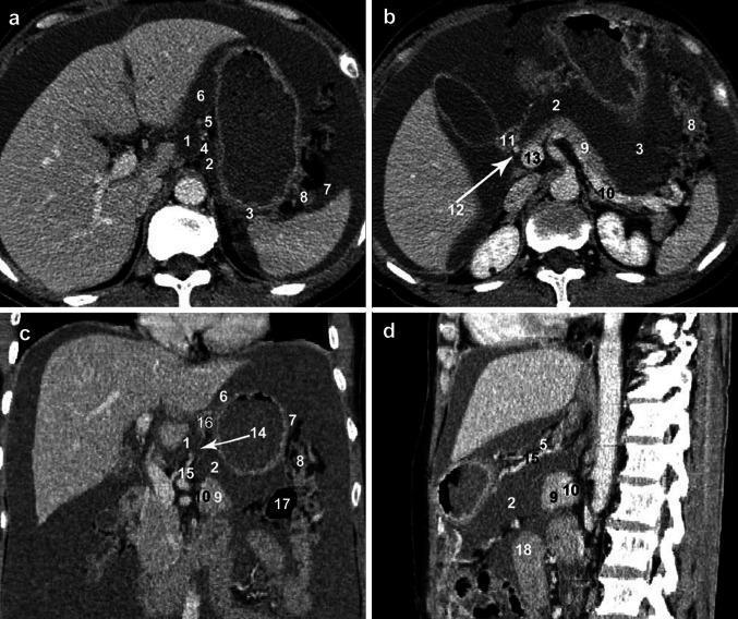
This set of diagnostic images consists of multiplanar computerized tomography (CT) scans—axial (a, b), coronal (c), and sagittal (d) views—demonstrating the complex anatomy of the omental bursa (lesser sac) and its recesses. The images utilize numerical labeling to identify 18 key anatomical structures. The superior recess (1) is visualized in the axial plane surrounding the caudate lobe of the liver, bounded anteriorly by the hepatogastric ligament (5). The inferior recess (2) and splenic recess (3) are shown in relation to the pancreas (9), stomach, and transverse colon (17). The coronal view highlights the foramen bursae omenti majoris (14), representing the communication between the superior and inferior compartments. Vasculature, including the splenic artery (10), common hepatic artery (12), portal vein (13), and left gastric artery (15), is clearly demarcated to show their proximity to the bursal spaces. These images are pedagogically significant for understanding the three-dimensional boundaries of the omental bursa, which is critical for identifying pathological fluid collections in conditions such as pancreatitis or metastatic ovarian carcinoma.
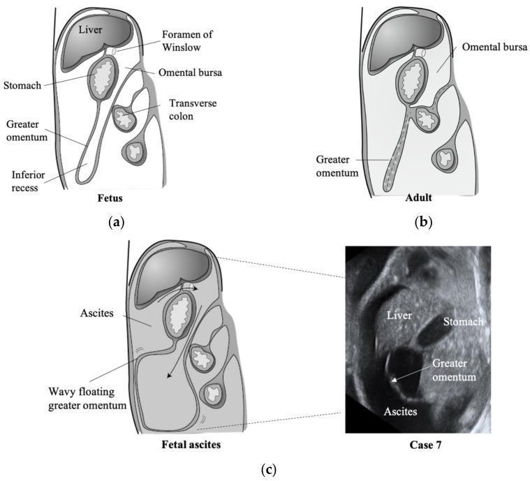
This educational resource features three sagittal schematic diagrams and one corresponding ultrasound image illustrating the embryological development and clinical presentation of the greater omentum and omental bursa. Diagram (a) shows the fetal configuration, where the greater omentum consists of a four-layered peritoneal fold with an open inferior recess extending deeply into the abdominal cavity, posterior to the stomach and anterior to the transverse colon. Diagram (b) demonstrates the adult configuration, where the anterior and posterior layers have fused (indicated by dashed lines), obliterating the inferior recess and limiting the vertical extent of the omental bursa. Diagram (c) and the accompanying grayscale ultrasound image (Case 7) depict fetal ascites. In the presence of free intraperitoneal fluid, ascites enters the omental bursa through the foramen of Winslow. This fluid separates the peritoneal layers, causing the greater omentum to appear as a 'wavy floating' structure within the fluid collection. Key labeled landmarks include the liver, stomach, transverse colon, and foramen of Winslow, highlighting the spatial relationship between the lesser sac and the greater peritoneal cavity.
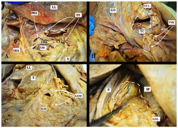
This medical anatomical composite consists of four clinical photographs of an embalmed cadaver dissection illustrating various surgical approaches to the omental bursa (lesser sac). Panel A shows the supragastric approach via the pars flaccida (PF) of the lesser omentum, revealing the celiac trunk (CT) and its branches: the common hepatic (CHA), splenic (SA), and proper hepatic (PHA) arteries, with the gastroduodenal artery (GDA) visible near the omental bursa (OB) and stomach (S). Panel B demonstrates the trans-mesocolic (TMC) approach and dissection of the gastrocolic ligament (GCL), highlighting the anatomical relationship between the transverse colon (TC) and greater omentum (GO). Panel C illustrates direct dissection through the greater omentum (GOD) inferior to the stomach's greater curvature. Panel D depicts the lateral approach via the gastrosplenic ligament (GSL), showing the spatial orientation of the spleen (SP) relative to the stomach. These panels serve as educational references for surgeons navigating the complex peritoneal attachments and vascular landmarks required for cytoreductive surgery in conditions such as advanced epithelial ovarian cancer.
The Lesser Sac (Omental Bursa) and Its Peritoneal Boundaries
3D Visual: Omental Bursa and Its Recesses
Multiplanar CT Showing All Boundaries
Cadaveric Dissection: Surgical Approaches to the Lesser Sac
How the Lesser Sac Forms - Embryology
"The embryonic stomach is invested by two mesenteries: dorsal (which becomes the gastrosplenic, gastrocolic, and gastrophrenic ligaments) and ventral (which becomes the hepatoduodenal and gastrohepatic ligaments of the lesser omentum and the falciform ligament). By the seventh week of gestation, the stomach rotates 90 degrees clockwise along its longitudinal axis... The 90-degree rotation results in the left vagus nerve being ventral and the right vagus nerve being dorsal; rotation also creates the omental bursa (lesser sac)." - Sabiston Textbook of Surgery
- Before rotation, the dorsal and ventral mesenteries are simple flat sheets.
- When the stomach rotates 90° clockwise (viewed from the front), the dorsal mesentery swings to the left and the ventral mesentery remains to the right.
- This rotation "traps" a blind peritoneal pocket behind the stomach - the lesser sac.
The Boundaries of the Lesser Sac
| Wall | Structure |
|---|---|
| Anterior | Lesser omentum (hepatogastric + hepatoduodenal ligaments), posterior wall of stomach, gastrocolic ligament |
| Posterior | Pancreas, left kidney/adrenal, aorta, diaphragm |
| RIGHT side | Hepatogastric ligament (part of lesser omentum) - this is the anterior wall of the entrance to the lesser sac |
| LEFT side | Gastrosplenic ligament - connects the greater curvature of the stomach to the spleen |
| Communication with greater sac | Foramen of Winslow (epiploic foramen) - bounded anteriorly by the hepatoduodenal ligament, posteriorly by the IVC |
On the Right: The Hepatogastric (Gastrohepatic) Ligament
- Right and left gastric arteries
- A variable accessory or replaced left hepatic artery (surgically important)
- Its thin upper portion is the pars flaccida; the thicker lower portion is the pars densa
On the Left: The Gastrosplenic Ligament
- Short gastric arteries
- Left gastroepiploic artery
Summary: How the Two Ligaments Relate to the Two Sacs
Greater Sac (general peritoneal cavity)
        |
        | <-- Foramen of Winslow (epiploic foramen) --
        |         [hepatoduodenal lig. ANTERIORLY]
        |         [IVC POSTERIORLY]
        |
     Lesser Sac (omental bursa)
        |
        |-- Anterior wall = hepatogastric ligament + posterior stomach
        |-- Right = foramen of Winslow (only opening)
        |-- Left = gastrosplenic ligament closes the splenic recess
        |-- Posterior = pancreas, retroperitoneum