Why left ventricle tract obstruction is called dynamic obstruction in hypertrophic cardiomyopathy

Reading File
Reading File
Finding Sources
Searching PubMed

"hypertrophic cardiomyopathy"[MeSH Terms] AND "outflow obstruction"

Searching the Web

hypertrophic cardiomyopathy dynamic left ventricular outflow tract obstruction SAM preload afterload

Reading File
LV outflow tract obstruction in hypertrophic cardiomyopathy is called dynamic because it is not a permanent, rigid narrowing like aortic stenosis or a subaortic membrane.
In HCM:
  • The basal interventricular septum is thickened.
  • During systole, the mitral valve moves abnormally forward toward this septum. This is systolic anterior motion (SAM).
  • The mitral leaflet and septum then transiently narrow the LV outflow tract, mainly in mid to late systole.
  • Therefore, the obstruction and pressure gradient vary from beat to beat and with the physiologic state.
It becomes worse when the left ventricular cavity becomes smaller or contraction stronger:
  • ↓ Preload: standing, Valsalva, dehydration
  • ↓ Afterload: vasodilators
  • ↑ Contractility: exercise, catecholamines
It becomes less severe with:
  • ↑ Preload: squatting, IV fluids where appropriate
  • ↑ Afterload
  • ↓ Contractility: beta-blockers
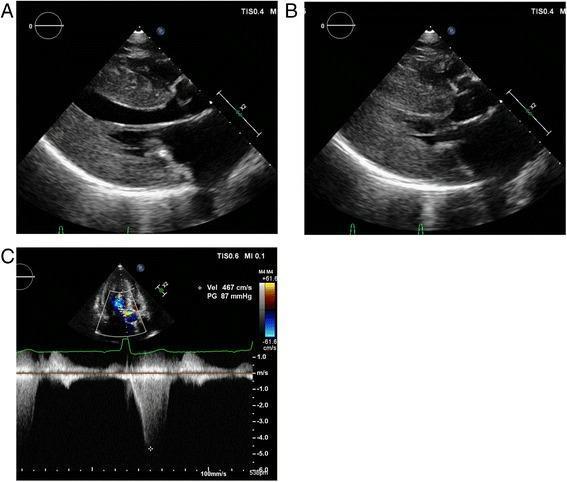
Thus, the obstruction is “dynamic” because the degree of LVOT narrowing changes according to ventricular loading and contractility, rather than being fixed throughout systole. It characteristically starts in mid-systole and peaks late in systole. This also explains the late-peaking, “dagger-shaped” Doppler profile and why the HCM murmur becomes louder during Valsalva or standing. Textbook of Clinical Echocardiography, p. 9-? (section shown in library at lines 3863-3875).

How do preload and afterload alter HCM obstruction?

Reading File
Finding Sources
Searching PubMed

"hypertrophic cardiomyopathy"[MeSH Terms] AND "outflow obstruction"

Searching the Web

hypertrophic cardiomyopathy LVOT obstruction preload afterload Valsalva squatting guideline

In obstructive HCM, the key is the size of the LV cavity during systole.
A smaller cavity brings the anterior mitral leaflet closer to the hypertrophied septum, making systolic anterior motion (SAM) and LVOT obstruction more likely.
Hemodynamic changeEffect on LV cavity / flowEffect on LVOT obstruction
↓ PreloadLess filling, smaller LV cavityIncreases obstruction
↑ PreloadMore filling, larger LV cavityDecreases obstruction
↓ AfterloadEasier, more vigorous emptying, smaller end-systolic LV cavityIncreases obstruction
↑ AfterloadLess complete emptying, larger systolic cavityDecreases obstruction

Preload

Reduced preload means less venous return and less LV filling. The septum and mitral leaflet are then closer together, so SAM worsens and the LVOT gradient rises.
Examples: standing suddenly, Valsalva strain, dehydration, hemorrhage, diuretics.
Increased preload expands the LV cavity and separates the mitral valve from the septum, reducing obstruction.
Example: squatting increases venous return.

Afterload

Reduced afterload lets the ventricle empty more forcefully. This reduces LV cavity size in systole and increases the high-velocity flow that draws/pushes the mitral leaflet anteriorly, worsening SAM and obstruction.
Examples: vasodilators, nitrates, ACE inhibitors/ARBs in susceptible obstructive HCM, and the transition from squatting to standing.
Increased afterload limits emptying, leaves a larger LV systolic cavity, and reduces SAM and the gradient.
Example: squatting increases systemic vascular resistance as well as preload.
Exam mnemonic:
HCM obstruction increases with “empty and hyperdynamic”: ↓ preload, ↓ afterload, ↑ contractility.
This is why the HCM murmur gets louder with Valsalva or standing and softer with squatting. Textbook of Clinical Echocardiography, lines 3863-3875.

Explain the concept of Systolic Anterior Motion and LV Outflow Tract Obstruction

Reading File
Finding Sources
Searching Images

hypertrophic cardiomyopathy echocardiography systolic anterior motion mitral valve septum left ventricular outflow tract obstruction
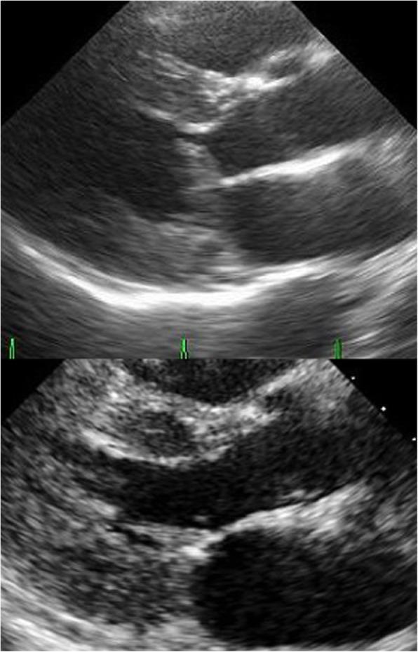
This diagnostic image comparison consists of two transthoracic echocardiography frames in the parasternal long-axis view, illustrating preoperative and postoperative states of the mitral valve. The top frame shows the preoperative condition characterized by Systolic Anterior Motion (SAM) of the anterior mitral leaflet. An elongated A2 segment is visible contacting the hypertrophied interventricular septum (17 mm thickness) during systole, resulting in significant left ventricular outflow tract (LVOT) obstruction. The bottom frame displays the postoperative result following surgical intervention. It demonstrates a clear, unobstructed LVOT with the absence of SAM. The mitral valve leaflets now exhibit normal coaptation and positioning, with no visible contact between the anterior leaflet and the septum. These images serve as an educational comparison for identifying mechanical LVOT obstruction in hypertrophic cardiomyopathy or related valvular pathologies, highlighting the anatomical markers of successful surgical repair.
This diagnostic image comparison consists of two transthoracic echocardiography frames in the parasternal long-axis view, illustrating preoperative and postoperative states of the mitral valve. The top frame shows the preoperative condition characterized by Systolic Anterior Motion (SAM) of the anterior mitral leaflet. An elongated A2 segment is visible contacting the hypertrophied interventricular septum (17 mm thickness) during systole, resulting in significant left ventricular outflow tract (LVOT) obstruction. The bottom frame displays the postoperative result following surgical intervention. It demonstrates a clear, unobstructed LVOT with the absence of SAM. The mitral valve leaflets now exhibit normal coaptation and positioning, with no visible contact between the anterior leaflet and the septum. These images serve as an educational comparison for identifying mechanical LVOT obstruction in hypertrophic cardiomyopathy or related valvular pathologies, highlighting the anatomical markers of successful surgical repair.

Transthoracic echocardiographic images demonstrating diagnostic features of hypertrophic obstructive cardiomyopathy (HOCM). Panels A and B present parasternal long-axis views. Panel A shows the heart in end-diastole with severe, diffuse left ventricular (LV) hypertrophy, particularly involving the interventricular septum and posterior wall. Panel B shows the heart in end-systole, highlighting significant chamber obliteration and the characteristic systolic anterior motion (SAM) of the anterior mitral valve leaflet. Panel C displays Doppler echocardiography, incorporating a color flow mapping sector and continuous-wave (CW) Doppler spectral tracing. The CW Doppler shows a high-velocity, late-peaking systolic jet characteristic of dynamic left ventricular outflow tract (LVOT) obstruction. Quantitative measurements indicate a peak velocity of 467 cm/s and a calculated peak pressure gradient of 87 mmHg, confirming severe outflow obstruction. These visuals are educationally significant for identifying phenotypic markers of cardiac hypertrophy and quantifying hemodynamic gradients in cardiomyopathy.
Transthoracic echocardiographic images demonstrating diagnostic features of hypertrophic obstructive cardiomyopathy (HOCM). Panels A and B present parasternal long-axis views. Panel A shows the heart in end-diastole with severe, diffuse left ventricular (LV) hypertrophy, particularly involving the interventricular septum and posterior wall. Panel B shows the heart in end-systole, highlighting significant chamber obliteration and the characteristic systolic anterior motion (SAM) of the anterior mitral valve leaflet. Panel C displays Doppler echocardiography, incorporating a color flow mapping sector and continuous-wave (CW) Doppler spectral tracing. The CW Doppler shows a high-velocity, late-peaking systolic jet characteristic of dynamic left ventricular outflow tract (LVOT) obstruction. Quantitative measurements indicate a peak velocity of 467 cm/s and a calculated peak pressure gradient of 87 mmHg, confirming severe outflow obstruction. These visuals are educationally significant for identifying phenotypic markers of cardiac hypertrophy and quantifying hemodynamic gradients in cardiomyopathy.

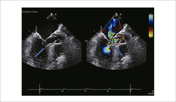
This diagnostic image displays a side-by-side comparison of intraprocedural transesophageal echocardiography (TEE) at approximately 150 degrees. The left panel is a grayscale B-mode image showing the mitral valve apparatus and left ventricular outflow tract (LVOT). An arrow highlights systolic anterior motion (SAM) of the mitral valve, where the anterior leaflet moves towards the hypertrophied interventricular septum. The right panel incorporates color Doppler imaging, revealing significant flow acceleration and turbulence (mosaic pattern/aliasing) at the point of obstruction in the LVOT, also indicated by an arrow. This turbulent jet is characteristic of dynamic outflow obstruction often seen in hypertrophic obstructive cardiomyopathy (HOCM). Additionally, the Doppler image suggests associated mitral regurgitation due to valve malcoaptation. An ECG tracing is visible at the bottom for cardiac cycle synchronization. This visual is used to identify the precise location of flow acceleration and to quantify the severity of subvalvular obstruction and regurgitation during septal ablation procedures.

This diagnostic image displays a side-by-side comparison of intraprocedural transesophageal echocardiography (TEE) at approximately 150 degrees. The left panel is a grayscale B-mode image showing the mitral valve apparatus and left ventricular outflow tract (LVOT). An arrow highlights systolic anterior motion (SAM) of the mitral valve, where the anterior leaflet moves towards the hypertrophied interventricular septum. The right panel incorporates color Doppler imaging, revealing significant flow acceleration and turbulence (mosaic pattern/aliasing) at the point of obstruction in the LVOT, also indicated by an arrow. This turbulent jet is characteristic of dynamic outflow obstruction often seen in hypertrophic obstructive cardiomyopathy (HOCM). Additionally, the Doppler image suggests associated mitral regurgitation due to valve malcoaptation. An ECG tracing is visible at the bottom for cardiac cycle synchronization. This visual is used to identify the precise location of flow acceleration and to quantify the severity of subvalvular obstruction and regurgitation during septal ablation procedures.

Searching PubMed

"hypertrophic cardiomyopathy"[MeSH Terms] AND "outflow obstruction"

Searching the Web

systolic anterior motion mitral valve left ventricular outflow tract obstruction hypertrophic cardiomyopathy mechanism

Systolic anterior motion (SAM)

SAM is the abnormal movement of the anterior mitral valve leaflet toward the interventricular septum during systole.
Normally, during systole the mitral valve remains closed and stays behind the LV outflow tract, while blood exits freely from the LV into the aorta.
In obstructive hypertrophic cardiomyopathy:
  1. The basal septum is hypertrophied, narrowing the LV outflow tract (LVOT).
  2. The mitral valve may also be elongated, and the papillary muscles may be abnormally positioned.
  3. When the LV contracts, blood accelerates through this narrowed channel.
  4. The rapid flow exerts predominantly drag forces on the mitral leaflet, drawing it forward toward the septum. This is SAM.
  5. The leaflet may touch the septum in mid-systole, creating a temporary obstruction below the aortic valve.
Echocardiographic appearance of SAM with anterior mitral leaflet approaching the hypertrophied septum and producing LVOT obstruction

How SAM produces LVOT obstruction

Think of the LVOT as a corridor between:
  • Anteriorly: hypertrophied interventricular septum
  • Posteriorly: anterior mitral leaflet
When SAM occurs, the leaflet moves anteriorly and narrows or briefly blocks that corridor:
Normal systole:
Septum              Open LVOT               Mitral leaflet
  |                     ↓                        \
  |------------------ blood to aorta ------------\

Obstructive HCM:
Hypertrophied septum       narrowed LVOT      SAM leaflet
       |                       ↓                  /
       |-----------------------X-----------------/
                         leaflet-septal contact
This produces:
  • accelerated, turbulent LVOT flow
  • a pressure difference between the LV and aorta, called the LVOT gradient
  • reduced forward cardiac output, particularly on exertion
  • a late-peaking systolic ejection murmur
  • possible exertional dyspnea, chest discomfort, presyncope, or syncope
The obstruction commonly begins in mid-systole and becomes greatest in late systole, hence the characteristic late-peaking, “dagger-shaped” continuous-wave Doppler signal. Textbook of Clinical Echocardiography, lines 3863-3875 and 3926-3948.

Why mitral regurgitation also occurs

When the mitral leaflet is pulled forward, the mitral leaflets cannot close properly, or coapt, in systole. This causes mitral regurgitation (MR), typically with a posteriorly directed MR jet. SAM therefore causes both:
SAM → LVOT obstruction + mitral leaflet malcoaptation → mitral regurgitation

Why it is dynamic

SAM becomes more likely when the LV is small and contracts forcefully:
  • Reduced preload: Valsalva, standing, dehydration
  • Reduced afterload: vasodilators
  • Increased contractility: exercise, catecholamines
These conditions increase flow acceleration and bring the mitral leaflet and septum closer together. Increasing preload or afterload, or reducing contractility, usually reduces SAM and the LVOT gradient.
SAM is strongly associated with obstructive HCM but is not exclusive to it. It can also arise transiently in other situations with a small, hyperdynamic LV, such as hypovolemia or after mitral valve repair.
This is a shared conversation. Sign in to Orris to start your own chat.