
Anti NMDA receptor encephalitis
"anti-NMDA receptor encephalitis"[MeSH Terms] OR "NMDAR encephalitis"
anti-NMDA receptor encephalitis MRI brain
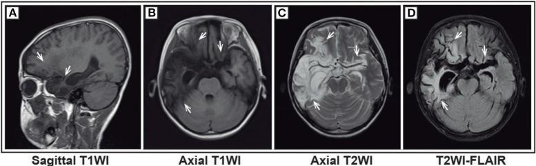
This set of diagnostic images features four brain MRI sequences of a pediatric patient, illustrating significant parenchymal abnormalities. Panel A (sagittal T1WI) and Panel B (axial T1WI) demonstrate diffuse, poorly defined regions of low signal intensity (hypointensity) localized to the bilateral frontal lobes, temporal lobes, and insula. Panel C (axial T2WI) and Panel D (axial T2-FLAIR) reveal these same anatomical regions as hyperintense (high signal), indicating widespread cortical and subcortical edema or tissue damage. The images show notable structural changes, including broadening and deepening of sulci and 'crack-like' patchy low signals in the adjacent brain tissue, suggesting atrophy or necrotic progression typical of post-viral encephalitis. The third ventricle is slightly expanded, and there is visible dilation of the right temporal horn of the lateral ventricle, though midline structures remain stable. These findings are clinically consistent with anti-NMDA receptor encephalitis secondary to Herpes Simplex Virus (HSV) encephalitis, highlighting the typical limbic and frontal involvement seen in pediatric neuro-inflammatory conditions.
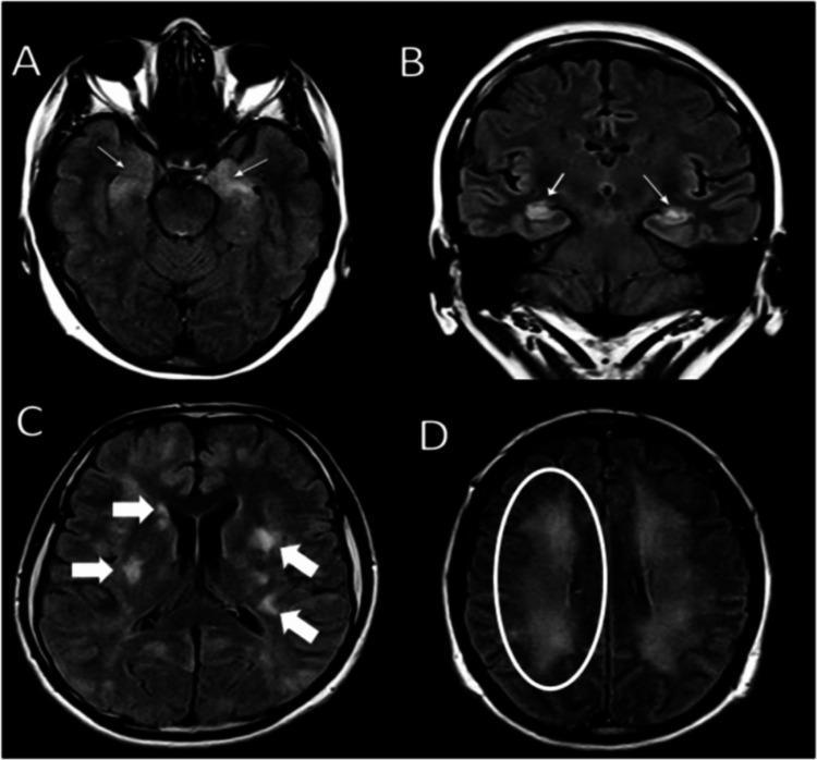
This diagnostic image contains four panels (A-D) displaying brain Magnetic Resonance Imaging (MRI) using the Fluid-Attenuated Inversion Recovery (FLAIR) sequence. The images highlight various hyperintense (bright) lesions associated with neuroinflammatory or demyelinating processes. Panel A (axial) and B (coronal) show symmetric, bilateral hyperintensities within the hippocampi, marked by thin white arrows. Panel C (axial) demonstrates multiple, asymmetrical multifocal hyperintensities involving the cortical, subcortical, and periventricular white matter, indicated by thick white arrows. Panel D (axial) reveals more diffuse, bilateral confluent hyperintense lesions within the frontal lobe white matter, specifically highlighted within a white circle. These findings are clinically significant for evaluating conditions such as autoimmune encephalitis (e.g., anti-NMDA receptor encephalitis) or acute disseminated encephalomyelitis (ADEM). The content is suitable for intermediate to advanced medical education in neurology and radiology, focusing on the visual identification of pathological signal changes in diverse brain regions.
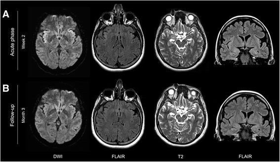
Comparative MRI neuroimaging panel showing brain evolution in a patient with anti-NMDA receptor encephalitis between the acute phase (week 2) and follow-up (month 3). The top row (A) demonstrates bilateral diffusion restriction on Diffusion-Weighted Imaging (DWI) and hyperintensity on axial and coronal Fluid-Attenuated Inversion Recovery (FLAIR) images within the insular cortex, with right-sided predominance. Slight T2 and FLAIR hyperintensities are also visible in both hippocampi. The bottom row (B) illustrates the follow-up phase where the insular diffusion restriction and FLAIR signal abnormalities have completely resolved. However, persistent hyperintensity remains visible in the bilateral hippocampi on both T2-weighted and FLAIR sequences. This diagnostic image sequence highlights the typical limbic involvement and dynamic cortical changes associated with autoimmune encephalitis, emphasizing the differentiation between transient cortical findings and potentially more permanent hippocampal signal changes.
![This diagnostic image set displays a longitudinal comparison of [18F]-FDG-PET and matching MRI-T2-FLAIR axial brain scans from three patients with anti-NMDA receptor (NMDAR) encephalitis. The panels are organized by patient (1, 2, and 3) across three time points: T0 (onset), T6 (6 months), and T18 (18 months). The visual content is divided into two columns: the Medial Temporal Lobe (left) and the Temporo-parieto-occipital region (right). Key findings include: (1) Patient 1 shows bilateral hypometabolism in lateral temporal and posterior parietal lobes at T0, peaking at T6 before resolving by T18. (2) Patient 2 exhibits bilateral occipital hypometabolism at T0 (white arrow) and transient basal ganglia hypermetabolism relative to cortical hypometabolism at T6. (3) Patient 3 demonstrates right-sided medial temporal lobe hypometabolism on PET (yellow arrow) corresponding to hyperintensity on MRI-FLAIR (yellow rectangle) at T0, with resolution at subsequent time points. The image illustrates the correlation between functional metabolic changes on PET and structural findings on MRI in autoimmune encephalitis progression and recovery.](https://cdn.orris.care/cdss_images/pmc_clinical_VQA_2160573defe2ee7efaccbc70050fcca9a1a01091098ad0ae4ad7ebba38356ebb.jpg)
This diagnostic image set displays a longitudinal comparison of [18F]-FDG-PET and matching MRI-T2-FLAIR axial brain scans from three patients with anti-NMDA receptor (NMDAR) encephalitis. The panels are organized by patient (1, 2, and 3) across three time points: T0 (onset), T6 (6 months), and T18 (18 months). The visual content is divided into two columns: the Medial Temporal Lobe (left) and the Temporo-parieto-occipital region (right). Key findings include: (1) Patient 1 shows bilateral hypometabolism in lateral temporal and posterior parietal lobes at T0, peaking at T6 before resolving by T18. (2) Patient 2 exhibits bilateral occipital hypometabolism at T0 (white arrow) and transient basal ganglia hypermetabolism relative to cortical hypometabolism at T6. (3) Patient 3 demonstrates right-sided medial temporal lobe hypometabolism on PET (yellow arrow) corresponding to hyperintensity on MRI-FLAIR (yellow rectangle) at T0, with resolution at subsequent time points. The image illustrates the correlation between functional metabolic changes on PET and structural findings on MRI in autoimmune encephalitis progression and recovery.
Anti-NMDA Receptor Encephalitis
Epidemiology
- Predominantly affects young women and children (~80% of patients), but men and older individuals can also be affected
- One of the most common causes of encephalitis in young adults
- ~40% of adult females over 18 years have an underlying ovarian teratoma; only <9% of girls under 14 have tumors
- Other associated tumors (rare): mediastinal teratoma, SCLC, Hodgkin lymphoma, neuroblastoma, breast cancer, testicular germ-cell tumor
- About 25% of cases arise after herpes simplex viral encephalitis (post-HSV autoimmune encephalitis)
- Immune checkpoint inhibitors can also trigger it
Pathogenesis
Clinical Features - Multistage Progression
| Stage | Features |
|---|---|
| Prodrome (days) | Flu-like illness, headache, fever, malaise, nausea/diarrhea |
| Psychiatric phase | Psychosis, hallucinations, agitation, delusions, anxiety - often initially misdiagnosed as primary psychiatric illness |
| Neurological phase | Seizures, memory loss, reduced verbal output/mutism, insomnia |
| Unresponsive/catatonic phase | Decreased consciousness, catatonia |
| Hyperkinetic phase | Orofacial dyskinesias, choreoathetosis, limb/trunk dyskinesias, dystonic postures |
| Autonomic instability | Tachycardia, bradycardia, hyperhidrosis, labile BP, hypersalivation, pupillary changes |
| Hypoventilation | Central hypoventilation requiring mechanical ventilation in severe cases |
- Children often present with behavioral change, new seizures, movement disorder, and insomnia/reduced speech
- Monosymptomatic episodes (e.g., pure psychosis) occur in ~5% of patients
- Atypical features (cerebellar ataxia, hemiparesis) are more common in children
- Autonomic dysfunction is well-described: episodes of hypertension, tachycardia, diaphoresis, excessive salivation
Investigations
CSF
- Lymphocytic pleocytosis in ~80% of patients
- Elevated protein in some
- Oligoclonal bands in some
- May be normal early on
- Diagnostic threshold: >5 cells/μL or protein >50 mg/dL counts as inflammatory
MRI Brain
EEG
- Abnormal in ~95% of cases
- Usually shows focal or generalized slow/disorganized activity
- Extreme delta brush (EDB): a pathognomonic EEG pattern seen in 10-30% - resembles neonatal delta brush; associated with prolonged illness and should strongly raise suspicion for anti-NMDAR encephalitis
Antibody Testing
- NMDAR antibodies should be tested in both serum AND CSF - CSF-only positivity occurs in up to 13% of cases (serum can be falsely negative)
- Testing CSF is mandatory; serum alone is insufficient
APE2 Score
- New-onset rapid mental status changes (1-6 weeks): +1
- Neuropsychiatric changes (agitation, emotional lability): +1
- Autonomic dysfunction: +1
- Viral prodrome: +1
- Faciobrachial dystonic seizures: +3
- Facial dyskinesias: +2
- Seizures refractory to ≥2 ASMs: +2
- CSF inflammation: +2
- MRI suggesting encephalitis: +2
Tumor Workup
- Vaginal ultrasound for ovarian teratoma
- CT/PET of chest, abdomen, pelvis for other tumors
- FDG-PET may show cortical hypo- and hypermetabolism patterns
Diagnosis
- Subacute onset (<3 months) of working memory deficits, altered consciousness, or psychiatric symptoms
- At least one of: CSF inflammation, MRI changes, EEG abnormality
- Reasonable exclusion of other causes
Treatment
First-line (Immunotherapy)
- Tumor removal - in ovarian teratoma or other underlying tumor: this is the single most important intervention; non-removal is associated with lower recovery rates and higher relapse risk
- IV methylprednisolone (high-dose corticosteroids)
- Intravenous immunoglobulin (IVIg) or plasma exchange (PLEX) - no data favoring one over the other; autonomic instability may favor IVIg
Second-line (for poor responders)
- Rituximab (anti-CD20, B-cell depletion) - reduces relapse risk; increasingly used early or upfront
- Cyclophosphamide - used in refractory cases
- Tocilizumab (anti-IL-6 receptor) - promising results in refractory cases
Supportive
- ICU monitoring for autonomic instability
- Mechanical ventilation for hypoventilation
- Antiseizure medications (though seizures may be refractory)
- Avoid prolonged antipsychotic use (dyskinesias may be misattributed to antipsychotics)
Prognosis
- Majority of patients respond to treatment, though recovery can be slow (months)
- ~80% achieve good functional recovery
- Clinical relapses occur in 12-24% of patients (most within 2 years)
- Patients who undergo tumor removal have better outcomes and lower relapse rates
- Worse prognosis if treatment is delayed or tumor not removed
- The antibody-mediated mechanism means neuronal damage is potentially reversible (unlike T-cell-mediated paraneoplastic syndromes)
Key Differentials
| Feature | Anti-NMDAR | Limbic Encephalitis (LGI1, CASPR2) | HSV Encephalitis |
|---|---|---|---|
| Age | Young women/children | Middle-aged/older | Any |
| MRI | Often normal | Medial temporal FLAIR | Temporal lobe, hemorrhage |
| EEG | Extreme delta brush | Temporal slowing | Temporal spikes/PLEDS |
| CSF | Lymphocytosis | May be normal | Lymphocytosis, RBCs |
| Tumor | Ovarian teratoma (40% adult F) | Rare (LGI1) | None |
| Psychiatric | Prominent early | Less prominent | Less prominent |