
Hi
Hi there! How can I help you today?
I’m an first year mbbs student,I have my cell, cell division and cycle viva tomorrow guide me or teach me in every way the teacher can ask question from basics to checking my understanding level
Reading File
Finding Sources
Finding Sources
Reading File
Finding Sources
Reading File
Reading File
Reading File
Reading File
Finding Sources
Finding Sources
Reading File
Reading File
I now have rich textbook content to build a thorough viva guide. Let me compile everything.
Loading Image
Loading Image
Here is your complete viva preparation guide, organized exactly the way a teacher would test you - from basic definitions to deeper understanding:
Cell, Cell Division & Cell Cycle - Complete Viva Guide
Sources: Histology: A Text and Atlas (Pawlina), Junqueira's Basic Histology, Thompson & Thompson Genetics & Genomics
PART 1: THE CELL (Basics)
Q: What is a cell?
The cell is the basic structural and functional unit of all living organisms. Eukaryotic cells have a membrane-bound nucleus; prokaryotic cells do not.
Q: What are the components of the nucleus?
- Chromatin - DNA + histone proteins, organized as euchromatin (active/light) or heterochromatin (inactive/dark)
- Nucleolus - site of rRNA synthesis; also contains regulatory cell cycle proteins
- Nuclear envelope - double membrane with inner + outer layers separated by perinuclear cisternal space; perforated by nuclear pores. The outer membrane is continuous with the rough ER (rER)
Q: What is interphase?
The period between two successive cell divisions. The cell is NOT dividing but is metabolically very active. It includes G1, S, and G2 phases.
PART 2: THE CELL CYCLE
Here is the cell cycle diagram with all checkpoints you must know:
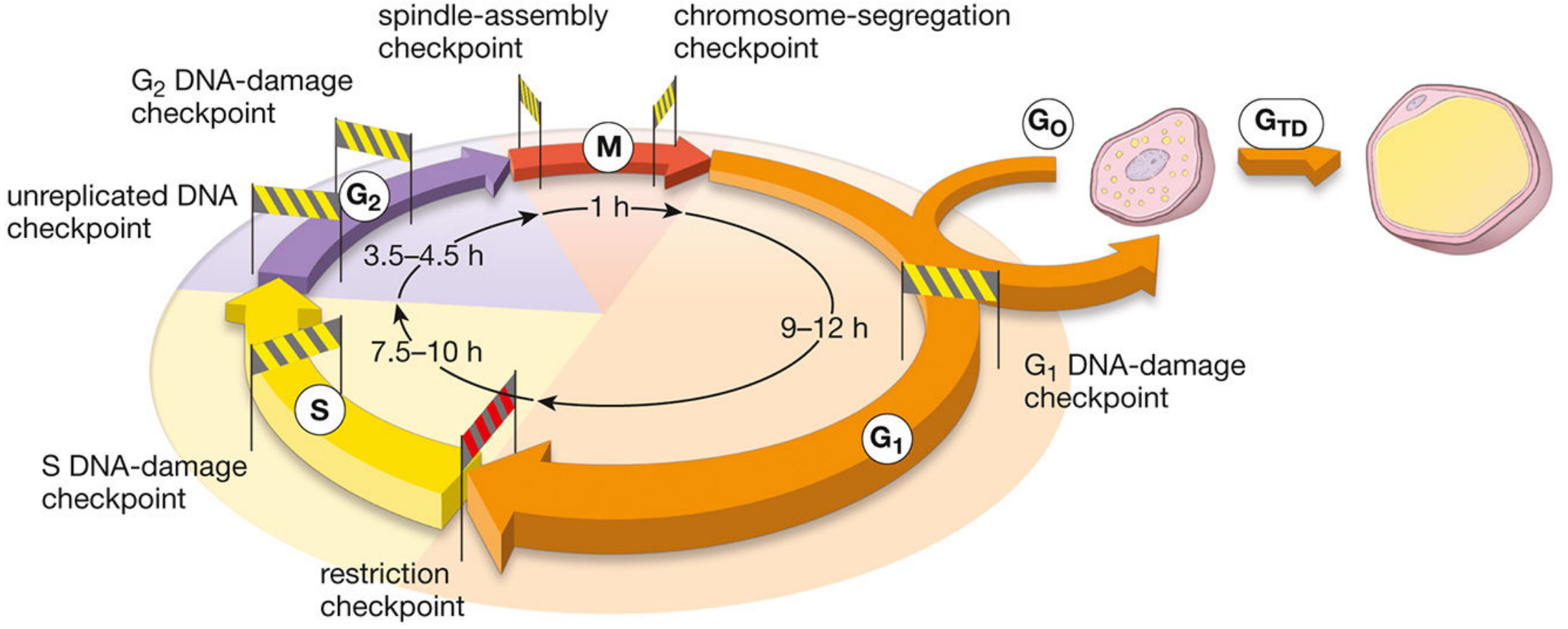
Phases and Their Duration
| Phase | Duration | What happens |
|---|---|---|
| G1 | 9-12 hours | Cell gathers nutrients, synthesizes RNA and proteins needed for DNA replication |
| S | 7.5-10 hours | DNA is replicated (chromosomes double) |
| G2 | 3.5-4.5 hours | Cell prepares for division, checks DNA quality |
| M (Mitosis) | ~1 hour | Chromosome segregation and cell division |
Q: What is G0 phase?
When a cell leaves the cycle from G1, it enters G0 ("outside" the cycle). These cells are non-dividing. They may re-enter the cycle with an appropriate stimulus, OR undergo terminal differentiation (GTD) and become permanently non-dividing (e.g., mature fat cells, neurons).
Q: What is the total duration of the human cell cycle?
Rapidly dividing human cells complete a full cycle in about 24 hours.
Checkpoints - The Most Important Topic!
Q: What are checkpoints? Why do they exist?
Checkpoints are internal quality-control mechanisms (biochemical pathways) that monitor whether the previous phase has been completed correctly and DNA is intact before allowing the cell to proceed.
Q: Name all checkpoints in the cell cycle:
- Restriction checkpoint (G1) - The most important checkpoint. The cell evaluates its own replicative potential - size, physiologic state, interactions with extracellular matrix. This is the "point of no return." Mediated by pRb (retinoblastoma protein) and E2F transcription factor.
- G1 DNA-damage checkpoint (G1) - Monitors integrity of newly replicated DNA. If DNA is irreparably damaged, p53 levels rise and block entry into S phase; cell undergoes apoptosis.
- S DNA-damage checkpoint (S phase) - Monitors quality of replicating DNA during synthesis.
- G2 DNA-damage checkpoint (G2) - Checks DNA quality after replication.
- Unreplicated DNA checkpoint (G2) - Prevents cell from entering M phase before DNA synthesis is complete.
- Spindle-assembly checkpoint (M phase) - Prevents premature entry into anaphase.
- Chromosome-segregation checkpoint (M phase) - Prevents cytokinesis until all chromosomes are correctly separated.
Q: What happens if checkpoints fail?
A mitotic catastrophe can occur - the cell may die OR develop into a tumor cell (cancer).
Q: What is p53?
p53 is a tumor suppressor protein. High p53 levels signal DNA damage and block the G1/S transition. It triggers either DNA repair or apoptosis.
Regulation - Cyclins and CDKs
Q: What drives cell cycle progression?
A complex of two proteins: Cyclin + Cyclin-Dependent Kinase (CDK). These are synthesized and degraded at regular intervals throughout the cycle.
| Phase | Cyclin-CDK Complex | Target |
|---|---|---|
| Early G1 | Cyclin D - CDK4/6 | Phosphorylates Rb protein, releases E2F |
| Late G1/entry of S | Cyclin E - CDK2 | Activates E2F-mediated transcription |
| Progression through S | Cyclin A - CDK2 | DNA polymerase and replication proteins |
| G2/entry of M | Cyclin A - CDK1 | Phosphatases, Cyclin B |
| Progression through M | Cyclin B - CDK1 | Nuclear lamin, histone H1, centrosome proteins |
Q: What is the Rb-E2F mechanism? (High-yield!)
- Normally, Rb protein binds to E2F transcription factor and BLOCKS cell cycle progression.
- A growth factor activates a kinase that phosphorylates Rb.
- Phosphorylated Rb releases E2F.
- Free E2F activates genes for G1 activities and Cyclin A, allowing the cell to enter S phase.
- Clinical link: Loss of Rb gene causes retinoblastoma (eye cancer in children).
PART 3: MITOSIS
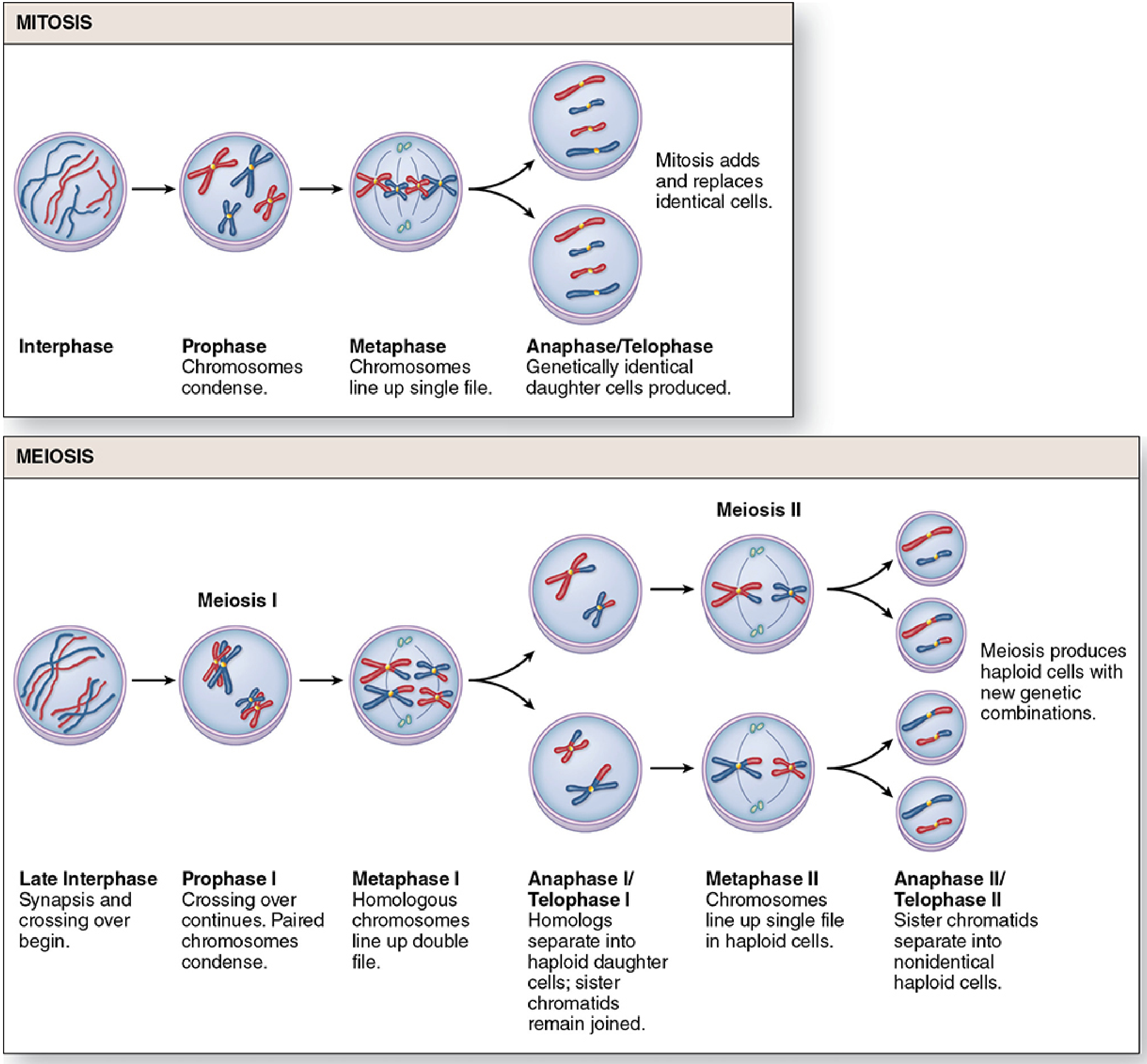
Q: Define mitosis.
Mitosis is a process of chromosome segregation, nuclear division (karyokinesis), and cell division (cytokinesis) that produces two daughter cells with the same chromosome number and DNA content as the parent cell (diploid → two diploid cells: 2n → 2n + 2n).
Q: Name and describe the phases of mitosis:
1. Prophase
- Chromosomes condense and become visible
- Nuclear envelope disassembles
- Mitotic spindle develops from microtubules
- Centrosomes move to poles of the cell
2. Prometaphase
- Nuclear membrane dissolves
- Chromosomes disperse into the cytoplasm
- Chromosomes attach by their kinetochores to spindle microtubules
3. Metaphase
- Chromosomes are maximally condensed
- Chromosomes align at the equatorial plate (metaphase plate)
- Best stage to study chromosomes (karyotyping is done here!)
4. Anaphase
- Chromosomes separate at the centromere
- Sister chromatids become independent daughter chromosomes
- Pulled to opposite poles of the cell
5. Telophase
- Chromosomes begin to decondense
- Nuclear membrane re-forms around each set of chromosomes
- Followed by cytokinesis (division of cytoplasm)
Q: What is karyokinesis vs cytokinesis?
- Karyokinesis = division of the nucleus
- Cytokinesis = division of the cytoplasm
Q: At what stage is karyotyping done and why?
At metaphase/prometaphase - chromosomes are maximally condensed and most visible.
Q: How many chromosomes does a cell in G2 have vs after mitosis?
- Cell in G2: 46 chromosomes, each as a pair of sister chromatids (4n DNA content)
- After mitosis: each daughter cell has 46 chromosomes, one copy of the genome (2n DNA content)
PART 4: MEIOSIS
Q: Define meiosis.
Meiosis involves two sequential nuclear divisions that produce gametes containing half the chromosome number (haploid) of somatic cells. It occurs only in germ cells (gonads).
Q: Why is meiosis important?
- Produces haploid cells (n=23) so that fertilization restores the diploid number (2n=46).
- Creates genetic diversity through crossing over (recombination).
Meiosis I (Reductional Division)
Separates homologous chromosomes.
Prophase I (longest phase - lasts 3 weeks in spermatogenesis!):
- Homologous chromosomes pair up (synapsis) - forming tetrads (4 chromatids)
- Crossing over / genetic recombination occurs between non-sister chromatids
- Attachment points = chiasmata
Metaphase I: Homologous chromosome pairs (bivalents) align double-file at the equatorial plate.
Anaphase I: Homologous chromosomes separate to opposite poles. Sister chromatids remain joined.
Telophase I: Two cells form, each haploid in chromosome number but still with 2 chromatids per chromosome.
Meiosis II (Equational Division - like mitosis)
Separates sister chromatids. Occurs without an intervening S phase.
Result: 4 haploid cells, each genetically unique.
PART 5: MITOSIS vs MEIOSIS - Comparison Table (Exam Favorite!)
| Feature | Mitosis | Meiosis |
|---|---|---|
| Where | All somatic cells | Gonads only (germ cells) |
| Purpose | Growth, repair, replacement | Gamete formation |
| Number of divisions | 1 | 2 |
| Daughter cells produced | 2 | 4 |
| Chromosome number | 2n → 2n (diploid) | 2n → n (haploid) |
| Genetic identity | Identical to parent | Genetically unique |
| Crossing over | Does NOT occur | Occurs (Prophase I) |
| Synapsis | No | Yes (Meiosis I) |
| S phase between divisions | N/A | No S phase between Meiosis I and II |
PART 6: UNDERSTANDING-LEVEL QUESTIONS (Teacher might ask these!)
Q: Why does a cell arrest at G0?
Lack of mitogenic stimulus, terminal differentiation, or nutrient stress. The cell evaluates at the restriction checkpoint and decides not to proceed.
Q: A cell has defective p53. What consequences would you expect?
Without p53, DNA damage cannot be detected at G1. The cell enters S phase with damaged DNA, replicates the defect, and can accumulate mutations - leading to cancer. p53 mutations are found in >50% of human cancers.
Q: Why is Prophase I of meiosis so prolonged?
Because complex events of synapsis and crossing over must be completed accurately. In females, oocytes arrest at this stage from fetal life until ovulation - potentially decades.
Q: What is nondisjunction? What is its clinical significance?
Nondisjunction = failure of chromosomes (or chromatids) to separate properly during meiosis. Results in gametes with abnormal chromosome numbers (aneuploidy).
- Examples: Trisomy 21 (Down syndrome), Turner syndrome (45, X), Klinefelter syndrome (47, XXY)
Q: What is the significance of crossing over?
Crossing over shuffles genetic information between maternal and paternal chromosomes, generating new allelic combinations. This is the main source of genetic diversity in sexually reproducing organisms.
Q: What is a proto-oncogene vs oncogene?
Proto-oncogenes are normal genes coding for proteins involved in cell cycle control (growth factors, receptors, kinases). Mutation or abnormal expression converts them to oncogenes, causing uncontrolled cell division and cancer.
Q: What happens at the spindle-assembly checkpoint? Why is it important?
It monitors whether all chromosomes have attached properly to spindle microtubules before anaphase begins. If even one chromosome is not properly attached, the checkpoint halts the process. Failure leads to unequal chromosome distribution to daughter cells (aneuploidy) - a feature of many tumor cells.
Quick Memory Tricks
- PMAT = Prophase, Metaphase, Anaphase, Telophase (mitosis phases)
- G1 → S → G2 → M = Growing 1, Synthesizing, Growing 2, Mitosis
- Mitosis: 1 parent → 2 identical diploid daughters
- Meiosis: 1 parent → 4 unique haploid daughters
- Crossing over only in Meiosis (not mitosis)
- Most important checkpoint = Restriction point (G1)
- p53 = guardian of the genome
- Cyclin D/CDK4,6 = pushes cell from G1 into S
Good luck in your viva tomorrow! If you want me to quiz you interactively or go deeper on any topic (like apoptosis, cell organelles, or chromosome structure), just ask.
What about the membranous and non membranous organelle
Finding Sources
Finding Sources
Reading File
Reading File
Reading File
Reading File
Reading File
Finding Sources
Finding Sources
Reading File
Reading File
Reading File
Loading Image
Reading File
I now have all the content needed. Let me compile the full organelle viva guide.
Membranous & Non-Membranous Organelles - Complete Viva Guide
Source: Histology: A Text and Atlas (Pawlina, 9th Ed)
CLASSIFICATION AT A GLANCE
| Membranous Organelles | Non-Membranous Organelles |
|---|---|
| Plasma Membrane | Ribosomes |
| Rough ER (rER) | Microtubules |
| Smooth ER (sER) | Actin Filaments (Microfilaments) |
| Golgi Apparatus | Intermediate Filaments |
| Mitochondria | Centrioles / MTOC |
| Lysosomes | Basal Bodies |
| Peroxisomes | Proteasomes |
| Endosomes | |
| Exosomes |
PART A: MEMBRANOUS ORGANELLES
1. PLASMA MEMBRANE
Q: What is the plasma membrane made of?
A phospholipid bilayer with embedded proteins. The fluid mosaic model (Singer & Nicolson) describes it best - proteins "float" in a fluid lipid sea.
Q: What are the types of membrane proteins?
- Integral proteins - embedded within the lipid bilayer (transmembrane proteins). Demonstrated by freeze-fracture technique.
- Peripheral proteins - loosely attached to the outer or inner surface.
Q: What are lipid rafts?
Specialized microdomains enriched in cholesterol and sphingolipids. Two types:
- Planar lipid rafts - contain flotillins (markers of lipid rafts), act as scaffolding proteins.
- Caveolae ("little caves") - flask-shaped (50-100 nm) invaginations containing caveolins. Rich in Ca²+ channels and G protein-coupled receptors. Prominent in smooth muscle cells.
Lipid rafts act as "signaling platforms" - all elements (receptors, effectors, substrates) are close together for rapid, efficient signal transduction.
Clinical link: Bacteria like Shigella and Salmonella hijack lipid rafts to enter cells.
Q: What are the functions of the plasma membrane?
- Selective permeability (controls what enters/exits the cell)
- Cell signaling
- Cell-to-cell recognition
- Endocytosis and exocytosis
2. ROUGH ENDOPLASMIC RETICULUM (rER)
Q: What makes rER "rough"?
Ribosomes studded on its cytoplasmic surface.
Q: What does rER do?
- Synthesizes proteins destined for export (secretion), lysosomes, or the plasma membrane
- Protein folding and quality control
- Post-translational modifications (glycosylation)
Q: How does a protein know to go to the rER?
The first 15-60 amino acids form a signal sequence (signal peptide). This is recognized by the Signal Recognition Particle (SRP), which arrests translation and brings the ribosome to the rER membrane. After docking, translation resumes and the polypeptide is threaded into the rER lumen. The signal peptide is then cleaved by signal peptidase.
Clinical link: Antibiotic action - aminoglycosides (streptomycin), macrolides (erythromycin), tetracyclines all inhibit protein synthesis by binding bacterial ribosomes. They exploit the difference between prokaryotic (70S) and eukaryotic (80S) ribosomes.
3. SMOOTH ENDOPLASMIC RETICULUM (sER)
Q: How does sER differ from rER?
No ribosomes - smooth surface.
Q: Functions of sER?
- Lipid and steroid hormone synthesis (abundant in cells of adrenal cortex, gonads)
- Detoxification of drugs and toxins (liver hepatocytes - cytochrome P450 enzymes)
- Calcium storage and release (sarcoplasmic reticulum in muscle cells is a specialized sER)
- Glycogen metabolism
Memory tip: sER = Steroids, detox (liver), calcium storage.
4. GOLGI APPARATUS
Q: Describe the structure of the Golgi apparatus.
A series of flattened, membrane-bound sacs (cisternae) stacked like plates. Has two faces:
- Cis face (forming face) - receives vesicles from the rER
- Trans face (maturing face / TGN = Trans-Golgi Network) - sorts and dispatches proteins to destinations
Q: What does the Golgi do?
- Receives proteins from rER
- Further modifies them (glycosylation, sulfation, phosphorylation)
- Sorts and packages them into transport vesicles for delivery to:
- Plasma membrane (exocytosis)
- Lysosomes
- Secretory vesicles
- Extracellular space
Q: What is M-6-P?
Mannose-6-phosphate (M-6-P) is a sorting signal added by the Golgi to enzymes destined for lysosomes. It acts as a "lysosome address tag."
Q: What is the difference between constitutive and regulated exocytosis?
- Constitutive pathway: Continuous secretion without stimulus (e.g., immunoglobulins by plasma cells).
- Regulated pathway: Secretion only upon a specific signal (e.g., neurotransmitter release, hormone secretion by endocrine cells).
5. MITOCHONDRIA
Q: What is unique about mitochondria compared to other organelles?
Mitochondria have TWO membranes - no other cytoplasmic organelle does.
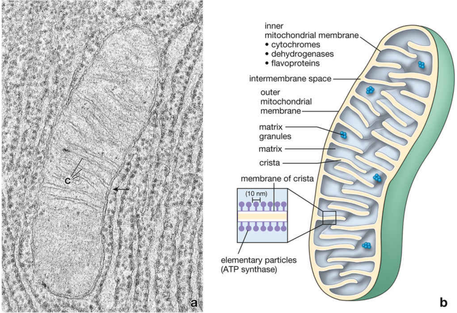
Q: Name the structural compartments of mitochondria:
- Outer mitochondrial membrane - smooth, contains porins (large channels permeable to molecules up to 5 kDa). Contains phospholipase A2, monoamine oxidase, acetyl-CoA synthase.
- Intermembrane space - environment similar to cytoplasm (ions and small molecules pass freely through outer membrane). Contains cytochrome c (important in apoptosis!).
- Inner mitochondrial membrane - forms cristae (infoldings that dramatically increase surface area). Rich in cardiolipin (makes it impermeable to ions). Contains:
- Respiratory electron transport chain enzymes
- ATP synthase (elementary particles) - tennis racquet-shaped structures seen on TEM, 10 nm diameter heads
- Matrix - contains mitochondrial DNA, ribosomes (70S), Krebs cycle enzymes, matrix granules (Ca²+ storage).
Q: What are cristae?
Infoldings of the inner mitochondrial membrane that dramatically increase its surface area, allowing more ATP synthesis.
Q: Which cells lack mitochondria?
Red blood cells (RBCs) and terminal keratinocytes.
Q: What is the endosymbiotic theory?
Mitochondria evolved from ancient bacteria engulfed by early eukaryotic cells. Evidence: they have their own circular DNA (like bacteria), 70S ribosomes (like bacteria), reproduce by binary fission, and are maternally inherited.
Q: Clinical significance - Mitochondrial inheritance?
Mitochondrial diseases are maternally inherited (e.g., MELAS, Leber's hereditary optic neuropathy).
Q: Why do steroid-secreting cells have more mitochondria with tubular cristae?
The inner membrane forms tubular/vesicular projections instead of flat cristae in steroid-metabolizing cells (adrenal cortex, Leydig cells) - more surface area for steroid biosynthesis.
6. LYSOSOMES
Q: What are lysosomes?
Membrane-bound organelles containing ~50 types of hydrolytic enzymes (acid hydrolases) that digest cellular debris, foreign material, and waste. pH inside is ~5 (acidic).
Q: How are lysosomal enzymes targeted to lysosomes?
Via the M-6-P (mannose-6-phosphate) tagging system by the Golgi apparatus.
Q: What is heterophagy vs autophagy?
- Heterophagy - digestion of material brought in from outside the cell (phagocytosis, receptor-mediated endocytosis)
- Autophagy - digestion of the cell's own worn-out organelles (self-eating)
Q: What are lysosomal storage diseases (LSDs)?
Genetic disorders where a lysosomal enzyme is absent or defective - the substrate accumulates inside lysosomes, causing cell damage. Symptoms: intellectual disability, hepatosplenomegaly, frequent infections.
| Disease | Enzyme Missing | Accumulates |
|---|---|---|
| Gaucher disease | Glucocerebrosidase | Glucosylceramide |
| Tay-Sachs disease | β-Hexosaminidase (α subunit) | GM2 ganglioside |
| Niemann-Pick disease | Sphingomyelinase | Sphingomyelin |
| Hurler syndrome (MPS I) | α-L-iduronidase | Dermatan/heparan sulfate |
| Pompe disease | Acid α-glucosidase | Glycogen |
7. PEROXISOMES
Q: What do peroxisomes do?
- Oxidation of very long chain fatty acids (β-oxidation)
- Detoxification using hydrogen peroxide (H₂O₂) - produced then neutralized by catalase
- Synthesis of bile acids and ether lipids (plasmalogens)
Q: What is Zellweger syndrome?
A severe peroxisomal biogenesis disorder (mutation in PEX genes). Causes craniofacial malformations, hepatomegaly, neurologic abnormalities, retinal degeneration, and deafness. Most infants do not survive past 1 year.
8. ENDOSOMES & EXOSOMES
Q: What are endosomes?
Membrane-bound vesicles formed by endocytosis. They sort internalized material - some recycled back to membrane, some sent to lysosomes for degradation.
Q: What is receptor-mediated endocytosis?
A selective process where specific molecules bind to surface receptors → receptor-molecule complexes gather in clathrin-coated pits → pinched off by dynamin to form coated vesicles → uncoated and fused with early endosomes.
Q: What are exosomes?
Small vesicles (30-150 nm) secreted by cells. They carry proteins, lipids, and RNA between cells - act as intercellular communication tools.
PART B: NON-MEMBRANOUS ORGANELLES
1. RIBOSOMES
Q: What are ribosomes made of?
rRNA + proteins. Two subunits:
- Eukaryotic: 80S = 60S (large) + 40S (small)
- Prokaryotic: 70S = 50S + 30S
Q: What is the difference between free ribosomes and membrane-bound ribosomes?
- Free ribosomes (polysomes) - in cytoplasm, make proteins for the cell's own use (cytosolic proteins, proteins for mitochondria and peroxisomes)
- Membrane-bound ribosomes (on rER) - make proteins for secretion, lysosomes, or plasma membrane
Clinical link: Antibiotics target 70S ribosomes (prokaryotic) - that's why they kill bacteria without harming human cells.
2. MICROTUBULES
Q: What are microtubules?
Rigid, hollow tubes of polymerized tubulin protein (α and β tubulin subunits). Diameter: ~25 nm. They are non-branching and can rapidly assemble and disassemble. They grow from the MTOC (Microtubule Organizing Center) near the nucleus, extending toward the cell periphery.
Q: Functions of microtubules:
- Intracellular vesicular transport (like railroad tracks)
- Movement of cilia and flagella
- Chromosome attachment to mitotic spindle - movement during mitosis/meiosis
- Maintenance of cell shape and asymmetry
- Cell elongation and migration
Q: What are motor proteins associated with microtubules?
- Dyneins - move toward the minus (-) end (toward cell center). Move chromosomes along the spindle during mitosis.
- Kinesins - move toward the plus (+) end (toward cell periphery). Separate spindle poles during cell division.
Q: What is the axoneme?
The microtubular framework inside cilia and flagella. Arrangement: 9+2 pattern (9 doublet microtubules around 2 central singlets). Dynein arms between doublets create sliding movement.
Clinical link - Kartagener syndrome (Primary Ciliary Dyskinesia): Defective dynein arms → immotile cilia → bronchiectasis, situs inversus, male infertility (immotile sperm).
3. ACTIN FILAMENTS (Microfilaments)
Q: What are actin filaments?
Thin (6-8 nm), flexible filaments made of polymerized actin (42 kDa protein). May constitute up to 20% of total protein in some cells.
Q: What is G-actin vs F-actin?
- G-actin (globular) - free, unpolymerized actin in cytoplasm
- F-actin (filamentous) - polymerized form making up microfilaments
Microfilaments are polarized: plus (barbed) end grows fast; minus (pointed) end grows slow.
Q: Functions of actin filaments:
- Cell motility and migration
- Formation of microvilli (core of brush border)
- Muscle contraction (with myosin)
- Cytokinesis (cleavage furrow)
- Maintenance of cell shape
4. INTERMEDIATE FILAMENTS
Q: What are intermediate filaments?
Fibrous proteins, ~10 nm diameter (between actin at 6-8 nm and microtubules at 25 nm - hence "intermediate"). Provide structural stability and mechanical strength to cells. Unlike actin and microtubules, they are not polarized and are very stable.
Q: Name the types and where they are found:
| Type | Protein | Location |
|---|---|---|
| Cytokeratins | Keratins | Epithelial cells |
| Vimentin | Vimentin | Mesenchymal cells (fibroblasts, endothelium) |
| Desmin | Desmin | Muscle cells |
| Neurofilaments | NF proteins | Neurons (axons) |
| GFAP | Glial fibrillary acidic protein | Astrocytes |
| Lamin | Lamins A, B, C | Nuclear lamina (in all cells) |
Clinical link: Immunohistochemistry uses intermediate filament antibodies to identify tumor origin. A tumor expressing cytokeratin = carcinoma. Expressing vimentin = sarcoma.
5. CENTRIOLES & MTOC
Q: What is a centriole?
A cylindrical structure made of 9 triplet microtubules arranged in a ring (9 x 3 pattern, no central pair). They come in pairs (diplosome) oriented at right angles.
Q: What is the MTOC (Centrosome)?
The microtubule-organizing center = pair of centrioles + surrounding pericentriolar material. It nucleates and organizes microtubules in the cell.
Q: What happens to centrioles during cell division?
They duplicate during S phase, then move to opposite poles of the cell in mitosis and form the mitotic spindle poles.
Clinical link: Abnormal duplication of centrioles is linked to cancer - extra centrioles cause multipolar spindles, leading to abnormal chromosome segregation.
SUMMARY TABLE: Organelle Quick Recall
| Organelle | Key Feature | Main Function | Viva High-Yield Point |
|---|---|---|---|
| Plasma membrane | Lipid bilayer, fluid mosaic | Selective permeability, signaling | Lipid rafts, caveolae |
| rER | Ribosomes on surface | Protein synthesis for export | Signal peptide, SRP |
| sER | No ribosomes | Lipid/steroid synthesis, detox, Ca²+ | Abundant in steroid cells, liver |
| Golgi | Cis/trans face | Modify, sort, package proteins | M-6-P tag for lysosomes |
| Mitochondria | 2 membranes, own DNA | ATP synthesis (powerhouse) | Cristae, elementary particles, endosymbiosis |
| Lysosomes | Acid pH (~5) | Intracellular digestion | M-6-P targeting, LSDs |
| Peroxisomes | Catalase | Fatty acid oxidation, detox | Zellweger syndrome |
| Ribosomes | 80S (eukaryote) | Protein synthesis | 70S in prokaryotes → antibiotic target |
| Microtubules | 25 nm, tubulin | Transport, spindle, cilia | 9+2 in cilia; Kartagener syndrome |
| Actin | 6-8 nm | Motility, shape, cytokinesis | G-actin vs F-actin |
| Intermediate filaments | 10 nm, stable | Structural support | Type identifies cell lineage (IHC) |
| Centrioles | 9 triplets | Spindle formation | Abnormal = cancer risk |
HIGH-YIELD COMPARISON QUESTIONS
Q: Mitochondria vs Peroxisomes - both oxidize fatty acids. What's the difference?
- Mitochondria oxidize short and long chain fatty acids via β-oxidation → generates ATP
- Peroxisomes oxidize very long chain fatty acids → generates H₂O₂ (neutralized by catalase), not directly ATP
Q: rER vs sER - how to tell them apart on EM?
rER has ribosomes (rough, dark granules on surface). sER is smooth. rER is more tubular/flat-saccular; sER is more vesicular.
Q: Why is the inner mitochondrial membrane impermeable to ions?
Rich in cardiolipin - a special phospholipid that creates a tight seal, essential for maintaining the proton gradient needed for ATP synthesis.
Q: What happens if lysosomes rupture inside the cell?
Acid hydrolases are released into cytoplasm → self-digestion (autolysis) → cell death. This is one mechanism in necrosis.
Good luck tomorrow! Ask me to quiz you on any section or go deeper on anything like endocytosis pathways, the Golgi in detail, or specific lysosomal diseases.
This is a shared conversation. Sign in to Orris to start your own chat.