
Tomorrow is my presentation on periodontal disease . Provide me concept on this along with figure . From standard books of dental
I'll start by reading the medical library skill to access dental textbooks on periodontal disease.
Searching sources...
Reading File
Now let me search the dental library for periodontal disease content in parallel.
Finding Sources
Finding Sources
Finding Sources
Reading File
Now let me read the most relevant dental sections in parallel - Robbins Pathology, Junqueira's Histology, Scott-Brown's, and the Goldman-Cecil full section:
Reading File
Reading File
Reading File
Reading File
Excellent content found. Now let me fetch the key figures and also get Park's preventive medicine content + microbiology content in parallel:
Loading Image
Loading Image
Reading File
Reading File
Let me get the remaining microbiology content and also fetch the Rosen's emergency medicine content on gingivitis/periodontitis treatment:
Reading File
Reading File
Finding Sources
Now let me fetch the two key figures from Sherris (normal vs diseased periodontium):
Loading Image
Loading Image
I now have comprehensive content and figures from multiple standard textbooks. Let me compile a thorough, well-illustrated presentation on periodontal disease.
🦷 Periodontal Disease - Complete Presentation Guide
Sources: Robbins & Cotran Pathologic Basis of Disease (10e), Junqueira's Basic Histology (17e), Sherris & Ryan's Medical Microbiology (8e), Goldman-Cecil Medicine, Scott-Brown's Otorhinolaryngology, Park's Preventive & Social Medicine, Rosen's Emergency Medicine
1. DEFINITION & OVERVIEW
Periodontal disease refers to a group of oral infections affecting the periodontium - the hard and soft tissues that support and maintain teeth in the jaws. It is the most common cause of tooth loss worldwide and was ranked the 11th most prevalent disease globally (2016).
"Periodontal diseases are a group of oral infections that affect the periodontium... A worldwide problem, it is the most common cause of tooth loss."
- Goldman-Cecil Medicine, International Edition
2. THE NORMAL PERIODONTIUM (Anatomy Foundation)
The periodontium comprises four structures:
| Structure | Description |
|---|---|
| Gingiva | Keratinized mucosa; free gingival (FG) overlies the tooth neck |
| Cementum | Covers dentin of root; avascular; bone-like; maintained by cementocytes |
| Periodontal Ligament (PDL) | Collagen (Sharpey) fibers binding cementum to alveolar bone; 150-350 μm thick; highly vascular and innervated |
| Alveolar Bone | Supports tooth sockets; continuously remodeled by osteoblasts/osteocytes |
Fig. 15-11 - Normal Periodontium (Junqueira's Basic Histology, 17e):
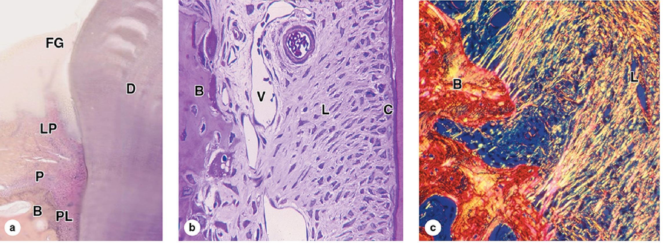
(a) Low-power H&E: Free gingiva (FG) over dentin (D), lamina propria (LP) continuous with periosteum (P) of alveolar bone (B), and PDL (PL). (b) The PDL (L) with abundant blood vessels (V) inserting into alveolar bone (B); cementum (C) is thin and bone-like. (c) Picrosirius/polarized light: collagen continuity from alveolar bone (B) periosteum into PDL (L).
3. ETIOLOGY & PATHOGENESIS
The Central Role of Dental Plaque
- Dental plaque is a sticky, colorless biofilm containing bacteria, salivary proteins, and desquamated epithelial cells
- Forms above (supragingival) and below (subgingival) the gingival margin
- If not removed, mineralizes into calculus (tartar)
- Subgingival plaque within the gingival crevice/sulcus is the primary etiologic agent
Microbial Shift in Disease
| Healthy Sites | Active Periodontitis |
|---|---|
| Facultative gram-positive organisms | Anaerobic & microaerophilic gram-negative flora |
Key Pathogens (from 300 oral species)
- Aggregatibacter (Actinobacillus) actinomycetemcomitans - produces leukotoxin; key in aggressive/juvenile periodontitis
- Porphyromonas gingivalis - key virulence factors; synergy with Treponema
- Prevotella intermedia
- Treponema denticola - cross-feeds with P. gingivalis; drives gingivitis → periodontitis progression
"Synergism between P. gingivalis, T. denticola, and other plaque members is felt to foster progression of gingivitis to chronic periodontitis."
- Sherris & Ryan's Medical Microbiology, 8e
Risk Factors
- Local: Poor oral hygiene, tobacco use, dental malocclusion
- Systemic: Diabetes mellitus (bidirectional relationship), HIV/AIDS, Down syndrome, Crohn's disease, sarcoidosis, neutrophil deficiency/dysfunction, pregnancy
- Genetic: Altered microbiome composition
4. CLASSIFICATION & STAGES
Stage 1 - GINGIVITIS (Reversible)
Definition: Inflammation limited to the marginal gingiva; no bone resorption.
Features:
- Develops within 2 weeks of cessation of effective oral hygiene
- Erythema, edema, bleeding on probing, contour changes
- Loss of soft tissue support without bone loss
- Fully reversible with oral hygiene restoration
"Gingivitis is inflammation of the oral mucosa surrounding the teeth, caused by accumulation of dental plaque and calculus... Fortunately, it is reversible."
- Robbins & Cotran Pathologic Basis of Disease, 10e
Histopathology of Gingivitis:
- Marked inflammatory infiltrate of PMNs, lymphocytes, and plasma cells in connective tissue adjacent to crevicular epithelium
- Collagen loss from inflamed connective tissue
- No direct bacterial invasion in early stages
Stage 2 - PERIODONTITIS (Irreversible)
Definition: Extension of inflammation to supporting structures - PDL, alveolar bone, cementum.
Key Events in Progression:
- Supragingival → subgingival plaque accumulation
- Deepening of gingival sulcus via apical migration of gingival attachment
- Loss of connective tissue attachment
- Alveolar bone resorption → periodontal pocket formation
- Loosening → eventual tooth loss
"Periodontitis is not considered to be a reversible disease in that the lost alveolar bone and periodontal ligament do not regenerate with cessation of inflammation, even though further progression may be halted."
- Sherris & Ryan's Medical Microbiology, 8e
Clinical Parameters Assessed:
- Pocket depth (periodontal probe in gingival sulcus)
- Bleeding on probing = active gingival inflammation
- Attachment loss
- Tooth mobility
5. CLINICAL PHOTOGRAPHS
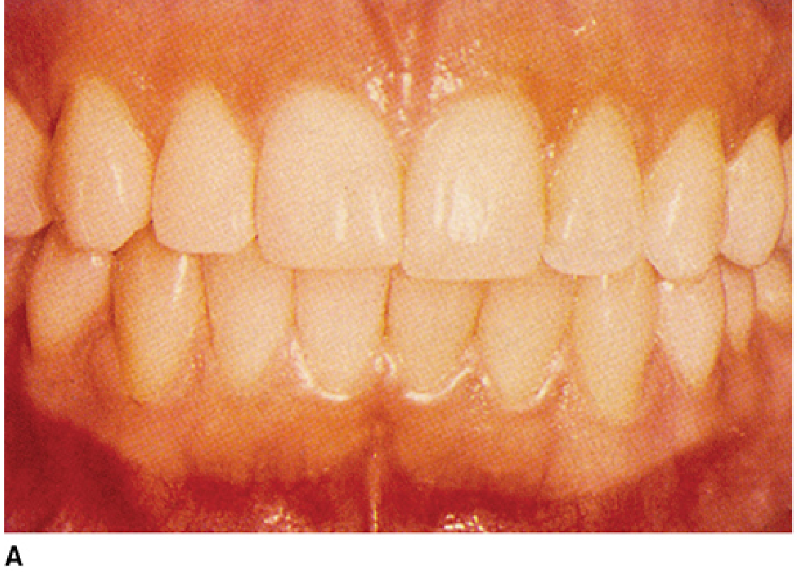
Fig. 41-5 - Normal vs. Periodontal Disease (Sherris & Ryan's Medical Microbiology, 8e):
A. Normal Gingiva - Pink, firm, well-adapted to teeth, no bleeding:
B. Periodontal Disease - Plaque, severe inflammation, bleeding, gingival recession with root exposure ("teeth look longer"):
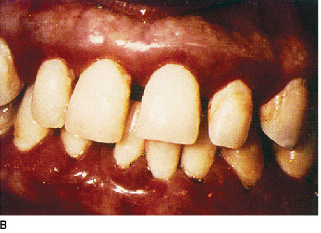
Fig. 42.1 - Chronic Periodontitis (Scott-Brown's Otorhinolaryngology, Head & Neck Surgery):

6. TYPES OF PERIODONTITIS
| Type | Age Group | Key Feature |
|---|---|---|
| Chronic Periodontitis | >35-40 years | Slow progressive destruction; most common cause of adult tooth loss |
| Localized Aggressive Periodontitis | Adolescents | Rapid bone loss; associated with A. actinomycetemcomitans leukotoxin |
| Generalized Aggressive Periodontitis | Young adults | Rapid widespread destruction |
| Necrotizing Periodontal Disease | Immunocompromised | HIV+, poorly controlled diabetes; requires debridement |
| Periodontal Abscess | Any | Acute exacerbation when pocket neck becomes constricted; requires drainage |
7. CAUSES OF GINGIVAL BLEEDING
(Scott-Brown's Table 42.2)
| Local Causes | Systemic Causes |
|---|---|
| Chronic gingivitis | Pregnancy (exacerbates gingivitis) |
| Chronic periodontitis | Leukaemia |
| Acute necrotizing gingivitis | HIV infection |
| Angioma/telangiectasia | Clotting defects |
| Anticoagulant drugs | |
| Scurvy (Vitamin C deficiency) |
8. SYSTEMIC ASSOCIATIONS
Periodontal disease is associated with multiple systemic conditions. When localized to the gums, it is linked to:
- Diabetes mellitus (bidirectional - each worsens the other)
- Atherosclerotic vascular disease (significant research exists; periodontitis is a source of bacteremia)
- Infective endocarditis (periodontal bacteria may seed cardiac valves)
- Brain and lung abscesses (hematogenous spread)
- AIDS/HIV
- Down syndrome, Crohn disease, sarcoidosis
Note: Despite ongoing concerns, Goldman-Cecil Medicine states there is currently "no evidence that periodontal disease is an independent risk factor for coronary artery disease."
9. GLOBAL BURDEN (Park's Preventive & Social Medicine)
- Severe periodontal disease was the 11th most prevalent disease globally in 2016
- Major causes: poor oral hygiene and tobacco use
- Dental caries + periodontal disease = leading causes of tooth loss
- Severe tooth loss ranked among top 10 causes of years lived with disability (YLD) in high-income countries with aging populations
10. MANAGEMENT
Non-Surgical (Mainstay)
| Treatment | Notes |
|---|---|
| Oral hygiene | Twice-daily brushing + flossing |
| Scaling & root planing | Removal of subgingival calculus and biofilm (gold standard) |
| Chlorhexidine rinse | 0.12%-0.2% preferred; antibacterial mouthwash for moderate/severe cases |
| 3% H₂O₂ | Diluted 1:1 warm water; alternative rinse |
| Smoking cessation | Most important risk factor modification in HIV-negative patients |
Analgesia
- Ibuprofen 400-600 mg every 6-8 hours, OR
- Acetaminophen 650 mg every 6 hours
- Topical: Viscous lidocaine (applied to small areas only)
Antibiotics (for severe/spreading disease, systemic symptoms, or immunocompromise)
(Rosen's Emergency Medicine, Table 56.2)
| Antibiotic | Dose | Duration |
|---|---|---|
| Penicillin V | 500 mg TID-QID | 10 days |
| Amoxicillin/clavulanate | 500/125 mg TID | 10 days |
| Metronidazole | 500 mg BID (PCN allergy) | 10 days |
| Clindamycin | 300 mg QID (PCN allergy) | 10 days |
| Nystatin | 100,000 U/mL swish/spit QID (immunocompromised) | 10 days |
(Goldman-Cecil first-line: Amoxicillin 250 mg TID × 3 days; alternative: Clindamycin 150 mg QID × 3 days)
Surgical
- Debridement of necrotic tissue (necrotizing disease)
- Periodontal abscess: drainage essential
- Referral to dentist for all cases
11. PREVENTION
- Regular oral hygiene (brushing ×2/day, flossing)
- Regular dental check-ups and professional scaling
- Smoking cessation
- Control of systemic risk factors (glycemic control in diabetics)
- Community water fluoridation (reduces caries which can co-contribute)
SUMMARY FLOW CHART
DENTAL PLAQUE/CALCULUS (poor hygiene, smoking, systemic risk factors)
         ↓
    GINGIVITIS
    (Reversible - inflammation of marginal gingiva only)
         ↓ (if unchecked - subgingival plaque, gram-negative anaerobes)
   PERIODONTITIS
    (Irreversible - PDL destruction + alveolar bone loss + pocket formation)
         ↓
   TOOTH LOOSENING → TOOTH LOSS / PERIODONTAL ABSCESS
Key Textbooks Referenced:
- Robbins & Cotran Pathologic Basis of Disease, 10e - Gingivitis & Periodontitis, p. 493-500
- Junqueira's Basic Histology, 17e - Periodontium, Fig. 15-11
- Sherris & Ryan's Medical Microbiology, 8e - Chronic Periodontitis, Fig. 41-5
- Goldman-Cecil Medicine, International Ed. - Periodontal Disease section
- Scott-Brown's Otorhinolaryngology Head & Neck Surgery - Fig. 42.1, Table 42.2
- Park's Textbook of Preventive & Social Medicine - Global burden data
- Rosen's Emergency Medicine - Management Table 56.2
Good luck with your presentation tomorrow!
Histological features of gingivitis and peridonitis in stages
The histological staging of gingivitis and periodontitis was classically described by Page & Schroeder (1976) into 4 stages: Initial, Early, Established, and Advanced lesions. Let me pull the detailed content from standard textbooks.
Searching sources...
Finding Sources
Finding Sources
The FTS search didn't find specific staging. Let me search more specifically with different terms and check Robbins and microbiology books:
Finding Sources
Finding Sources
Finding Sources
Good findings. Let me now read the Histology textbook (Wheater's/Ross) content on periodontitis and the Tintinalli's normal periodontium, and also search for the Page & Schroeder staging:
Reading File
Reading File
Finding Sources
Let me read the Tintinalli's periodontal pathology section and also the Sherris microbiology section in full detail, plus search for images:
Reading File
Reading File
Finding Sources
Now let me get the Tintinalli's diagram image and also the Robbins pathology full gingivitis/periodontitis section with any images:
Loading Image
Reading File
Reading File
I now have comprehensive content from multiple standard textbooks. The classic Page & Schroeder (1976) staging is the gold standard histological classification - let me now compile the full, detailed answer combining all sources.
Histological Features of Gingivitis & Periodontitis - By Stages
Sources: Robbins & Cotran Pathologic Basis of Disease (10e), Sherris & Ryan's Medical Microbiology (8e), Ross & Pawlina Histology: A Text and Atlas (8e), Junqueira's Basic Histology (17e), Tintinalli's Emergency Medicine
FOUNDATION: Normal Gingival Histology
Before understanding disease, grasp the normal architecture:
Fig. 245-3 - The Dental Anatomic Unit & Attachment Apparatus (Tintinalli's Emergency Medicine):
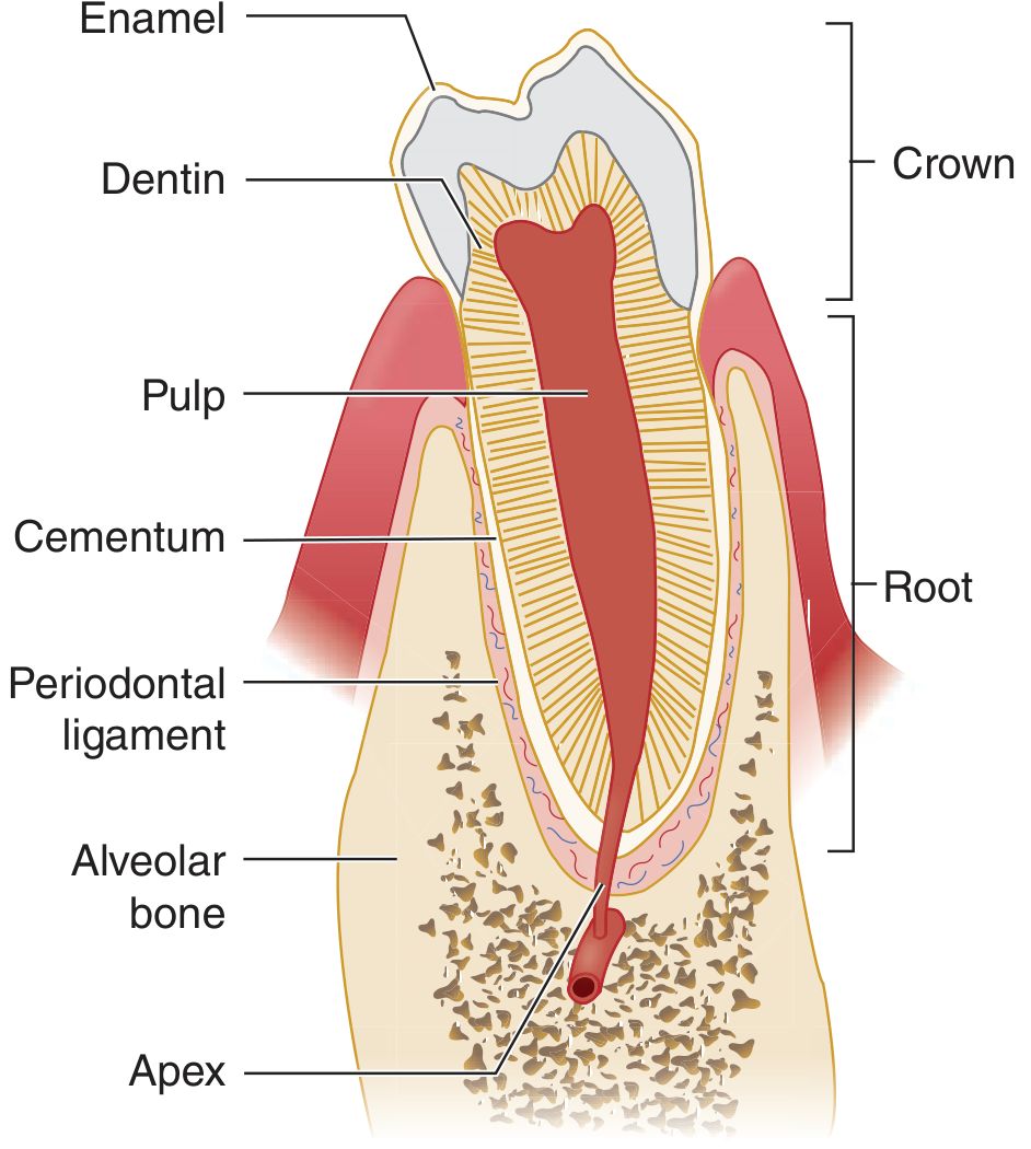
Normal Gingival Tissue Layers (from surface inward):
| Layer | Histology |
|---|---|
| Gingival mucosa | Keratinized stratified squamous epithelium over lamina propria |
| Junctional (attachment) epithelium | Attaches firmly to tooth via basal lamina-like material + hemidesmosomes; "epithelial attachment" |
| Crevicular epithelium | Lines the 2-3 mm gingival sulcus; continuous with junctional epithelium; non-keratinized |
| Periodontal ligament | Dense collagen (Sharpey) fibers; fibroblasts that both produce AND resorb collagen; blood vessels + nerve endings + oxytalan fibers |
| Alveolar bone | Woven bone pattern; continuous osteoblast/osteoclast remodeling |
"The free gingiva is the portion that forms the 2- to 3-mm-deep gingival sulcus in the disease-free state." - Tintinalli's Emergency Medicine
THE PAGE & SCHROEDER (1976) HISTOLOGICAL STAGING
The classic classification describes 4 sequential histological stages of periodontal disease:
STAGE 1 - INITIAL LESION (2-4 days after plaque accumulation)
Clinical appearance:
- Clinically silent - no visible signs
- Subclinical vascular changes only
Histological features:
| Feature | Detail |
|---|---|
| Vascular changes | Vasodilation of vessels in gingival plexus beneath sulcular epithelium |
| Increased vascular permeability | Exudation begins |
| Polymorphonuclear neutrophils (PMNs) | First cells to migrate - neutrophils emigrate through the junctional epithelium into the sulcus |
| Gingival crevicular fluid (GCF) | Increased flow of GCF into sulcus |
| Serum proteins | Extravasation - fibrinogen, complement, immunoglobulins leak into perivascular tissue |
| Collagen | Minimal perivascular collagen degradation begins |
Key cell: PMN/Neutrophil (acute inflammatory response)
Reversibility: Fully reversible
STAGE 2 - EARLY LESION (4-7 days; peaks at 14 days)
Clinical appearance:
- Early redness (erythema) appears
- Bleeding on probing begins
- Gingival edema starts
Histological features:
| Feature | Detail |
|---|---|
| Accentuated neutrophil infiltration | Dense band of PMNs in junctional and sulcular epithelium |
| Lymphocytes predominate | Predominantly T lymphocytes (>75%) in connective tissue infiltrate - hallmark of this stage |
| Fibroblast alteration | Fibroblasts show cytopathic changes - altered morphology, reduced function |
| Collagen loss | 60-70% collagen loss in the infiltrated connective tissue zone beneath the junctional epithelium |
| Epithelial proliferation | Rete pegs of junctional epithelium begin to elongate and proliferate laterally |
| Increased GCF | Continues to increase |
| Vasodilation | Persistent; capillary loops become prominent |
Key cell: T lymphocyte
Reversibility: Still reversible with plaque removal
STAGE 3 - ESTABLISHED LESION (Chronic Gingivitis; weeks to months)
Clinical appearance:
- Frank gingivitis - bright red/bluish-red gingiva
- Edematous, spongy, swollen gingiva
- Spontaneous or easy bleeding
- Sulcus depth may increase slightly
- No bone loss yet - gingiva only
Histological features:
| Feature | Detail |
|---|---|
| Plasma cells DOMINATE | B lymphocytes and plasma cells become the predominant infiltrating cells - hallmark of established lesion |
| Dense chronic inflammatory infiltrate | Mixed: PMNs + lymphocytes + plasma cells + macrophages in connective tissue |
| Collagen loss continues | Further degradation of perivascular and subepithelial collagen |
| Pocket epithelium formation | Junctional epithelium continues to proliferate; becomes "pocket epithelium" - thin, ulcerated, permeable |
| Epithelial migration | Coronal epithelium spreads apically |
| Vascular proliferation | Increased vascularity (explains bleeding) |
| No alveolar bone loss | Inflammation still confined to soft tissue |
| Immunoglobulin-producing cells | Plasma cells actively secreting IgG, IgA, IgM into tissues |
Key cell: Plasma cell (B cell lineage)
"The characteristic histopathologic picture of gingivitis is of a marked inflammatory infiltrate of polymorphonuclear leukocytes, lymphocytes, and plasma cells in the connective tissue that lies immediately adjacent to the epithelium lining the gingival crevice... Collagen is lost from the inflamed connective tissue." - Sherris & Ryan's Medical Microbiology, 8e
Reversibility: Reversible - returns to Stage 0 with effective oral hygiene
STAGE 4 - ADVANCED LESION (Periodontitis; Irreversible)
Clinical appearance:
- Deep periodontal pockets (>3 mm)
- Alveolar bone loss (horizontal and/or vertical)
- Gingival recession
- Tooth mobility
- Suppuration possible
- Eventual tooth loss
Histological features:
| Feature | Detail |
|---|---|
| Extension into alveolar bone | Inflammatory infiltrate extends from soft tissue into alveolar bone and periodontal ligament |
| Alveolar bone resorption | Osteoclastic resorption of alveolar bone is the defining event - distinguishes periodontitis from gingivitis |
| Loss of Sharpey fiber insertions | Periodontal ligament (collagen Sharpey fibers) detach from cementum and bone |
| Periodontal pocket | Deepening of sulcus due to: (1) apical migration of junctional epithelium along root surface, and (2) coronal displacement of gingival margin |
| Pocket epithelium | Thin, non-keratinized, ulcerated; highly permeable to bacteria and bacterial products |
| Continued plasma cell dominance | Dense plasma cell-rich infiltrate; immunoglobulin deposition |
| Fibroblast destruction | Extensive fibroblast and collagen destruction by bacterial enzymes (e.g., P. gingivalis proteases) |
| Cementum changes | Bacterial invasion of cementum surface; necrotic cementum |
| Bacterial invasion | Evidence of direct bacterial penetration at advanced stages (unlike early gingivitis) |
| Periodontal abscess | When pocket becomes occluded: acute neutrophilic abscess within periodontal pocket |
Key cells: Plasma cells + Osteoclasts (bone resorption)
"Periodontitis is not considered to be a reversible disease in that the lost alveolar bone and periodontal ligament do not regenerate with cessation of the inflammation, even though further progression may be halted." - Sherris & Ryan's Medical Microbiology, 8e
"Periodontitis is an inflammatory oral disease that leads to the destruction of periodontal tissue involved in the attachment of the tooth... conventional therapies are unable to restore damaged periodontal structures." - Ross & Pawlina Histology: A Text and Atlas, 8e
SUMMARY TABLE - Histological Stages at a Glance
| Feature | Stage 1: Initial | Stage 2: Early | Stage 3: Established (Gingivitis) | Stage 4: Advanced (Periodontitis) |
|---|---|---|---|---|
| Time | 2-4 days | 4-14 days | Weeks-months | Months-years |
| Dominant cells | PMNs | T-lymphocytes | Plasma cells | Plasma cells + Osteoclasts |
| Collagen loss | Minimal (perivascular) | 60-70% in infiltrated zone | Progressive | Extensive |
| Junctional epithelium | Intact | Rete peg proliferation | Pocket epithelium forming | Deep pocket epithelium, ulcerated |
| Alveolar bone | Normal | Normal | Normal | Osteoclastic resorption |
| PDL | Intact | Intact | Intact | Destroyed |
| Reversibility | Yes | Yes | Yes | NO |
| Clinical signs | None | Early erythema | Frank gingivitis | Pocketing, mobility, bone loss |
PROGRESSION DIAGRAM
PLAQUE accumulation
       ↓ (2-4 days)
STAGE 1 - Initial Lesion
[Vasodilation → PMN migration → GCF ↑]
       ↓ (4-14 days)
STAGE 2 - Early Lesion
[T-lymphocyte infiltrate → collagen loss 60-70% → rete peg proliferation]
       ↓ (weeks to months)
STAGE 3 - Established Lesion = GINGIVITIS
[Plasma cells dominate → pocket epithelium → no bone loss → REVERSIBLE]
       ↓ (susceptible individuals)
STAGE 4 - Advanced Lesion = PERIODONTITIS
[Bone resorption → PDL destruction → true pocket → IRREVERSIBLE]
MICROBIAL HISTOPATHOLOGY NOTES
The bacteria do not directly invade tissues in early stages - they cause destruction via:
- Bacterial enzymes - e.g., P. gingivalis extracellular proteases degrade collagen and tissue proteins
- LPS-driven TLR activation - gram-negative cell wall components trigger host inflammatory cascade
- Leukotoxin (A. actinomycetemcomitans) - kills PMNs, evades immune defense
- Complement evasion - T. denticola binds serum factors to block complement deposition
- Host-mediated destruction - matrix metalloproteinases (MMPs), prostaglandins, and cytokines (IL-1β, TNF-α) from host cells cause most of the connective tissue and bone destruction
"There does not seem to be any direct invasion of the gingival tissues by large numbers of intact bacteria, at least in the early stages of the disease." - Sherris & Ryan's Medical Microbiology, 8e
Key Textbooks Referenced:
- Robbins & Kumar Basic Pathology + Robbins & Cotran Pathologic Basis of Disease - Gingivitis & Periodontitis sections
- Sherris & Ryan's Medical Microbiology, 8e - Chronic Periodontitis chapter
- Ross & Pawlina Histology: A Text and Atlas, 8e - Supporting Tissues of the Teeth, p. 1462-63
- Junqueira's Basic Histology, 17e - Periodontium, Fig. 15-11
- Tintinalli's Emergency Medicine - Normal Periodontium & Periodontal Pathology sections
Generate a quick reference flashcard PDF for histological features of gingivitis and periodontitis along with stages
Reading File
Running Command
ls -la /tmp/workspace/ 2>/dev/null || echo "workspace empty or missing"
Running Command
mkdir -p /tmp/workspace/periodontal-flashcards
Writing File
~/periodontal-flashcards/make_flashcards.py
from reportlab.lib.pagesizes import A4
from reportlab.lib import colors
from reportlab.lib.units import mm
from reportlab.platypus import (
SimpleDocTemplate, Paragraph, Spacer, Table, TableStyle,
HRFlowable, KeepTogether
)
from reportlab.lib.styles import getSampleStyleSheet, ParagraphStyle
from reportlab.lib.enums import TA_CENTER, TA_LEFT, TA_RIGHT
from reportlab.platypus import PageBreak
OUTPUT = "/tmp/workspace/periodontal-flashcards/Periodontal_Histology_Flashcards.pdf"
# ── Colour palette ──────────────────────────────────────────────────
C_DARK_TEAL = colors.HexColor("#0D4F5C") # header bg
C_TEAL = colors.HexColor("#1A7A8A") # card header band
C_STAGE1 = colors.HexColor("#2E86AB") # Stage 1 – blue
C_STAGE2 = colors.HexColor("#4CAF82") # Stage 2 – green
C_STAGE3 = colors.HexColor("#E67E22") # Stage 3 – orange (gingivitis)
C_STAGE4 = colors.HexColor("#C0392B") # Stage 4 – red (periodontitis)
C_LIGHT_BLUE = colors.HexColor("#EAF6FB")
C_LIGHT_GREEN = colors.HexColor("#EAFAF1")
C_LIGHT_ORANGE= colors.HexColor("#FEF9E7")
C_LIGHT_RED = colors.HexColor("#FDEDEC")
C_LIGHT_TEAL = colors.HexColor("#E8F6F8")
C_GOLD = colors.HexColor("#F1C40F")
C_WHITE = colors.white
C_DARK = colors.HexColor("#1C1C1C")
C_GREY = colors.HexColor("#666666")
C_LIGHT_GREY = colors.HexColor("#F5F5F5")
C_MID_GREY = colors.HexColor("#DDDDDD")
W, H = A4 # 595.27 x 841.89 pt
styles = getSampleStyleSheet()
def ps(name, **kw):
return ParagraphStyle(name, **kw)
# ── custom styles ────────────────────────────────────────────────────
COVER_TITLE = ps("CoverTitle", fontName="Helvetica-Bold",
fontSize=28, textColor=C_WHITE, alignment=TA_CENTER, leading=34)
COVER_SUB = ps("CoverSub", fontName="Helvetica",
fontSize=14, textColor=colors.HexColor("#AADDE6"),
alignment=TA_CENTER, leading=20)
COVER_NOTE = ps("CoverNote", fontName="Helvetica-Oblique",
fontSize=10, textColor=colors.HexColor("#CCCCCC"),
alignment=TA_CENTER)
CARD_TITLE = ps("CardTitle", fontName="Helvetica-Bold",
fontSize=13, textColor=C_WHITE, alignment=TA_CENTER, leading=16)
CARD_SUB = ps("CardSub", fontName="Helvetica-Bold",
fontSize=9.5, textColor=C_WHITE, alignment=TA_CENTER)
SEC_TITLE = ps("SecTitle", fontName="Helvetica-Bold",
fontSize=10, textColor=C_DARK_TEAL, leading=14)
BODY = ps("Body", fontName="Helvetica",
fontSize=8.5, textColor=C_DARK, leading=12)
BODY_B = ps("BodyB", fontName="Helvetica-Bold",
fontSize=8.5, textColor=C_DARK, leading=12)
SMALL = ps("Small", fontName="Helvetica",
fontSize=7.5, textColor=C_GREY, leading=10)
TABLE_HDR = ps("TblHdr", fontName="Helvetica-Bold",
fontSize=8, textColor=C_WHITE, alignment=TA_CENTER, leading=11)
TABLE_CELL = ps("TblCell", fontName="Helvetica",
fontSize=7.8, textColor=C_DARK, alignment=TA_LEFT, leading=11)
TABLE_CELL_B= ps("TblCellB", fontName="Helvetica-Bold",
fontSize=7.8, textColor=C_DARK, alignment=TA_LEFT, leading=11)
BULLET = ps("Bullet", fontName="Helvetica",
fontSize=8.5, textColor=C_DARK, leading=12, leftIndent=12,
bulletIndent=0, bulletFontName="Helvetica", bulletFontSize=8.5)
FOOT = ps("Foot", fontName="Helvetica-Oblique",
fontSize=7, textColor=C_GREY, alignment=TA_CENTER)
KEYBOX_TXT = ps("KeyboxTxt", fontName="Helvetica-Bold",
fontSize=9, textColor=C_DARK_TEAL, alignment=TA_CENTER, leading=13)
# ── helper: coloured card header table ──────────────────────────────
def card_header(stage_num, stage_name, subtext, bg_color, light_bg):
data = [[Paragraph(f"STAGE {stage_num} · {stage_name}", CARD_TITLE)],
[Paragraph(subtext, CARD_SUB)]]
t = Table(data, colWidths=[W - 28*mm])
t.setStyle(TableStyle([
("BACKGROUND", (0, 0), (-1, 0), bg_color),
("BACKGROUND", (0, 1), (-1, 1), bg_color),
("TOPPADDING", (0, 0), (-1, -1), 5),
("BOTTOMPADDING", (0, 0), (-1, -1), 5),
("LEFTPADDING", (0, 0), (-1, -1), 8),
("RIGHTPADDING", (0, 0), (-1, -1), 8),
("ROUNDEDCORNERS", [5]),
]))
return t
# ── helper: two-column feature table ────────────────────────────────
def feature_table(rows, col1_w=52*mm, col2_w=None, hdr_bg=C_TEAL):
if col2_w is None:
col2_w = W - 28*mm - col1_w
header_row = [Paragraph("FEATURE", TABLE_HDR),
Paragraph("DETAIL", TABLE_HDR)]
table_data = [header_row]
for feat, detail in rows:
table_data.append([
Paragraph(feat, TABLE_CELL_B),
Paragraph(detail, TABLE_CELL),
])
t = Table(table_data, colWidths=[col1_w, col2_w])
style = TableStyle([
("BACKGROUND", (0, 0), (-1, 0), hdr_bg),
("ROWBACKGROUNDS",(0, 1), (-1, -1), [C_WHITE, C_LIGHT_GREY]),
("GRID", (0, 0), (-1, -1), 0.4, C_MID_GREY),
("TOPPADDING", (0, 0), (-1, -1), 4),
("BOTTOMPADDING", (0, 0), (-1, -1), 4),
("LEFTPADDING", (0, 0), (-1, -1), 5),
("RIGHTPADDING", (0, 0), (-1, -1), 5),
("VALIGN", (0, 0), (-1, -1), "TOP"),
])
t.setStyle(style)
return t
# ── helper: key cell callout ─────────────────────────────────────────
def key_cell_box(cell_name, bg):
data = [[Paragraph(f"🔑 KEY CELL: {cell_name}", KEYBOX_TXT)]]
t = Table(data, colWidths=[W - 28*mm])
t.setStyle(TableStyle([
("BACKGROUND", (0, 0), (-1, -1), bg),
("TOPPADDING", (0, 0), (-1, -1), 6),
("BOTTOMPADDING", (0, 0), (-1, -1), 6),
("LEFTPADDING", (0, 0), (-1, -1), 8),
("RIGHTPADDING", (0, 0), (-1, -1), 8),
("BOX", (0, 0), (-1, -1), 1, C_TEAL),
]))
return t
# ── helper: reversibility badge ──────────────────────────────────────
def rev_badge(text, bg):
data = [[Paragraph(text, ps("Rev", fontName="Helvetica-Bold",
fontSize=9, textColor=C_WHITE,
alignment=TA_CENTER))]]
t = Table(data, colWidths=[W - 28*mm])
t.setStyle(TableStyle([
("BACKGROUND", (0, 0), (-1, -1), bg),
("TOPPADDING", (0, 0), (-1, -1), 5),
("BOTTOMPADDING", (0, 0), (-1, -1), 5),
]))
return t
# ── helper: section label ────────────────────────────────────────────
def sec(txt, color=C_DARK_TEAL):
return Paragraph(f"<b><font color='#{color.hexval()[2:]}'>▸ {txt}</font></b>", SEC_TITLE)
def sp(h=4): return Spacer(1, h)
def hr(color=C_MID_GREY): return HRFlowable(width="100%", thickness=0.5, color=color)
# ════════════════════════════════════════════════════════════════════
# Build content
# ════════════════════════════════════════════════════════════════════
story = []
# ── PAGE 1: COVER ────────────────────────────────────────────────────
def cover_page(canvas, doc):
canvas.saveState()
# Full-page gradient-ish background
canvas.setFillColor(C_DARK_TEAL)
canvas.rect(0, 0, W, H, fill=1, stroke=0)
# Decorative top stripe
canvas.setFillColor(C_TEAL)
canvas.rect(0, H - 20*mm, W, 20*mm, fill=1, stroke=0)
# Bottom stripe
canvas.setFillColor(colors.HexColor("#0A3A45"))
canvas.rect(0, 0, W, 18*mm, fill=1, stroke=0)
canvas.restoreState()
# Cover content as a table (centred vertically)
cover_data = [
[Paragraph("🦷", ps("Em", fontName="Helvetica", fontSize=40,
textColor=C_WHITE, alignment=TA_CENTER))],
[Spacer(1, 6)],
[Paragraph("PERIODONTAL DISEASE", COVER_TITLE)],
[Paragraph("Histological Features & Staging", COVER_SUB)],
[Spacer(1, 10)],
[HRFlowable(width="60%", thickness=1.5, color=C_GOLD)],
[Spacer(1, 10)],
[Paragraph("Quick Reference Flashcard Set", ps("QR", fontName="Helvetica-Bold",
fontSize=13, textColor=C_GOLD, alignment=TA_CENTER))],
[Spacer(1, 6)],
[Paragraph("Page & Schroeder (1976) Classification", COVER_SUB)],
[Spacer(1, 30)],
[Paragraph("Based on: Robbins & Cotran Pathologic Basis of Disease · Sherris & Ryan's Medical Microbiology<br/>"
"Ross & Pawlina Histology · Junqueira's Basic Histology · Tintinalli's Emergency Medicine",
COVER_NOTE)],
[Spacer(1, 8)],
[Paragraph("July 2026", COVER_NOTE)],
]
cover_tbl = Table(cover_data, colWidths=[W - 40*mm])
cover_tbl.setStyle(TableStyle([
("ALIGN", (0, 0), (-1, -1), "CENTER"),
("VALIGN", (0, 0), (-1, -1), "MIDDLE"),
("TOPPADDING", (0, 0), (-1, -1), 2),
("BOTTOMPADDING", (0, 0), (-1, -1), 2),
]))
story.append(Spacer(1, 55*mm))
story.append(cover_tbl)
story.append(PageBreak())
# ── PAGE 2: NORMAL ANATOMY + OVERVIEW ────────────────────────────────
story.append(sp(4))
# Title banner
anat_hdr = Table([[Paragraph("NORMAL PERIODONTIUM — ANATOMY FLASHCARD", CARD_TITLE)]],
colWidths=[W - 28*mm])
anat_hdr.setStyle(TableStyle([
("BACKGROUND", (0, 0), (-1, -1), C_DARK_TEAL),
("TOPPADDING", (0, 0), (-1, -1), 7),
("BOTTOMPADDING", (0, 0), (-1, -1), 7),
("LEFTPADDING", (0, 0), (-1, -1), 8),
]))
story.append(anat_hdr)
story.append(sp(6))
anatomy_rows = [
("Gingiva",
"Keratinized stratified squamous epithelium over lamina propria. "
"Free gingiva forms 2–3 mm sulcus. Attached gingiva adheres to alveolar bone."),
("Junctional (Attachment) Epithelium",
"Adheres to tooth via basal lamina + hemidesmosomes (= epithelial attachment). "
"In young: to enamel. In older: migrates to cementum."),
("Crevicular Epithelium",
"Lines the gingival sulcus; non-keratinized; continuous with junctional epithelium. "
"First site of bacterial contact."),
("Periodontal Ligament (PDL)",
"Short collagen bundles (Sharpey fibers) binding cementum to alveolar bone. "
"Highly vascular + innervated. Width: 150–350 µm. Fibroblasts both produce AND resorb collagen."),
("Cementum",
"Covers root dentin. Avascular. Bone-like (cementocytes in lacunae). "
"Thickest at root apices."),
("Alveolar Bone",
"Woven (non-lamellar) bone pattern. Continuous osteoblast/osteoclast remodeling. "
"Surrounds root sockets."),
]
story.append(feature_table(anatomy_rows, col1_w=58*mm, hdr_bg=C_DARK_TEAL))
story.append(sp(8))
# Disease overview box
story.append(sec("DISEASE CONTINUUM"))
story.append(sp(3))
flow_data = [[
Paragraph("PLAQUE<br/>ACCUMULATION", ps("F0", fontName="Helvetica-Bold",
fontSize=8, textColor=C_DARK, alignment=TA_CENTER)),
Paragraph("→", ps("AR", fontName="Helvetica-Bold", fontSize=14,
textColor=C_TEAL, alignment=TA_CENTER)),
Paragraph("STAGE 1–2<br/><font color='#2E86AB'>Initial/Early</font>",
ps("F1", fontName="Helvetica-Bold", fontSize=8,
textColor=C_DARK, alignment=TA_CENTER)),
Paragraph("→", ps("AR2", fontName="Helvetica-Bold", fontSize=14,
textColor=C_TEAL, alignment=TA_CENTER)),
Paragraph("STAGE 3<br/><font color='#E67E22'>Gingivitis</font>",
ps("F2", fontName="Helvetica-Bold", fontSize=8,
textColor=C_DARK, alignment=TA_CENTER)),
Paragraph("→", ps("AR3", fontName="Helvetica-Bold", fontSize=14,
textColor=C_TEAL, alignment=TA_CENTER)),
Paragraph("STAGE 4<br/><font color='#C0392B'>Periodontitis</font>",
ps("F3", fontName="Helvetica-Bold", fontSize=8,
textColor=C_DARK, alignment=TA_CENTER)),
]]
flow_col = (W - 28*mm) / 7
flow_tbl = Table(flow_data, colWidths=[flow_col]*7)
flow_tbl.setStyle(TableStyle([
("BACKGROUND", (0, 0), (0, 0), C_LIGHT_GREY),
("BACKGROUND", (2, 0), (2, 0), C_LIGHT_BLUE),
("BACKGROUND", (4, 0), (4, 0), C_LIGHT_ORANGE),
("BACKGROUND", (6, 0), (6, 0), C_LIGHT_RED),
("ALIGN", (0, 0), (-1, -1), "CENTER"),
("VALIGN", (0, 0), (-1, -1), "MIDDLE"),
("TOPPADDING", (0, 0), (-1, -1), 8),
("BOTTOMPADDING", (0, 0), (-1, -1), 8),
("BOX", (0, 0), (0, 0), 0.5, C_MID_GREY),
("BOX", (2, 0), (2, 0), 0.5, C_STAGE1),
("BOX", (4, 0), (4, 0), 0.5, C_STAGE3),
("BOX", (6, 0), (6, 0), 0.5, C_STAGE4),
]))
story.append(flow_tbl)
story.append(PageBreak())
# ── PAGE 3: STAGE 1 ──────────────────────────────────────────────────
story.append(card_header(1, "INITIAL LESION", "2–4 Days After Plaque Accumulation · Subclinical",
C_STAGE1, C_LIGHT_BLUE))
story.append(sp(6))
story.append(sec("CLINICAL FEATURES", C_STAGE1))
story.append(sp(3))
story.append(Paragraph("● Clinically <b>silent</b> — no visible signs to patient or clinician", BULLET))
story.append(Paragraph("● Only <b>subclinical vascular changes</b> detectable histologically", BULLET))
story.append(sp(6))
story.append(sec("HISTOLOGICAL FEATURES", C_STAGE1))
story.append(sp(3))
s1_rows = [
("Vascular changes",
"Vasodilation of vessels in gingival plexus beneath sulcular epithelium; acute inflammatory response begins"),
("Vascular permeability ↑",
"Plasma proteins (fibrinogen, complement, immunoglobulins) extravasate into perivascular connective tissue"),
("Dominant cells",
"PMNs (polymorphonuclear neutrophils) — FIRST cells to migrate through junctional epithelium into sulcus"),
("GCF (Gingival Crevicular Fluid)",
"Increased flow of GCF into gingival sulcus; early warning marker"),
("Collagen",
"Minimal perivascular collagen degradation begins — earliest structural change"),
("Bacterial invasion",
"NO direct bacterial invasion of tissues; bacteria remain in sulcus"),
]
story.append(feature_table(s1_rows, hdr_bg=C_STAGE1))
story.append(sp(8))
story.append(key_cell_box("PMN / Neutrophil (Polymorphonuclear Leucocyte)", C_LIGHT_BLUE))
story.append(sp(6))
story.append(rev_badge("✔ FULLY REVERSIBLE — removal of plaque returns tissue to normal", C_STAGE1))
story.append(PageBreak())
# ── PAGE 4: STAGE 2 ──────────────────────────────────────────────────
story.append(card_header(2, "EARLY LESION", "4–14 Days · Peaks at ~14 Days",
C_STAGE2, C_LIGHT_GREEN))
story.append(sp(6))
story.append(sec("CLINICAL FEATURES", C_STAGE2))
story.append(sp(3))
story.append(Paragraph("● Early <b>erythema</b> (redness) — first visible sign", BULLET))
story.append(Paragraph("● <b>Bleeding on probing</b> begins", BULLET))
story.append(Paragraph("● Mild gingival edema", BULLET))
story.append(sp(6))
story.append(sec("HISTOLOGICAL FEATURES", C_STAGE2))
story.append(sp(3))
s2_rows = [
("Dominant cells",
"T-LYMPHOCYTES predominate (>75% of infiltrate) in the connective tissue — hallmark of this stage"),
("PMN infiltration",
"Accentuated neutrophil band persists in junctional + sulcular epithelium"),
("Collagen loss",
"60–70% collagen loss in the INFILTRATED CONNECTIVE TISSUE zone beneath junctional epithelium"),
("Fibroblasts",
"Cytopathic changes: fibroblasts show altered morphology, reduced collagen-producing function"),
("Junctional epithelium",
"Rete pegs begin to ELONGATE and proliferate laterally — early structural remodelling"),
("GCF",
"Continues to increase; now contains inflammatory mediators and enzymes"),
("Vasodilation",
"Persistent; capillary loops become prominent — explains visible erythema"),
("Bacterial invasion",
"Still NO direct tissue invasion by bacteria"),
]
story.append(feature_table(s2_rows, hdr_bg=C_STAGE2))
story.append(sp(8))
story.append(key_cell_box("T-Lymphocyte (CD4+ helper T cells predominate)", C_LIGHT_GREEN))
story.append(sp(6))
story.append(rev_badge("✔ REVERSIBLE — effective oral hygiene restores normal histology", C_STAGE2))
story.append(PageBreak())
# ── PAGE 5: STAGE 3 ──────────────────────────────────────────────────
story.append(card_header(3, "ESTABLISHED LESION", "Weeks to Months · Chronic GINGIVITIS",
C_STAGE3, C_LIGHT_ORANGE))
story.append(sp(6))
story.append(sec("CLINICAL FEATURES", C_STAGE3))
story.append(sp(3))
story.append(Paragraph("● <b>Frank gingivitis</b> — bright red or bluish-red, oedematous, spongy gingiva", BULLET))
story.append(Paragraph("● Spontaneous or easy <b>bleeding</b>", BULLET))
story.append(Paragraph("● Gingival contour changes; pseudo-pocket may form", BULLET))
story.append(Paragraph("● <b>No alveolar bone loss</b> — inflammation confined to soft tissue only", BULLET))
story.append(sp(6))
story.append(sec("HISTOLOGICAL FEATURES", C_STAGE3))
story.append(sp(3))
s3_rows = [
("Dominant cells",
"PLASMA CELLS dominate — hallmark of established lesion; B lymphocytes → plasma cells producing IgG, IgA, IgM"),
("Overall infiltrate",
"Dense mixed chronic infiltrate: PMNs + lymphocytes + PLASMA CELLS + macrophages in connective tissue"),
("Collagen loss",
"Continued progressive degradation of perivascular and subepithelial collagen"),
("Pocket epithelium",
"Junctional epithelium proliferates apically → becomes 'pocket epithelium': thin, ulcerated, highly permeable"),
("Epithelial migration",
"Coronal epithelium spreads apically along root surface — sulcus deepens slightly"),
("Vascular proliferation",
"Increased vascularity and capillary loops — explains spontaneous bleeding"),
("Alveolar bone",
"STILL NORMAL — no bone resorption; this is the critical distinction from Stage 4"),
("Immunoglobulins",
"Plasma cells actively secrete Ig into tissues and GCF — attempts at humoral immunity"),
]
story.append(feature_table(s3_rows, hdr_bg=C_STAGE3))
story.append(sp(8))
story.append(key_cell_box("Plasma Cell (B-lymphocyte lineage — IgG/IgA/IgM secreting)", C_LIGHT_ORANGE))
story.append(sp(6))
story.append(rev_badge("✔ REVERSIBLE — returns to Stage 0 with effective plaque control", C_STAGE3))
story.append(PageBreak())
# ── PAGE 6: STAGE 4 ──────────────────────────────────────────────────
story.append(card_header(4, "ADVANCED LESION", "Months to Years · PERIODONTITIS · IRREVERSIBLE",
C_STAGE4, C_LIGHT_RED))
story.append(sp(6))
story.append(sec("CLINICAL FEATURES", C_STAGE4))
story.append(sp(3))
story.append(Paragraph("● Deep <b>periodontal pockets</b> (>3 mm on probing)", BULLET))
story.append(Paragraph("● <b>Alveolar bone loss</b> — horizontal and/or vertical", BULLET))
story.append(Paragraph("● Gingival recession; roots exposed; 'teeth appear longer'", BULLET))
story.append(Paragraph("● <b>Tooth mobility</b>; possible suppuration", BULLET))
story.append(Paragraph("● Eventual <b>tooth loss</b> if untreated", BULLET))
story.append(sp(6))
story.append(sec("HISTOLOGICAL FEATURES", C_STAGE4))
story.append(sp(3))
s4_rows = [
("Defining event",
"OSTEOCLASTIC ALVEOLAR BONE RESORPTION — distinguishes periodontitis from gingivitis absolutely"),
("Infiltrate extension",
"Inflammatory cells extend from soft tissue INTO alveolar bone and periodontal ligament"),
("PDL destruction",
"Loss of Sharpey fiber insertions from cementum and alveolar bone; ligament destroyed"),
("Periodontal pocket",
"True pocket: apical migration of junctional epithelium + coronal displacement of gingival margin"),
("Pocket epithelium",
"Thin, non-keratinized, ulcerated; permeable to bacteria and their products; may show microulcerations"),
("Cementum changes",
"Bacterial invasion of cementum surface; necrotic/infected cementum (requires root planing)"),
("Fibroblast destruction",
"Extensive fibroblast + collagen destruction by P. gingivalis proteases and MMPs"),
("Plasma cells",
"Dense plasma cell-rich infiltrate continues; immunoglobulin deposition in connective tissue"),
("Periodontal abscess",
"Acute complication: when pocket occludes → acute neutrophilic abscess forms in periodontal pocket"),
("Regeneration",
"LOST BONE AND PDL DO NOT REGENERATE — even if inflammation halted (irreversible)"),
]
story.append(feature_table(s4_rows, hdr_bg=C_STAGE4))
story.append(sp(8))
story.append(key_cell_box("Plasma Cells + Osteoclasts (bone-resorbing cells)", C_LIGHT_RED))
story.append(sp(6))
story.append(rev_badge("✘ IRREVERSIBLE — lost bone & PDL do not regenerate; only progression can be halted", C_STAGE4))
story.append(PageBreak())
# ── PAGE 7: SUMMARY COMPARISON TABLE ─────────────────────────────────
story.append(sp(4))
summary_hdr = Table([[Paragraph("MASTER SUMMARY — ALL 4 STAGES AT A GLANCE", CARD_TITLE)]],
colWidths=[W - 28*mm])
summary_hdr.setStyle(TableStyle([
("BACKGROUND", (0, 0), (-1, -1), C_DARK_TEAL),
("TOPPADDING", (0, 0), (-1, -1), 8),
("BOTTOMPADDING", (0, 0), (-1, -1), 8),
("LEFTPADDING", (0, 0), (-1, -1), 8),
]))
story.append(summary_hdr)
story.append(sp(6))
TH = ps("SumHdr", fontName="Helvetica-Bold", fontSize=7.5,
textColor=C_WHITE, alignment=TA_CENTER, leading=10)
TC = ps("SumCell", fontName="Helvetica", fontSize=7.5,
textColor=C_DARK, alignment=TA_CENTER, leading=10)
TC_L = ps("SumCellL", fontName="Helvetica", fontSize=7.5,
textColor=C_DARK, alignment=TA_LEFT, leading=10)
TC_B = ps("SumCellB", fontName="Helvetica-Bold", fontSize=7.5,
textColor=C_DARK, alignment=TA_LEFT, leading=10)
cw = (W - 28*mm) / 5
sum_data = [
[Paragraph("PARAMETER", TH),
Paragraph("STAGE 1\nInitial", TH),
Paragraph("STAGE 2\nEarly", TH),
Paragraph("STAGE 3\nEstablished\n(Gingivitis)", TH),
Paragraph("STAGE 4\nAdvanced\n(Periodontitis)", TH)],
[Paragraph("Time onset", TC_B),
Paragraph("2–4 days", TC), Paragraph("4–14 days", TC),
Paragraph("Weeks–months", TC), Paragraph("Months–years", TC)],
[Paragraph("Dominant cells", TC_B),
Paragraph("PMNs", TC), Paragraph("T-lymphocytes", TC),
Paragraph("Plasma cells", TC), Paragraph("Plasma cells +\nOsteoclasts", TC)],
[Paragraph("Collagen loss", TC_B),
Paragraph("Minimal\n(perivascular)", TC), Paragraph("60–70%\n(infiltrated zone)", TC),
Paragraph("Progressive\n(subepithelial)", TC), Paragraph("Extensive +\nPDL destroyed", TC)],
[Paragraph("Junctional\nEpithelium", TC_B),
Paragraph("Intact", TC), Paragraph("Rete peg\nproliferation", TC),
Paragraph("Pocket epithelium\nforming", TC), Paragraph("Deep pocket,\nulcerated, thin", TC)],
[Paragraph("Alveolar bone", TC_B),
Paragraph("Normal", TC), Paragraph("Normal", TC),
Paragraph("Normal\n(no loss)", TC), Paragraph("OSTEOCLASTIC\nRESORP-TION ⚠", TC)],
[Paragraph("PDL", TC_B),
Paragraph("Intact", TC), Paragraph("Intact", TC),
Paragraph("Intact", TC), Paragraph("DESTROYED", TC)],
[Paragraph("Vasculature", TC_B),
Paragraph("Vasodilation\nbegins", TC), Paragraph("Capillary loops\nprominent", TC),
Paragraph("Proliferation\n(explains bleeding)", TC), Paragraph("Continued\nproliferation", TC)],
[Paragraph("Clinical signs", TC_B),
Paragraph("NONE\n(subclinical)", TC), Paragraph("Early erythema,\nbleeding on probing", TC),
Paragraph("Frank gingivitis:\nred, oedematous,\nbleeding", TC),
Paragraph("Pockets, bone\nloss, mobility,\ntooth loss", TC)],
[Paragraph("Reversible?", TC_B),
Paragraph("✔ YES", TC), Paragraph("✔ YES", TC),
Paragraph("✔ YES", TC), Paragraph("✘ NO", TC)],
]
sum_tbl = Table(sum_data, colWidths=[cw]*5)
sum_tbl.setStyle(TableStyle([
("BACKGROUND", (0, 0), (-1, 0), C_DARK_TEAL),
("BACKGROUND", (1, 1), (1, -1), C_LIGHT_BLUE),
("BACKGROUND", (2, 1), (2, -1), C_LIGHT_GREEN),
("BACKGROUND", (3, 1), (3, -1), C_LIGHT_ORANGE),
("BACKGROUND", (4, 1), (4, -1), C_LIGHT_RED),
("BACKGROUND", (0, 1), (0, -1), C_LIGHT_GREY),
("ROWBACKGROUNDS",(0, 1), (0, -1), [C_LIGHT_GREY]),
# Alternate row tinting on feature column
("GRID", (0, 0), (-1, -1), 0.4, C_MID_GREY),
("TOPPADDING", (0, 0), (-1, -1), 4),
("BOTTOMPADDING", (0, 0), (-1, -1), 4),
("LEFTPADDING", (0, 0), (-1, -1), 4),
("RIGHTPADDING", (0, 0), (-1, -1), 4),
("VALIGN", (0, 0), (-1, -1), "MIDDLE"),
("FONTNAME", (4, 9), (4, 9), "Helvetica-Bold"),
("TEXTCOLOR", (4, 9), (4, 9), C_STAGE4),
("FONTNAME", (1, 9), (3, 9), "Helvetica-Bold"),
("TEXTCOLOR", (1, 9), (3, 9), C_STAGE2),
]))
story.append(sum_tbl)
story.append(PageBreak())
# ── PAGE 8: MICROBIOLOGY + PATHOMECHANISM ────────────────────────────
story.append(sp(4))
micro_hdr = Table([[Paragraph("MICROBIOLOGY & PATHOMECHANISM FLASHCARD", CARD_TITLE)]],
colWidths=[W - 28*mm])
micro_hdr.setStyle(TableStyle([
("BACKGROUND", (0, 0), (-1, -1), C_DARK_TEAL),
("TOPPADDING", (0, 0), (-1, -1), 7),
("BOTTOMPADDING", (0, 0), (-1, -1), 7),
("LEFTPADDING", (0, 0), (-1, -1), 8),
]))
story.append(micro_hdr)
story.append(sp(6))
story.append(sec("KEY PERIODONTAL PATHOGENS"))
story.append(sp(3))
micro_rows = [
("Aggregatibacter\nactinomycetemcomitans",
"Gram-negative capnophilic rod. Produces LEUKOTOXIN (kills PMNs). "
"Key in localized aggressive periodontitis (adolescents)."),
("Porphyromonas\ngingivalis",
"Gram-negative anaerobe. Potent extracellular PROTEASES (tissue injury). "
"Central to chronic periodontitis. Works synergistically with T. denticola."),
("Prevotella intermedia",
"Gram-negative anaerobe. Associated with adult chronic periodontitis "
"and pregnancy gingivitis (metabolizes progesterone)."),
("Treponema denticola",
"Anaerobic spirochete. Binds serum factors → INTERFERES WITH COMPLEMENT. "
"Synergistic growth with P. gingivalis; drives progression from gingivitis to periodontitis."),
]
story.append(feature_table(micro_rows, col1_w=52*mm, hdr_bg=C_DARK_TEAL))
story.append(sp(8))
story.append(sec("MECHANISMS OF TISSUE DESTRUCTION"))
story.append(sp(3))
mech_rows = [
("Bacterial enzymes",
"P. gingivalis proteases (gingipains) degrade collagen, fibrinogen, complement factors"),
("Leukotoxin",
"A. actinomycetemcomitans leukotoxin kills PMNs → immune evasion"),
("Complement evasion",
"T. denticola binds serum complement inhibitors → survives in tissue"),
("TLR activation",
"LPS (gram-negative cell walls) trigger Toll-like receptors → IL-1β, TNF-α, prostaglandins"),
("Host MMPs",
"Matrix metalloproteinases from host cells degrade collagen and ECM (most destruction is HOST-mediated)"),
("Osteoclast activation",
"IL-1β, TNF-α, RANKL → osteoclast differentiation and activation → alveolar bone resorption"),
("Direct invasion",
"Minimal in early stages; bacteria remain in sulcus/pocket. Direct invasion occurs in ADVANCED disease only."),
]
story.append(feature_table(mech_rows, col1_w=52*mm, hdr_bg=C_TEAL))
story.append(sp(8))
# Microbial shift box
shift_data = [
[Paragraph("HEALTHY SITES", ps("SHdr", fontName="Helvetica-Bold",
fontSize=9, textColor=C_WHITE, alignment=TA_CENTER)),
Paragraph(" → ", ps("SAr", fontName="Helvetica-Bold", fontSize=14,
textColor=C_DARK, alignment=TA_CENTER)),
Paragraph("DISEASED SITES", ps("SDHdr", fontName="Helvetica-Bold",
fontSize=9, textColor=C_WHITE, alignment=TA_CENTER))],
[Paragraph("Facultative gram-positive\norganisms dominate", TABLE_CELL),
Paragraph("", TABLE_CELL),
Paragraph("Anaerobic + microaerophilic\ngram-negative flora dominate", TABLE_CELL)],
]
shift_tbl = Table(shift_data, colWidths=[(W-28*mm)*0.44, (W-28*mm)*0.12, (W-28*mm)*0.44])
shift_tbl.setStyle(TableStyle([
("BACKGROUND", (0, 0), (0, 0), C_STAGE2),
("BACKGROUND", (2, 0), (2, 0), C_STAGE4),
("BACKGROUND", (0, 1), (0, 1), C_LIGHT_GREEN),
("BACKGROUND", (2, 1), (2, 1), C_LIGHT_RED),
("ALIGN", (0, 0), (-1, -1), "CENTER"),
("VALIGN", (0, 0), (-1, -1), "MIDDLE"),
("TOPPADDING", (0, 0), (-1, -1), 6),
("BOTTOMPADDING", (0, 0), (-1, -1), 6),
("GRID", (0, 0), (-1, -1), 0.5, C_MID_GREY),
]))
story.append(shift_tbl)
story.append(PageBreak())
# ── PAGE 9: CLINICAL ASSESSMENT + TREATMENT ──────────────────────────
story.append(sp(4))
tx_hdr = Table([[Paragraph("CLINICAL ASSESSMENT & TREATMENT FLASHCARD", CARD_TITLE)]],
colWidths=[W - 28*mm])
tx_hdr.setStyle(TableStyle([
("BACKGROUND", (0, 0), (-1, -1), C_DARK_TEAL),
("TOPPADDING", (0, 0), (-1, -1), 7),
("BOTTOMPADDING", (0, 0), (-1, -1), 7),
("LEFTPADDING", (0, 0), (-1, -1), 8),
]))
story.append(tx_hdr)
story.append(sp(6))
story.append(sec("CLINICAL ASSESSMENT PARAMETERS"))
story.append(sp(3))
assess_rows = [
("Periodontal probe",
"Gold standard. Probe inserted into sulcus; pocket depth measured in mm. Normal ≤3 mm."),
("Bleeding on probing (BOP)",
"Reflects ACTIVE gingival inflammation. Present from Stage 2 onwards."),
("Attachment loss",
"Distance from CEJ to base of pocket. Key indicator of periodontitis severity."),
("Tooth mobility",
"Graded I–III. Reflects extent of PDL and bone destruction. Stage 4 finding."),
("Radiographs",
"Detect alveolar bone loss (horizontal/vertical patterns). Stage 4 prerequisite."),
]
story.append(feature_table(assess_rows, col1_w=52*mm, hdr_bg=C_TEAL))
story.append(sp(7))
story.append(sec("TREATMENT OVERVIEW BY STAGE"))
story.append(sp(3))
tx_rows = [
("Stage 1–2", "Oral hygiene instruction (brushing ×2/day, flossing). Plaque removal → REVERSIBLE."),
("Stage 3\n(Gingivitis)",
"Scaling + root planing (supragingival). Chlorhexidine rinse 0.12–0.2%. "
"Hydrogen peroxide (3%, diluted 1:1). NSAIDs for pain. REVERSIBLE."),
("Stage 4\n(Periodontitis)",
"Subgingival scaling + root planing (SRP). Referral to periodontist. "
"Antibiotics for severe/spreading disease. Periodontal surgery in refractory cases. IRREVERSIBLE."),
("Antibiotics\n(severe disease)",
"Penicillin V 500mg TID-QID × 10d; Amoxicillin/Clavulanate 500/125mg TID × 10d; "
"Metronidazole 500mg BID × 10d (PCN allergy); Clindamycin 300mg QID × 10d (PCN allergy)"),
("Abscess",
"Periodontal abscess requires DRAINAGE. Acute exacerbation needs urgent intervention."),
("Risk modification",
"Smoking cessation (most important in HIV-negative). Glycaemic control in diabetics. "
"Hormone management. Regular recall every 3–6 months."),
]
story.append(feature_table(tx_rows, col1_w=36*mm, hdr_bg=C_DARK_TEAL))
story.append(sp(7))
# Systemic associations box
story.append(sec("SYSTEMIC ASSOCIATIONS (BIDIRECTIONAL RELATIONSHIPS)"))
story.append(sp(3))
sys_data = [
[Paragraph("DISEASE", TABLE_HDR), Paragraph("RELATIONSHIP", TABLE_HDR)],
[Paragraph("Diabetes mellitus", TABLE_CELL_B),
Paragraph("Bidirectional: poor glycaemic control → worse periodontitis; periodontitis → worse glycaemia", TABLE_CELL)],
[Paragraph("HIV/AIDS", TABLE_CELL_B),
Paragraph("Increased severity; necrotizing periodontal disease; linear gingival erythema", TABLE_CELL)],
[Paragraph("Cardiovascular disease", TABLE_CELL_B),
Paragraph("Associated but no proven independent causal link (Goldman-Cecil)", TABLE_CELL)],
[Paragraph("Pregnancy", TABLE_CELL_B),
Paragraph("Pregnancy gingivitis: oestrogen/progesterone alter gingival response to plaque", TABLE_CELL)],
[Paragraph("Infective endocarditis", TABLE_CELL_B),
Paragraph("Periodontal bacteria (P. gingivalis, Streptococci) can seed cardiac valves", TABLE_CELL)],
]
sys_tbl = Table(sys_data, colWidths=[42*mm, W - 28*mm - 42*mm])
sys_tbl.setStyle(TableStyle([
("BACKGROUND", (0, 0), (-1, 0), C_TEAL),
("ROWBACKGROUNDS",(0, 1), (-1, -1), [C_WHITE, C_LIGHT_TEAL]),
("GRID", (0, 0), (-1, -1), 0.4, C_MID_GREY),
("TOPPADDING", (0, 0), (-1, -1), 4),
("BOTTOMPADDING", (0, 0), (-1, -1), 4),
("LEFTPADDING", (0, 0), (-1, -1), 5),
("RIGHTPADDING", (0, 0), (-1, -1), 5),
("VALIGN", (0, 0), (-1, -1), "TOP"),
]))
story.append(sys_tbl)
story.append(PageBreak())
# ── PAGE 10: HIGH-YIELD RAPID RECALL ─────────────────────────────────
story.append(sp(4))
hyr_hdr = Table([[Paragraph("HIGH-YIELD RAPID RECALL FLASHCARD", CARD_TITLE)]],
colWidths=[W - 28*mm])
hyr_hdr.setStyle(TableStyle([
("BACKGROUND", (0, 0), (-1, -1), C_GOLD),
("TOPPADDING", (0, 0), (-1, -1), 7),
("BOTTOMPADDING", (0, 0), (-1, -1), 7),
("LEFTPADDING", (0, 0), (-1, -1), 8),
]))
story.append(hyr_hdr)
story.append(sp(6))
hy_items = [
("Q: First cell to arrive in periodontal inflammation?",
"A: PMN (Neutrophil) — Stage 1 Initial Lesion"),
("Q: Dominant cell in Early Lesion (Stage 2)?",
"A: T-Lymphocyte (>75% of connective tissue infiltrate)"),
("Q: Dominant cell in Established Lesion (Stage 3 = Gingivitis)?",
"A: PLASMA CELL — hallmark of chronic gingivitis"),
("Q: What DEFINES periodontitis histologically?",
"A: Osteoclastic alveolar bone resorption + PDL destruction — Stage 4"),
("Q: Collagen loss % in Stage 2 (Early Lesion)?",
"A: 60–70% in the infiltrated connective tissue zone"),
("Q: Is gingivitis reversible?",
"A: YES (Stages 1–3 all reversible with plaque removal)"),
("Q: Is periodontitis reversible?",
"A: NO — lost alveolar bone and PDL do NOT regenerate"),
("Q: Normal gingival sulcus depth?",
"A: 2–3 mm (disease-free state)"),
("Q: Which organism produces leukotoxin?",
"A: Aggregatibacter actinomycetemcomitans → kills PMNs"),
("Q: 'Red complex' bacteria in periodontitis?",
"A: P. gingivalis, T. denticola, T. forsythia (Socransky classification)"),
("Q: Key synergistic pair in periodontitis progression?",
"A: Porphyromonas gingivalis + Treponema denticola"),
("Q: What separates gingivitis from periodontitis?",
"A: BONE LOSS — no bone loss = gingivitis; bone loss = periodontitis"),
("Q: Histology of gingival sulcus lining?",
"A: Non-keratinized crevicular epithelium (→ ulcerated pocket epithelium in disease)"),
("Q: Global rank of severe periodontal disease?",
"A: 11th most prevalent disease globally (2016) — Park's Preventive Medicine"),
]
col1 = (W - 28*mm) * 0.45
col2 = (W - 28*mm) * 0.55
for i, (q, a) in enumerate(hy_items):
bg1 = C_LIGHT_TEAL if i % 2 == 0 else C_LIGHT_GREY
bg2 = C_WHITE if i % 2 == 0 else C_LIGHT_GREY
row = Table(
[[Paragraph(q, ps("QQ", fontName="Helvetica-Bold", fontSize=8,
textColor=C_DARK_TEAL, leading=11)),
Paragraph(a, ps("AA", fontName="Helvetica", fontSize=8,
textColor=C_DARK, leading=11))]],
colWidths=[col1, col2]
)
row.setStyle(TableStyle([
("BACKGROUND", (0, 0), (0, 0), bg1),
("BACKGROUND", (1, 0), (1, 0), bg2),
("GRID", (0, 0), (-1, -1), 0.3, C_MID_GREY),
("TOPPADDING", (0, 0), (-1, -1), 4),
("BOTTOMPADDING", (0, 0), (-1, -1), 4),
("LEFTPADDING", (0, 0), (-1, -1), 5),
("RIGHTPADDING", (0, 0), (-1, -1), 5),
("VALIGN", (0, 0), (-1, -1), "TOP"),
]))
story.append(row)
story.append(sp(8))
story.append(HRFlowable(width="100%", thickness=1, color=C_MID_GREY))
story.append(sp(4))
story.append(Paragraph(
"Sources: Robbins & Cotran Pathologic Basis of Disease 10e · Sherris & Ryan's Medical Microbiology 8e · "
"Ross & Pawlina Histology 8e · Junqueira's Basic Histology 17e · Tintinalli's Emergency Medicine · "
"Park's Textbook of Preventive & Social Medicine · Page & Schroeder (1976) Histological Classification",
FOOT))
# ════════════════════════════════════════════════════════════════════
# Build PDF
# ════════════════════════════════════════════════════════════════════
def on_first_page(canvas, doc):
cover_page(canvas, doc)
canvas.saveState()
# Footer on cover
canvas.setFont("Helvetica", 8)
canvas.setFillColor(colors.HexColor("#AAAAAA"))
canvas.drawCentredString(W/2, 12*mm, "Periodontal Histology Flashcards · Quick Reference")
canvas.restoreState()
def on_later_pages(canvas, doc):
canvas.saveState()
canvas.setFont("Helvetica", 7)
canvas.setFillColor(C_GREY)
canvas.drawString(14*mm, 10*mm, "Periodontal Disease — Histology Flashcards")
canvas.drawRightString(W - 14*mm, 10*mm, f"Page {doc.page}")
canvas.setStrokeColor(C_MID_GREY)
canvas.setLineWidth(0.5)
canvas.line(14*mm, 13*mm, W - 14*mm, 13*mm)
canvas.restoreState()
doc = SimpleDocTemplate(
OUTPUT,
pagesize=A4,
leftMargin=14*mm, rightMargin=14*mm,
topMargin=14*mm, bottomMargin=18*mm,
title="Periodontal Disease — Histological Flashcards",
author="Orris Medical AI",
subject="Histology of Gingivitis and Periodontitis — Page & Schroeder Staging",
)
doc.build(story, onFirstPage=on_first_page, onLaterPages=on_later_pages)
print("PDF created successfully:", OUTPUT)
Running Command
cd /tmp/workspace/periodontal-flashcards && python make_flashcards.py 2>&1
Running Command
ls -lh /tmp/workspace/periodontal-flashcards/Periodontal_Histology_Flashcards.pdf
Your flashcard PDF is ready! Here's what's inside:
Periodontal Histology Flashcards
PDF Document · PDF
What's in the PDF (10 pages):
| Page | Content |
|---|---|
| 1 | Cover page — title, classification source, textbook references |
| 2 | Normal Periodontium anatomy + disease continuum flow diagram |
| 3 | Stage 1 - Initial Lesion (2-4 days; PMN, subclinical, reversible) |
| 4 | Stage 2 - Early Lesion (4-14 days; T-lymphocytes, 60-70% collagen loss) |
| 5 | Stage 3 - Established Lesion / Gingivitis (plasma cells, no bone loss, reversible) |
| 6 | Stage 4 - Advanced Lesion / Periodontitis (osteoclasts, bone resorption, irreversible) |
| 7 | Master summary table - all 4 stages compared side-by-side |
| 8 | Microbiology card - key pathogens, virulence factors, healthy vs diseased microbial shift |
| 9 | Clinical assessment + treatment by stage + systemic associations |
| 10 | High-yield rapid recall Q&A (14 exam-style questions with answers) |
Each stage card includes:
- Colour-coded header (blue → green → orange → red)
- Histological feature table
- "Key Cell" callout box
- Reversibility badge
This is a shared conversation. Sign in to Orris to start your own chat.