
Describe the Chicago Classification for achalasia. How does achalasia subtype influence the choice of treatment and prognosis?(8 marks) 8. Discuss complications of achalasia (6 marks)
Reading File
Now let me search the medical library and PubMed simultaneously for achalasia content.
Finding Sources
Searching PubMed
achalasia Chicago classification subtypes treatment outcomes
Excellent - I found rich content in Sleisenger & Fordtran's and Current Surgical Therapy. Let me read the key sections in parallel.
Reading File
Reading File
Finding Sources
I found a great HRM classification figure in Sabiston. Let me now read the key detailed sections on treatment by subtype, complications, and the manometry section, and also fetch the classification image.
Reading File
Reading File
Reading File
Excellent data. Now let me read the complications section and the treatment details by subtype, including POEM.
Reading File
Finding Sources
Finding Sources
Let me now read the POEM section and the complications/esophagectomy section for completeness.
Reading File
Reading File
Finding Sources
Let me get the complications section from Fischer's and also the achalasia treatment algorithm image, plus the full HRM image.
Reading File
Finding Sources
I now have comprehensive content to write a thorough answer. Let me also fetch the key manometric images to include in the response.
Loading Image
Loading Image
I now have everything needed for a comprehensive, well-sourced answer. Here is the full response:
Question 7: Chicago Classification for Achalasia - Subtypes, Treatment Choice & Prognosis
Background: What is Achalasia?
Achalasia is characterized by impaired LES relaxation with swallowing combined with aperistalsis of the smooth muscle esophagus. The underlying defect is loss of inhibitory ganglion cells (nitric oxide/VIP neurons) within the myenteric (Auerbach) plexus, likely through an autoimmune process in genetically susceptible individuals, possibly triggered by latent HSV-1 infection.
The disease is progressive: EGJ outflow obstruction → Type II achalasia → Type I achalasia → end-stage achalasia (megaesophagus). Type III appears to have a distinct pathogenesis involving myenteric plexus inflammation and altered function without destruction.
The Chicago Classification (v3.0/4.0)
High-resolution manometry (HRM) generates esophageal pressure topography plots (EPT) that allow subclassification. All three types share the mandatory features:
- Elevated integrated relaxation pressure (IRP) - the 4-second mean nadir EGJ pressure during a swallow exceeds the upper limit of normal (>15 mmHg in supine position)
- Aperistalsis - complete absence of normal peristalsis in the smooth muscle esophagus
The three subtypes are distinguished by the pattern of esophageal body pressurization:
Type I - Classic Achalasia ("minimal pressurization")
- Incomplete LES relaxation + aperistalsis + no significant esophageal pressurization
- Esophageal body is essentially isobaric at atmospheric/gastric pressure
- Typically represents later disease with an already dilated, atonic esophagus
- The manometric tracing shows a "quiet" esophageal body between a persistently elevated EGJ pressure band
Type II - Achalasia with Panesophageal Pressurization
- Incomplete LES relaxation + aperistalsis + panesophageal pressurization in ≥20% of swallows (pressurization ≥30 mmHg extending from UES to EGJ)
- Represents fluid/air trapped between the UES and non-relaxing LES being compressed as the esophagus shortens
- Most common subtype in clinical practice
- Represents earlier disease before the esophagus dilates
Type III - Spastic Achalasia
- Incomplete LES relaxation + premature (spastic) lumen-obliterating contractions in ≥20% of swallows
- Contractions are rapidly propagated but are not true peristalsis; they represent distal esophageal spasm superimposed on aperistalsis
- Distinct pathogenesis: myenteric plexus inflammation/dysfunction without ganglion cell destruction (unlike Types I/II)
- Patients typically present with chest pain in addition to dysphagia
The HRM pressure topography plots for all three types (IRP shown: Type I = 22.3 mmHg, Type II = 24.2 mmHg, Type III = 29.8 mmHg):
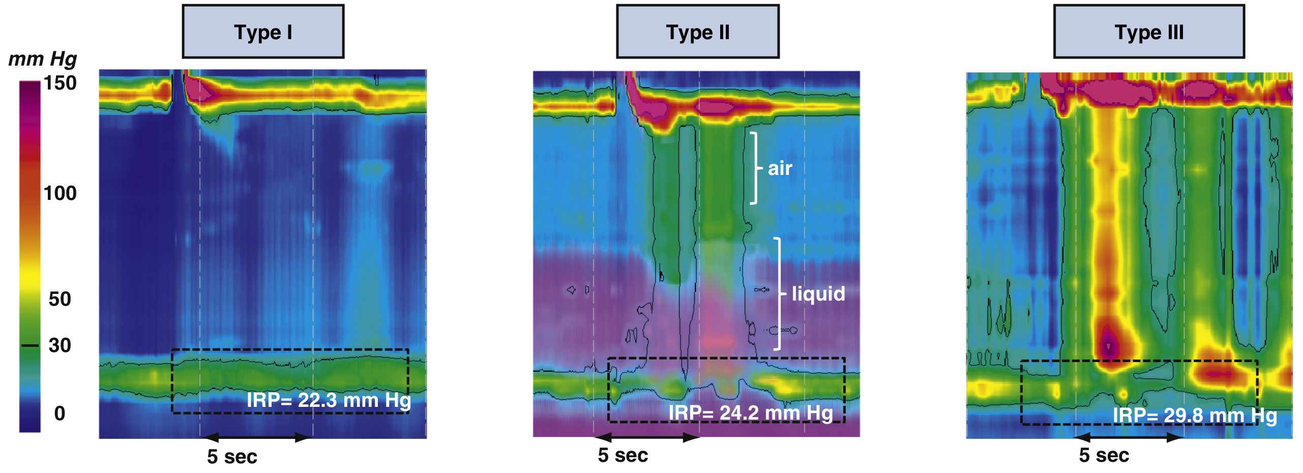
Fig. 44.14 - Sleisenger & Fordtran's: Type I shows quiet aperistaltic esophagus; Type II shows pan-esophageal pressurization trapping air and liquid; Type III shows high-amplitude spastic contractions with elevated IRP
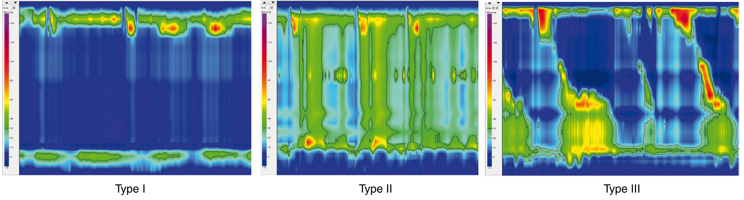
Figure 83.5 (Sabiston Textbook of Surgery): Achalasia types according to the Chicago Classification
How Subtype Influences Treatment Choice and Prognosis
The significance of subtyping lies entirely in how differently the subtypes respond to the available therapies. The treatment options are:
| Treatment | Mechanism |
|---|---|
| Pharmacologic (nitrates, CCa blockers, sildenafil) | Reduce LES pressure transiently |
| Botulinum toxin injection | Blocks ACh release from presynaptic terminals; temporary |
| Pneumatic dilation (PD) | Disrupts circular muscle of LES via 3.0-4.0 cm balloon |
| Laparoscopic Heller myotomy (LHM) + partial fundoplication | Surgical division of LES muscle |
| Peroral endoscopic myotomy (POEM) | Endoscopic submucosal myotomy; no fundoplication |
Type I (Classic Achalasia)
- Treatment: LHM with partial fundoplication (Dor or Toupet) or POEM are the standard approaches. Pneumatic dilation is less effective here compared to Type II.
- Prognosis: Intermediate. Response to therapy is generally good but not as reliable as Type II. The dilated, atonic esophagus may not recover normal dimensions even after successful LES disruption.
Type II (Achalasia with Compression)
- Treatment: This subtype has the best prognosis and responds to virtually all modalities. The pivotal European RCT (multicenter) found that pneumatic dilation achieved 100% efficacy for Type II versus 93% for LHM (p = 0.03). Given comparable perforation risk (~1%) and significantly lower cost, pneumatic dilation is the preferred initial treatment for Type II achalasia.
- POEM is equally effective and a valid alternative.
- Prognosis: Best of the three subtypes. Excellent response rates with PD, LHM, and POEM.
Type III (Spastic Achalasia)
- Treatment: This subtype has a unique therapeutic niche for POEM. Because Type III involves spastic contractions that may extend well above the EGJ, the myotomy must be longer (average 16 cm with POEM vs. 8 cm with LHM). POEM is not anatomically limited by the diaphragm and allows extension of the myotomy proximally along the entire spastic segment.
- In a retrospective analysis of 49 Type III patients treated with POEM vs. 26 with LHM, POEM achieved clinical success in 98% vs. 80% with surgical myotomy.
- Both LHM and POEM are effective for Types I and II, but POEM is superior to LHM for Type III (Sabiston).
- Pneumatic dilation is notably least effective for Type III (40% success in one series vs. 86% for LHM).
- Prognosis: Historically the worst prognosis with conventional therapy; substantially improved with POEM.
Summary Table: Subtype, Treatment of Choice, and Prognosis
| Subtype | Best Treatment | 2nd Line | Prognosis |
|---|---|---|---|
| Type I | LHM + fundoplication or POEM | PD (less effective) | Moderate; good with surgery |
| Type II | Pneumatic dilation (1st line), or POEM/LHM | All modalities effective | Best prognosis |
| Type III | POEM (long myotomy, 15-20 cm) | LHM (shorter myotomy) | Worst with conventional Tx; good with POEM |
Note on POEM and Reflux
POEM's major drawback is a ~50% rate of pathologic GERD on pH monitoring (compared to ~9-11% after LHM with partial fundoplication). This must be considered, particularly in young patients, given the long-term risks of GERD - esophagitis, Barrett's esophagus, and adenocarcinoma. Post-POEM patients require long-term PPI therapy and surveillance endoscopy.
Sources: Sleisenger & Fordtran's Gastrointestinal and Liver Disease, 10e; Sabiston Textbook of Surgery, 21e; Clinical Gastrointestinal Endoscopy, 3e; Fischer's Mastery of Surgery, 8e
Question 8: Complications of Achalasia (6 marks)
Complications arise from two main mechanisms: (1) stasis of esophageal contents due to functional obstruction, and (2) sequelae of treatment.
1. Aspiration Pneumonia / Pulmonary Complications
- Retained food and saliva in a dilated esophagus pool overnight and aspirate into the tracheobronchial tree
- Leads to recurrent aspiration pneumonia, lung abscess, bronchiectasis, and progressive pulmonary fibrosis in severe/longstanding cases
- Aspiration is especially common at night (supine position) and in Type I with a massively dilated esophagus
2. Esophageal Squamous Cell Carcinoma (SCC)
- The most feared long-term complication
- Chronic stasis leads to fermentation of retained food, mucosal irritation, and chronic esophagitis, promoting malignant transformation
- Risk of SCC in long-standing achalasia is estimated at 1-33% of patients (up to 15 times the general population)
- SCC typically develops in the mid-esophagus (site of maximal stasis)
- No consensus exists on optimal surveillance endoscopy intervals
3. Adenocarcinoma (Post-treatment)
- Can develop as a consequence of Barrett's esophagus following treatment-related GERD (especially post-POEM or post-LHM without adequate fundoplication)
- Represents a treatment complication rather than a disease-intrinsic complication
4. Megaesophagus / End-Stage Achalasia
- Progressive dilation leads to a massively dilated, tortuous, sigmoid-shaped esophagus
- Esophageal function is irreversibly lost
- Management becomes very challenging; esophagectomy may be the only viable option
- Indications for esophagectomy: pronounced esophageal dilation, severe dysphagia refractory to LES-directed therapy, esophageal sump, peptic stricture not amenable to dilation, recurrent aspiration, and perforation during dilation
5. Esophageal Perforation (Iatrogenic)
- Occurs in approximately 1% of cases with either pneumatic dilation or surgical myotomy
- Perforation during PD is the most feared acute complication of endoscopic treatment
- May require emergency surgical repair or esophageal stenting
6. Gastroesophageal Reflux Disease (GERD) and Peptic Stricture
- Paradoxically, over-treatment (excessive LES disruption without adequate antireflux protection) leads to GERD
- Pathologic acid exposure occurs in ~50% after POEM, ~20% after LHM without fundoplication, and ~9% after LHM with partial fundoplication
- Complications of GERD include: peptic stricture (may require surgical intervention), Barrett's esophagus, and esophageal adenocarcinoma
- All achalasia patients treated with POEM or myotomy should undergo pH testing 3-6 months post-procedure
7. Candida Esophagitis
- Stasis of food in the esophagus creates an ideal environment for fungal overgrowth
- Candida esophagitis is a recognized complication of untreated or poorly treated achalasia
- May also complicate post-treatment surveillance
8. Epiphrenic Diverticulum
- A pulsion diverticulum that can form from the raised intraluminal pressure proximal to the non-relaxing LES
- May cause additional dysphagia, regurgitation, and aspiration
- Treated with diverticulectomy + myotomy (laparoscopic or D-POEM)
Summary of Complications
| Category | Complication |
|---|---|
| Stasis-related | Aspiration pneumonia, megaesophagus, Candida esophagitis, SCC |
| Treatment-related | Perforation (PD/surgery), GERD, Barrett's, adenocarcinoma, peptic stricture |
| Structural | Epiphrenic diverticulum |
Sources: Fischer's Mastery of Surgery, 8e; Clinical Gastrointestinal Endoscopy, 3e; Sleisenger & Fordtran's Gastrointestinal and Liver Disease; Yamada's Textbook of Gastroenterology, 7e
This is a shared conversation. Sign in to Orris to start your own chat.