
Lens related glaucoma refer kansikis clinical ophthalmology and give answers
Reading File
Now let me search the library for Kanski's Clinical Ophthalmology and lens-related glaucoma content.
Finding Sources
Finding Sources
Found it. Let me now read the Kanski lens-related glaucoma section and also search for specific subtypes.
Reading File
Finding Sources
Good, I can see the content starts at line 2001. Let me read more of this section to get all the subtypes.
Reading File
Reading File
I now also need to check the block2 section for any additional lens-related glaucoma content and fetch the key images.
Reading File
Loading Image
Loading Image
I now have all the content needed. The table of contents confirms the three subtypes covered: Phacolytic glaucoma (p.393), Phacomorphic glaucoma (p.393), and Pupillary block from disruption of lens position (p.394). Let me fetch the remaining important image (Fig 11.50 - dislocated lens) as well.
Loading Image
I now have all the content from Kanski's 10th edition. Let me compile the complete, well-structured answer.
Lens-Related Glaucoma
Kanski's Clinical Ophthalmology: A Systematic Approach, 10th Edition (pp. 393-394)
Kanski classifies lens-related glaucoma into three distinct entities:
- Phacolytic glaucoma
- Phacomorphic glaucoma
- Pupillary block from disruption of lens position
1. Phacolytic Glaucoma
Pathogenesis
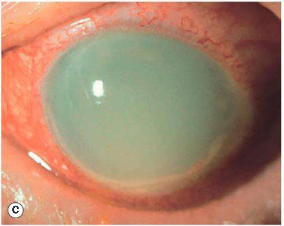
Phacolytic glaucoma is a secondary open-angle glaucoma occurring in association with a hypermature cataract. High molecular-weight lens proteins leak through the intact capsule into the aqueous humour, causing trabecular obstruction. Macrophages that have engulfed these lens proteins may also contribute to the blockage.
Important distinction: Phacolytic glaucoma must NOT be confused with phacogenic (phacoanaphylactic) uveitis, which is an autoimmune granulomatous reaction to exposed lens proteins occurring when the capsule is compromised (not intact).
Diagnosis
- Symptoms: Pain; poor vision due to cataract
- Slit lamp findings:
- Corneal oedema
- Hypermature cataract with a deep anterior chamber
- Large floating white particles in the AC (lens protein + protein-containing macrophages)
- Dense milky aqueous if particles are very abundant
- Pseudohypopyon may form (Fig. 11.48C and D)
- Gonioscopy: Open angle with lens-derived material and inflammatory cells, most prominent inferiorly
Treatment
- Medical IOP control first (to reduce inflammation and corneal oedema)
- Wash out proteinaceous material from the AC
- Cataract extraction (definitive treatment)
- Note: The zonule is likely more fragile than usual - handle with care during surgery
2. Phacomorphic Glaucoma
Pathogenesis
Phacomorphic glaucoma is an acute secondary angle-closure glaucoma precipitated by an intumescent (swollen) cataractous lens. The mechanism is:
- Equatorial age-related lens growth slackens the suspensory ligament → lens moves anteriorly
- Anteroposterior growth increases iridolenticular contact
- This potentiates pupillary block and iris bombé
Diagnosis
- Presentation similar to acute primary angle-closure glaucoma (PACG): shallow AC and mid-dilated pupil
- A dense white (intumescent) cataract is evident
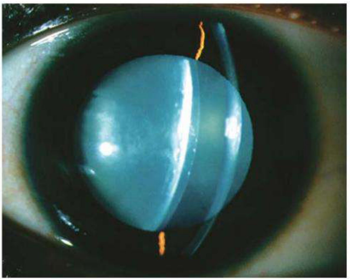
- Fellow eye: More likely in eyes with shorter axial length and shallower AC; the fellow eye may paradoxically show a deep AC and open angle
- Anterior segment OCT or US biomicroscopy may be useful
Treatment
- Medical treatment initially similar to acute PACG
- Miotics are OMITTED - they increase iris-lens apposition and shift the lens anteriorly
- Dilatation may be helpful but should be used with caution
- Systemic hyperosmotic agents may be required more commonly than in PACG
- Laser iridotomy - may be worthwhile but often not possible (corneal oedema or lens-cornea proximity) or ineffective
- Laser iridoplasty - useful temporizing measure
- Cataract extraction - definitive treatment, ideally after IOP is normalized and the eye is quiet
- Surgery is difficult and carries a higher risk of complications
- Consider a similar prophylactic procedure for the fellow eye
3. Pupillary Block from Disruption of Lens Position
Causes
- Blunt ocular trauma (even relatively trivial)
- Congenitally small lens (microspherophakia), e.g., Weill-Marchesani syndrome
Pathogenesis
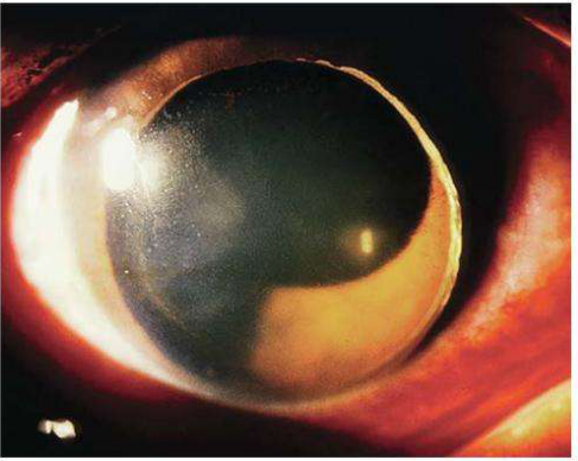
- Dislocation may be into the AC
- Zonules may be stretched, or only partial disruption with the intact part acting as a hinge
- The lens may remain fully or partially in the posterior chamber
- Vitreous herniation may be contributory
Diagnosis
- A lens fully or partially dislocated into the anterior chamber is usually evident on slit lamp
- Acute pupillary block causes sudden severe IOP elevation with visual impairment
- Ultrasound biomicroscopy is often diagnostic
Treatment
This is an ophthalmic emergency - prolonged lenticular corneal contact, especially with high IOP, can cause permanent endothelial damage.
- IOP reduction with osmotic agents (reduce vitreous volume) - urgent
- Initial repositioning attempt: Patient adopts supine posture with pupil dilated → attempt to reposition lens in posterior chamber → miotic can then be used with caution
- Bilateral laser iridotomy may provide extended control in some cases
- Definitive treatment: Surgical lens extraction
- An anterior chamber IOL, iris-fixated IOL, or sclera-fixated IOL will be necessary
Summary Comparison Table
| Feature | Phacolytic | Phacomorphic | Pupillary Block (Dislocation) |
|---|---|---|---|
| Type | Secondary open-angle | Secondary angle-closure | Secondary angle-closure |
| Lens status | Hypermature (intact capsule) | Intumescent cataract | Dislocated/subluxated |
| AC depth | Deep | Shallow | Variable |
| Mechanism | Lens protein + macrophage trabecular blockage | Pupillary block/iris bombé from swollen lens | Direct pupillary block by displaced lens |
| Key finding | Floating white particles, pseudohypopyon | Dense white cataract, iris bombé | Visible dislocation on slit lamp |
| Definitive Rx | Cataract extraction | Cataract extraction | Surgical lens extraction + IOL |
| Special caution | Fragile zonules at surgery | Avoid miotics | Urgent - risk of corneal endothelial damage |
Source: Kanski's Clinical Ophthalmology: A Systematic Approach, 10th Edition, pp. 393-394
Treatment for snake bite
Reading File
This is a broad clinical topic. Let me search the library and PubMed in parallel.
Finding Sources
Finding Sources
Searching PubMed
snakebite envenomation treatment management
Excellent - Tintinalli's Emergency Medicine and Sabiston Surgery have the most relevant content. Let me read both simultaneously.
Reading File
Reading File
I have very rich content. Let me also read the Tintinalli section from the beginning (pre-2820) to get the full clinical picture including initial assessment and types of snakes, plus the Elapid treatment section.
Reading File
Reading File
I now have comprehensive content from both Tintinalli's Emergency Medicine and Sabiston's Surgery. Let me compile the full, authoritative answer.
Treatment of Snakebite
Sources: Tintinalli's Emergency Medicine (Comprehensive Study Guide) & Sabiston Textbook of Surgery, 7th Edition
Overview
Venomous snakebites are a medical emergency. Mortality with modern treatment is <0.5% (compared to ~25% historically), largely due to antivenom availability and advances in critical care. Venomous snakes fall into two major groups:
| Group | Examples | Venom Type | Predominant Injury |
|---|---|---|---|
| Crotalinae (Pit Vipers) | Rattlesnakes, copperheads, water moccasins | Cytotoxic, hemotoxic | Local tissue necrosis + coagulopathy |
| Elapidae (Coral snakes, cobras, mambas) | Coral snake, king cobra, mamba | Neurotoxin | Neuromuscular paralysis |
Up to 25% of crotaline bites are "dry bites" - no venom is injected.
STEP 1: First Aid / Pre-Hospital
DO these:
- Remove jewelry and tight clothing from the affected extremity (anticipate swelling)
- Immobilize and splint the limb in a functional position (reduces discomfort; may slow venom spread)
- Limit exertion - especially walking for lower extremity bites
- Transport to a specialized centre with snakebite experience as soon as possible
- Do NOT wash the bite wound if in Australia/PNG - a venom identification swab kit may be used
DO NOT do these (historical treatments to AVOID):
- No incision and suction - not recommended since 1980; only tiny amounts of venom extracted and risk of injury to vessels/tendons/nerves
- No ice or cryotherapy - associated with a high rate of amputation
- No tourniquets or ligation
- No massage of the wound
- No electrical current (some have tried outboard motor generators - ineffective and dangerous)
Pressure bandage / constrictor band is species-specific:
- Elapid bites (neurotoxic - e.g., Australia, coral snakes): Apply pressure bandage between wound and heart at ~55 mmHg - shown to reduce systemic toxicity
- Crotalid bites (US pit vipers): Constrictor band concentrates venom locally → increased necrosis - DO NOT use
STEP 2: Emergency Department Assessment
History and Identification
- Do not risk a second bite to catch the snake; do not handle dead snakes (reflexive bites possible)
- Contact regional poison control for identification, expected toxicity, and antivenom location
- Document time of bite, species if known, first aid given
Clinical Assessment
Local signs:
- Pain, edema, erythema at bite site
- Distance between fang marks correlates with snake size
- Mark the advancing border of edema with a pen every 30 minutes
Systemic signs:
- Muscle twitching, perioral paresthesias, metallic taste
- Crotalid: hypovolemia, coagulopathy, hemolysis
- Elapid: confusion, muscle spasm, nausea/vomiting, blurred vision, speech difficulty, respiratory failure
Investigations
- CBC, coagulation profile (PT, aPTT, fibrinogen), blood type and crossmatch
- Metabolic panel, renal function
- Repeat labs every 4 hours or after each antivenom course
STEP 3: Antivenom - The Definitive Treatment
Antivenom is the cornerstone of treatment. Administer in a critical care setting (ED or ICU) under direct physician supervision with resuscitative drugs (including epinephrine) and equipment immediately available.
Indications for Antivenom
- Any evidence of local envenomation
- Worsening significant thrombocytopenia (<100,000/μL) or hypofibrinogenemia (<100 mg/dL)
- Progressive edema despite initial treatment
- Systemic envenomation signs
Crotalid (Pit Viper) Antivenom
- FabAV (Crotalidae Polyvalent Immune Fab, Ovine) or equivalent
- Goal: establish initial control = cessation of progression of all components - local effects, systemic effects, and coagulopathy
- Additional doses if condition worsens
- FabAV is also effective for copperhead envenomation where progression occurs
- Measure limb circumference at multiple sites above and below bite every 30 minutes as guide
Elapid (Coral Snake) Antivenom
- Neurotoxicity onset: immediate (15-30 min for mamba/Australian brown snake) or delayed (2-5 hours for coral snakes)
- No longer any antivenom available for North American coral snakes - local zoos and other countries may help for exotic species
- Neurotoxic symptoms: confusion, muscle spasm, respiratory failure, cranial nerve palsies
Antivenom Reactions
- Incidence of acute reactions to modern antivenoms is low
- If allergic reaction occurs: stop infusion immediately, administer H1 and H2 antihistamines, have epinephrine ready for anaphylaxis
STEP 4: Supportive Care
- Fluid resuscitation with isotonic fluids for hypovolemia; vasopressors for refractory hypotension
- Blood component replacement (only if active bleeding AND after antivenom has been given - antivenom is the best treatment for hematologic abnormalities)
- Wound care: clean wound, tetanus prophylaxis
- Pain management
STEP 5: Compartment Syndrome Management
A critical complication - venom injected directly into a compartment causes increased pressure.
| Step | Action |
|---|---|
| 1 | Measure intracompartmental pressure |
| 2 | If not elevated: continue standard management |
| 3 | If signs present AND pressure >30 mmHg: elevate limb + mannitol 1-2 g/kg IV over 30 min + additional antivenom over 60 min simultaneously |
| 4 | If elevated pressure persists another 60 min: consider fasciotomy |
Key principle: The most effective treatment for snakebite-associated compartment syndrome is neutralizing the venom with antivenom - not fasciotomy. Antivenom reduces compartment pressure. Prophylactic fasciotomy has no role. Fasciotomy may actually increase severity of local tissue loss.
STEP 6: Surgical Considerations (Crotalid)
- Wound debridement may be required for necrotic tissue
- Fasciotomy only when compartment pressures do not respond to antivenom and mannitol
- Post-fasciotomy: consider negative-pressure wound therapy
- Do NOT do elective early debridement of tissue that may be salvageable with antivenom
Summary Algorithm
Snakebite
↓
First Aid: Remove jewelry, immobilize, limit exertion
↓
Transport to ER → Identify snake type, call poison control
↓
Assess: Local + systemic signs, serial labs
↓
Dry bite (no signs)? Yes → Observe 8-12h, discharge if no progression
↓ No
Antivenom (species-specific) → critical care setting
↓
Monitor progression (mark edema, serial labs every 4h)
↓
Compartment syndrome? → Mannitol + more antivenom → Fasciotomy if unresponsive
↓
Supportive: Fluids, vasopressors, blood products if needed
Tintinalli's Emergency Medicine: A Comprehensive Study Guide - Ch. 212 | Sabiston Textbook of Surgery, 7th Ed. - Ch. 44
This is a shared conversation. Sign in to Orris to start your own chat.