Here is your complete, MUHS exam-oriented, Apurba Shastri-style answer covering all the topics you listed. Each section is exam-ready with tables, mnemonics, and key diagrams in ASCII/text format.
🧫 MEDICAL MYCOLOGY - COMPLETE MUHS EXAM NOTES
2nd MBBS Microbiology | Based on Apurba Shastri & Standard Textbooks
SECTION 1: CLASSIFICATION OF MEDICALLY IMPORTANT FUNGI
A. MORPHOLOGICAL CLASSIFICATION
Fungi are classified morphologically based on their physical form in culture and tissue:
┌─────────────────────────────────────────────────────────────────────┐
│           MORPHOLOGICAL CLASSIFICATION OF FUNGI                      │
├──────────────────┬───────────────────────────────┬──────────────────┤
│ MORPHOLOGY       │ DESCRIPTION                   │ EXAMPLE          │
├──────────────────┼───────────────────────────────┼──────────────────┤
│ YEASTS           │ Unicellular, reproduce by      │ Cryptococcus     │
│                  │ budding; round/oval cells      │ neoformans       │
├──────────────────┼───────────────────────────────┼──────────────────┤
│ MOULDS (Hyphae)  │ Multicellular; grow as hyphae  │ Aspergillus      │
│                  │ - Septate hyphae               │ fumigatus        │
│                  │ - Aseptate/coenocytic hyphae   │ Mucor spp.       │
├──────────────────┼───────────────────────────────┼──────────────────┤
│ DIMORPHIC FUNGI  │ Yeast in tissue (37°C);        │ Histoplasma      │
│                  │ Mould in environment (25°C)    │ capsulatum       │
├──────────────────┼───────────────────────────────┼──────────────────┤
│ YEAST-LIKE FUNGI │ Predominantly yeast but        │ Candida          │
│                  │ produce pseudohyphae/hyphae    │ albicans         │
└──────────────────┴───────────────────────────────┴──────────────────┘
Exam Mnemonic for Dimorphic Fungi: "BASHED"
Blastomyces dermatitidis | Aspergillus (not truly dimorphic) | Sporothrix schenckii | Histoplasma capsulatum | Epidermophyton (actually dermatophyte) | Dimorphic fungi = Coccidioides + Paracoccidioides
True Dimorphic Fungi (MUHS Favourite):
- Histoplasma capsulatum
- Coccidioides immitis
- Blastomyces dermatitidis
- Paracoccidioides brasiliensis
- Sporothrix schenckii
B. MEDICAL / CLINICAL CLASSIFICATION OF FUNGI
┌─────────────────────────────────────────────────────────────────────────────┐
│                MEDICAL CLASSIFICATION OF FUNGI                               │
├──────────────────────┬──────────────────────────────────┬───────────────────┤
│ CATEGORY             │ DESCRIPTION                      │ EXAMPLE           │
├──────────────────────┼──────────────────────────────────┼───────────────────┤
│ SUPERFICIAL MYCOSES  │ Outermost keratinized layers of  │ Malassezia furfur  │
│                      │ skin/hair; no host reaction       │ (Pityriasis       │
│                      │                                  │ versicolor)       │
├──────────────────────┼──────────────────────────────────┼───────────────────┤
│ CUTANEOUS MYCOSES    │ Deeper epidermis, hair, nails;   │ Trichophyton       │
│                      │ keratinophilic fungi              │ rubrum             │
├──────────────────────┼──────────────────────────────────┼───────────────────┤
│ SUBCUTANEOUS MYCOSES │ Dermis, subcutaneous tissue,     │ Sporothrix         │
│                      │ muscle, fascia                   │ schenckii          │
├──────────────────────┼──────────────────────────────────┼───────────────────┤
│ SYSTEMIC MYCOSES     │ Deep-seated infection of viscera │ Histoplasma        │
│ (Primary)            │ in immunocompetent hosts         │ capsulatum         │
├──────────────────────┼──────────────────────────────────┼───────────────────┤
│ SYSTEMIC MYCOSES     │ Occur in immunocompromised       │ Candida albicans,  │
│ (Opportunistic)      │ hosts; otherwise non-pathogenic  │ Aspergillus,       │
│                      │                                  │ Cryptococcus       │
└──────────────────────┴──────────────────────────────────┴───────────────────┘
SECTION 2: SUBCUTANEOUS MYCOSES
Subcutaneous mycoses involve dermis, subcutaneous tissue, fascia, and bone. Organisms are introduced by traumatic implantation (thorns, splinters, soil).
┌────────────────────────────────────────────────────────────────────────┐
│                   SUBCUTANEOUS MYCOSES                                  │
├────────────────────┬───────────────────────────────────────────────────┤
│ DISEASE            │ CAUSATIVE AGENT(S)                                │
├────────────────────┼───────────────────────────────────────────────────┤
│ Mycetoma           │ Eumycetoma: Madurella mycetomatis,                │
│                    │ Scedosporium apiospermum                           │
│                    │ Actinomycetoma: Nocardia, Streptomyces             │
├────────────────────┼───────────────────────────────────────────────────┤
│ Chromoblastomycosis│ Fonsecaea pedrosoi, Phialophora verrucosa,        │
│                    │ Cladosporium carrionii                             │
├────────────────────┼───────────────────────────────────────────────────┤
│ Sporotrichosis     │ Sporothrix schenckii (dimorphic)                  │
├────────────────────┼───────────────────────────────────────────────────┤
│ Phaeohyphomycosis  │ Dematiaceous (melanized) moulds - Exophiala,      │
│                    │ Alternaria, Bipolaris spp.                         │
└────────────────────┴───────────────────────────────────────────────────┘
SECTION 3: MYCETOMA
Definition
Mycetoma is a chronic, granulomatous subcutaneous infection characterized by:
- Painless subcutaneous swelling
- Multiple draining sinuses
- Discharge of grains/granules
- Spread to underlying bone and muscle
Also called "Madura Foot" (most commonly affects foot/hand; named after Madura, India).
Causative Agents (Enumerate)
A. Eumycetoma (Fungal - True Fungal Grain):
| Grain Color | Organism |
|---|
| Black grain | Madurella mycetomatis (most common worldwide) |
| Black grain | Madurella grisea |
| White/pale grain | Scedosporium apiospermum (most common USA) |
| White grain | Acremonium falciforme |
| White grain | Pseudallescheria boydii |
| Yellow grain | Aspergillus nidulans |
| Red grain | Neotestudina rosatii |
B. Actinomycetoma (Bacterial - Branching filamentous bacteria):
| Grain Color | Organism |
|---|
| Yellow grain | Nocardia brasiliensis, Nocardia asteroides |
| White grain | Streptomyces somaliensis |
| Red grain | Actinomadura madurae |
| Black grain | Madurella species |
Key Differentiator - MUHS Exam:
- Eumycetoma: broader hyphae (2-5 μm), septate, eukaryotic
- Actinomycetoma: thinner filaments (<1 μm), prokaryotic - respond to antibiotics
Eumycetoma - Laboratory Diagnosis
1. Specimen Collection
- Pus from sinus tracts
- Tissue biopsy (most reliable)
- Aspirated material from unopened lesion
2. Gross Examination - Look for GRAINS
┌──────────────────────────────────────────────────────────────┐
│              GRAIN CHARACTERISTICS                            │
├────────────────┬─────────────────────────────────────────────┤
│ Eumycetoma     │ Hard, large grains (0.5-2 mm)               │
│ Grain          │ Black (dematiaceous) or white (hyaline)     │
│                │ Composed of broad septate hyphae            │
├────────────────┼─────────────────────────────────────────────┤
│ Actinomycetoma │ Smaller, soft grains                        │
│ Grain          │ Composed of narrow bacterial filaments      │
└────────────────┴─────────────────────────────────────────────┘
3. Direct Microscopy (KOH preparation)
- Crush grain on glass slide in 10% KOH
- Eumycetoma: broad, septate hyphae 2-5 μm in diameter; may show melanin (brown pigment) in black grain fungi
- Hyphae lined with Splendore-Hoeppli material (eosinophilic hyaline sheath) on Gram/H&E
4. Histopathology (Tissue Section)
- Granule surrounded by neutrophilic infiltrate and outer zone of granulomatous reaction (epithelioid cells, giant cells)
- PAS stain or GMS (Gomori Methenamine Silver) - fungal cell walls stain positive
- Black grains: melanin pigment visible on H&E
5. Culture
- Medium: Sabouraud Dextrose Agar (SDA) at 25-30°C
- Growth may take 4-8 weeks
- Species identified by colony morphology + microscopy of reproductive structures
6. Radiological (X-Ray/MRI)
- Bone involvement: cortical erosion, sclerosis, "bag of marbles" or "dot in circle" sign on MRI
7. Molecular Methods
- PCR/ITS sequencing for species identification when culture fails
SECTION 4: DERMATOPHYTES ⭐⭐ (Most Important MUHS Topic)
Definition
Dermatophytes are a group of closely related filamentous fungi capable of invading keratinized tissues (skin, hair, nails). They produce keratinases that digest keratin.
Three genera:
          DERMATOPHYTES
               │
    ┌──────────┼──────────┐
    │          │          │
Trichophyton  Epidermophyton  Microsporum
(skin, hair,  (skin, nails   (skin, hair
nails)        only)          only)
20+ species   1-2 species    Many species
Classification of Dermatophytes
A. By Genus
| Genus | Species (Examples) | Infects |
|---|
| Trichophyton | T. rubrum (MC worldwide), T. mentagrophytes, T. violaceum, T. tonsurans, T. verrucosum | Skin, Hair, Nails |
| Microsporum | M. canis, M. audouinii, M. gypseum | Skin, Hair (not nails) |
| Epidermophyton | E. floccosum | Skin, Nails (not hair) |
Mnemonic: "THE MAN"
Trichophyton - Hair, skin, nails
Epidermophyton - Nails and skin (Not hair)
Microsporum - Mainly skin & hair (Avoid Nails)
B. Ecological Classification
| Category | Habitat | Inflammatory Reaction | Example |
|---|
| Geophilic | Soil | Strong (acute) | M. gypseum |
| Zoophilic | Animals | Moderate-severe | M. canis, T. verrucosum |
| Anthropophilic | Humans | Mild (chronic) | T. rubrum, T. violaceum |
Pathogenicity
- Infection route: Direct/indirect contact; fomites (infected towels, floors)
- Keratinases & proteases degrade keratin - allowing invasion
- Mannan in cell wall inhibits cell-mediated immunity
- Only outermost keratinized layers invaded - never penetrates living tissue in immunocompetent hosts
- Hypersensitivity reaction (dermatophytid/"id reaction") - distant vesicular eruption
Clinical Forms (Tineas):
┌──────────────────────────────────────────────────────────────────┐
│                   TINEA (DERMATOPHYTOSIS)                         │
├─────────────────────┬────────────────────┬──────────────────────┤
│ CLINICAL NAME       │ SITE               │ MAIN ORGANISM        │
├─────────────────────┼────────────────────┼──────────────────────┤
│ Tinea capitis       │ Scalp/hair         │ Microsporum, Tricho. │
│ Tinea corporis      │ Body (ringworm)    │ T. rubrum, M. canis  │
│ Tinea pedis         │ Foot (Athlete's   │ T. rubrum, T. ment.  │
│                     │ foot)              │                      │
│ Tinea unguium       │ Nails (Onycho-    │ T. rubrum            │
│ (onychomycosis)     │ mycosis)           │                      │
│ Tinea cruris        │ Groin (Jock itch) │ T. rubrum, E. flocc. │
│ Tinea barbae        │ Beard region      │ T. verrucosum        │
│ Tinea manuum        │ Hands             │ T. rubrum            │
│ Tinea favosa        │ Scalp (favus)     │ T. schoenleinii      │
└─────────────────────┴────────────────────┴──────────────────────┘
Dermatophytosis - Laboratory Diagnosis ⭐⭐
Step 1: Specimen Collection
- Skin: Scrapings from active edge of lesion (not center)
- Hair: Epilated/broken hair stubs from margin; Fluorescent hair under Wood's lamp
- Nails: Scrapings from subungual debris + clippings
Step 2: Direct Microscopy (KOH Preparation) ⭐
┌──────────────────────────────────────────────────────────────────────┐
│             KOH PREPARATION - METHOD                                  │
├──────────────────────────────────────────────────────────────────────┤
│ 1. Place specimen on glass slide                                       │
│ 2. Add 1 drop of 10-20% KOH (+ DMSO for rapid clearing)              │
│ 3. Gently heat, apply coverslip                                        │
│ 4. Wait 15-30 min for clearing                                         │
│ 5. Examine under 10x then 40x                                          │
└──────────────────────────────────────────────────────────────────────┘
Microscopic Findings in Dermatophytosis:
| Specimen | KOH Finding |
|---|
| Skin scraping | Septate, branching hyphae of uniform width (2-4 μm) running through keratinocytes ("broken spaghetti" appearance) |
| Scalp - Endothrix (T. violaceum, T. tonsurans) | Spores INSIDE hair shaft; no fluorescence with Wood's lamp |
| Scalp - Ectothrix (M. canis, M. audouinii) | Chains of spores OUTSIDE hair shaft (arthroconidia around hair); fluoresce green under Wood's lamp |
| Nail | Hyphae and arthroconidia in nail keratin |
| Favus (T. schoenleinii) | Air spaces + tortuous hyphae within hair shaft; Wood's lamp - dull green fluorescence |
ENDOTHRIX vs ECTOTHRIX (MUHS Exam Distinction)

  ENDOTHRIX                        ECTOTHRIX
  ─────────                        ─────────
  Spores INSIDE hair shaft         Spores OUTSIDE shaft (sheath)
  No Wood's lamp fluorescence      Green fluorescence (most)
  T. violaceum, T. tonsurans       M. canis, M. audouinii
  Black dot tinea capitis          Gray patch tinea capitis
Step 3: Culture (Gold Standard)
- Medium: Sabouraud Dextrose Agar (SDA) with cycloheximide + chloramphenicol (DTM = Dermatophyte Test Medium)
- Temperature: 25-28°C
- Duration: 1-4 weeks
- Colony & Microscopy Characteristics:
| Organism | Colony (25°C) | Macroconidia | Microconidia |
|---|
| T. rubrum | White/red reverse | Cigar-shaped | Tear-drop, single |
| T. mentagrophytes | White to cream | Cigar-shaped | Round, clustered (grapes) |
| M. canis | Yellow/orange-reverse | Thick-walled, spindle, rough | Rare |
| E. floccosum | Yellowish-green, powdery | Club-shaped, smooth, no microconidia | Absent |
Exam Key: Epidermophyton has NO microconidia - only club-shaped macroconidia
Step 4: Wood's Lamp Examination (UV 365 nm)
| Organism | Fluorescence |
|---|
| M. canis, M. audouinii | Bright green |
| T. schoenleinii (Favus) | Dull blue-green |
| T. violaceum, T. tonsurans | No fluorescence |
| Pityriasis versicolor | Yellow-green |
Step 5: Hair Perforation Test
- Positive in T. mentagrophytes (perforates hair in vitro)
- Negative in T. rubrum - helps differentiate the two
SECTION 5: HISTOPLASMA CAPSULATUM ⭐⭐
Morphology
Dimorphic fungus:
┌──────────────────────────────────────────────────────────────┐
│              DIMORPHISM OF H. CAPSULATUM                      │
├─────────────────────────────┬───────────────────────────────┤
│ MOULD FORM (25°C / SDA)     │ YEAST FORM (37°C / In tissue) │
├─────────────────────────────┼───────────────────────────────┤
│ Cottony white/brown colony  │ Small oval yeast cells         │
│ Hyphae with:                │ 2-4 μm diameter               │
│ • Macroconidia (tuberculate │ Found INSIDE macrophages       │
│   chlamydospores) - large,  │ (obligate intracellular)       │
│   round, thick-walled,      │ Single narrow-based buds       │
│   with finger-like spines   │                                │
│   ("lollipop conidia")      │                                │
│ • Microconidia (2-5 μm) -   │                                │
│   infective propagules      │                                │
└─────────────────────────────┴───────────────────────────────┘
Growth Characters
- Natural habitat: Soil enriched with bat/bird droppings (Ohio and Mississippi River valleys endemic area)
- SDA at 25°C: Slow growing (2-6 weeks); white cottony mould
- Tuberculate macroconidia are PATHOGNOMONIC (diagnostic)
- At 37°C on BHI (Brain Heart Infusion agar): small budding yeasts
Pathogenesis
- Inhalation of microconidia from contaminated soil
- Microconidia deposited in alveoli → transform to yeast form within hours
- Phagocytosed by alveolar macrophages but resist killing:
- Modulate phagosomal pH to 6.0-6.5 (inhibits lysosomal enzymes)
- Conceal β-glucans (evades Dectin-1 receptor)
- Express superoxide dismutase and catalase (resist ROS)
- Acquire iron from host macrophage environment
- Yeasts multiply inside macrophages → spread via lymphatics → hematogenous dissemination
- In immunocompetent host: granuloma formation controls infection (latent/primary pulmonary)
- In immunocompromised (AIDS, transplant): Progressive Disseminated Histoplasmosis (PDH) - involves liver, spleen, bone marrow, adrenals
Key Feature: H. capsulatum is an obligate intracellular pathogen residing in macrophage phagolysosomes
SECTION 6: OPPORTUNISTIC MYCOSES ⭐
Definition
Opportunistic mycoses are fungal infections that primarily affect immunocompromised individuals (otherwise normal hosts are resistant).
Risk Factors (Host Factors)
┌──────────────────────────────────────────────────────────┐
│         RISK FACTORS FOR OPPORTUNISTIC MYCOSES            │
├──────────────────────────────────────────────────────────┤
│ • HIV/AIDS (CD4 < 200 cells/μL)                          │
│ • Prolonged corticosteroid therapy                       │
│ • Chemotherapy / Cytotoxic drugs                         │
│ • Organ transplantation (BMT/Solid organ)                │
│ • Neutropenia (WBC < 500/mm³)                           │
│ • Hematological malignancies (Leukemia, Lymphoma)        │
│ • Broad-spectrum antibiotic therapy                      │
│ • Central venous catheters                               │
│ • Total parenteral nutrition                             │
│ • Premature infants / Extremes of age                    │
│ • Diabetes mellitus (especially Mucormycosis)            │
└──────────────────────────────────────────────────────────┘
Major Opportunistic Fungi
| Organism | Disease | Host Risk Factor |
|---|
| Candida albicans | Candidiasis (oral, vaginal, systemic) | Antibiotics, immunosuppression |
| Cryptococcus neoformans | Cryptococcal meningitis | HIV/AIDS (CD4<100) |
| Aspergillus fumigatus | Invasive aspergillosis | Neutropenia |
| Mucor/Rhizopus spp. | Mucormycosis | Diabetes (DKA), neutropenia |
| Pneumocystis jirovecii | PCP pneumonia | AIDS |
| Fusarium spp. | Fusariosis | Neutropenia, burns |
| Trichosporon spp. | Disseminated trichosporonosis | Neutropenia |
SECTION 7: CANDIDA ALBICANS ⭐⭐
Morphology
Candida albicans is a yeast-like fungus with the unique ability to form multiple morphological forms:
┌───────────────────────────────────────────────────────────────┐
│                   MORPHOLOGY OF C. ALBICANS                    │
├────────────────────┬──────────────────────────────────────────┤
│ FORM               │ DESCRIPTION                              │
├────────────────────┼──────────────────────────────────────────┤
│ Yeast (blastoconia)│ Oval, 3-5 μm, thin-walled, budding      │
├────────────────────┼──────────────────────────────────────────┤
│ Pseudohyphae       │ Elongated buds that remain attached;     │
│                    │ constriction at septa                     │
├────────────────────┼──────────────────────────────────────────┤
│ True hyphae        │ Parallel walls, no constriction           │
├────────────────────┼──────────────────────────────────────────┤
│ Germ tube          │ Short, narrow tube emerging from yeast    │
│                    │ (no constriction at base) - seen at 37°C  │
│                    │ in serum after 2-3 hours                  │
├────────────────────┼──────────────────────────────────────────┤
│ Chlamydoconidia    │ Large (8-12 μm), thick-walled, terminal  │
│                    │ spores on cornmeal agar - PATHOGNOMONIC   │
│                    │ for C. albicans                           │
└────────────────────┴──────────────────────────────────────────┘
Reynolds-Braude Phenomenon (Germ Tube Test) ⭐
Definition: When Candida albicans is incubated in human/animal serum at 37°C for 2-3 hours, it forms germ tubes - short hyphal outgrowths with no constriction at their base.
Significance:
- Presumptive identification of C. albicans
- Positive in ~95% of C. albicans strains
- C. dubliniensis also positive (differentiated by growth at 45°C - C. albicans grows, C. dubliniensis does not)
- Quick, cheap, simple test done directly from colony
GERM TUBE vs PSEUDOHYPHA

 GERM TUBE             PSEUDOHYPHA
 ─────────             ───────────
 No constriction       Constriction present
 at base               at each septum

     ┌───────                ┌───────
  ○──┤       vs          ○─┤ ────)─────
     └───────                └───────
Laboratory Diagnosis of Candidiasis ⭐⭐
Specimen
- Oral swab, vaginal swab, urine, blood, BAL, CSF (meningitis), tissue biopsy
1. Direct Microscopy
- KOH prep / Gram stain (Candida is Gram-positive)
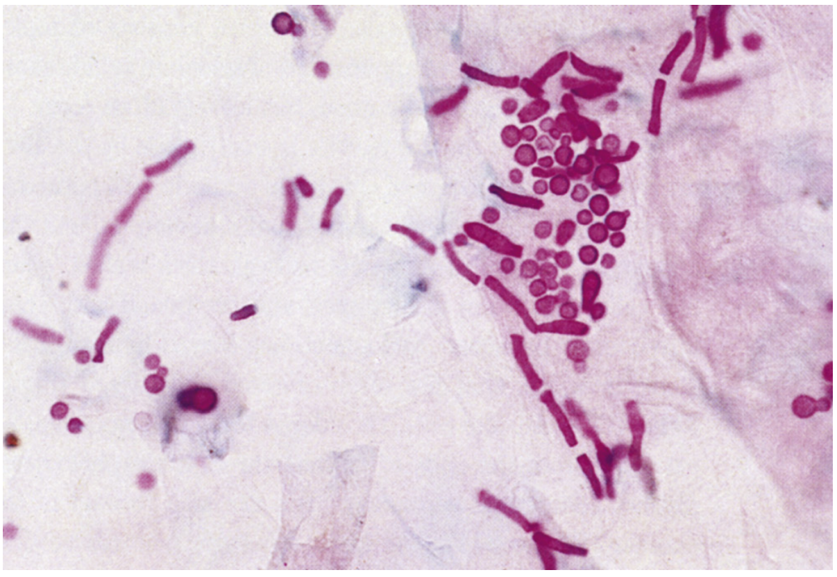
- See: budding yeast cells + pseudohyphae (this combination is diagnostic)
- In tissue: PAS, GMS staining
2. Culture
- SDA at 25°C and 37°C: Cream-colored, smooth, pasty, yeast-like colonies with "yeast odor"
- Chromagar Candida: Differentiate species by color:
- C. albicans → GREEN colonies
- C. tropicalis → Blue-grey
- C. krusei → Pink, rough
- C. glabrata → Purple/white, tiny
3. Germ Tube Test (Reynolds-Braude Phenomenon)
- Incubate colony in human serum at 37°C for 2-3 hours
- Positive (germ tube formation) = C. albicans
4. Chlamydoconidia on Cornmeal Agar
- Inoculate on Cornmeal + Tween-80 agar, 25°C, 48-72h
- C. albicans: Terminal chlamydoconidia + pseudohyphae = pathognomonic
5. Biochemical Tests
- Carbohydrate assimilation/fermentation (API 20C AUX system)
- C. albicans: Ferments glucose, maltose; does NOT ferment lactose
6. Serology
- Mannan antigen detection (ELISA)
- Anti-mannan antibodies
- Beta-D-glucan assay (non-specific for all fungi)
7. Molecular
- PCR, MALDI-TOF MS (gold standard for rapid ID in BSI)
SECTION 8: ASPERGILLOSIS IN HUMANS ⭐
Causative Organisms
- Aspergillus fumigatus (most common, 70-80%)
- A. flavus, A. niger, A. terreus
Morphology
- Septate hyphae with acute angle branching (45°) - diagnostic feature
- Conidiophore: swells into a vesicle → bears phialides → conidia ("aspergillum" = holy water sprinkler - hence the name)
ASPERGILLUS CONIDIOPHORE STRUCTURE:
(Diagnostic on microscopy)

        ○○○○○○○○○  ← Conidia (spores)
        ||||||||
        ||||||||   ← Phialides (bottle-shaped cells)
       [vesicle]   ← Swollen tip
         |
         |         ← Conidiophore (stalk)
      ───────
      Foot cell
Clinical Spectrum
┌───────────────────────────────────────────────────────────────────────┐
│                   CLINICAL FORMS OF ASPERGILLOSIS                      │
├──────────────────────┬────────────────────────────────────────────────┤
│ TYPE                 │ CLINICAL FEATURES & HOST                       │
├──────────────────────┼────────────────────────────────────────────────┤
│ Allergic Broncho-    │ Asthma, eosinophilia, bronchiectasis           │
│ pulmonary Aspergillo-│ Immunocompetent with allergic predisposition   │
│ sis (ABPA)           │ IgE elevated, fleeting pulmonary infiltrates   │
├──────────────────────┼────────────────────────────────────────────────┤
│ Aspergilloma         │ "Fungus ball" in pre-existing cavity (TB cavity│
│ (Mycetoma)           │ or bronchiectasis); "monod sign" on X-ray      │
│                      │ (crescent of air around fungal ball)           │
├──────────────────────┼────────────────────────────────────────────────┤
│ Invasive Pulmonary   │ Neutropenic patients; fever, cough, haemoptysis│
│ Aspergillosis (IPA)  │ Angioinvasion → infarction; high mortality     │
├──────────────────────┼────────────────────────────────────────────────┤
│ Chronic Necrotizing  │ Semi-invasive; elderly/mildly immunocompromised│
│ Aspergillosis        │                                                 │
├──────────────────────┼────────────────────────────────────────────────┤
│ Disseminated         │ Brain, liver, kidney, heart affected; profound │
│ Aspergillosis        │ immunosuppression                               │
└──────────────────────┴────────────────────────────────────────────────┘
Laboratory Diagnosis
- Microscopy (BAL/tissue): Septate hyphae with 45° branching, GMS/PAS stain
- Culture: SDA - fast-growing granular colonies; green (A. fumigatus), yellow-green (A. flavus), black (A. niger)
- Galactomannan assay (serum/BAL) - highly specific for Aspergillus
- Beta-D-glucan - non-specific, used as adjunct
- CT Chest: "Halo sign" (early) and "Air crescent sign" (later) in IPA
SECTION 9: CRYPTOCOCCAL MENINGITIS ⭐⭐
Organism: Cryptococcus neoformans
- Encapsulated yeast - hallmark is large polysaccharide capsule
- Habitat: pigeon/bird droppings
- Risk: HIV/AIDS (CD4 < 100 cells/μL)
Laboratory Diagnosis of Cryptococcal Meningitis ⭐⭐
Specimen: CSF (LP), Blood, Urine
1. Direct Microscopy - INDIA INK PREPARATION ⭐
┌─────────────────────────────────────────────────────────────┐
│              INDIA INK PREPARATION                           │
├─────────────────────────────────────────────────────────────┤
│ Principle: India ink particles cannot penetrate the         │
│ polysaccharide capsule → capsule appears as a clear halo   │
│ (NEGATIVE stain)                                            │
│                                                             │
│          ░░░░░░░░░░░░░░░░░░ ← India Ink background         │
│          ░░░░░░░░░░░░░░░░░░                                 │
│          ░░░░[  ●  ]░░░░░░░ ← Clear halo = capsule         │
│          ░░░░░░░░░░░░░░░░░░   ● = yeast cell               │
│                                                             │
│ Sensitivity: 50-80% in AIDS patients                        │
└─────────────────────────────────────────────────────────────┘
- CSF + drop of India ink on slide → look for encapsulated yeasts with narrow-based buds
- "Ping pong ball in ink" or "halo appearance"
2. Culture
- SDA at 37°C: Mucoid, cream/tan colonies (encapsulation → mucoid appearance)
- Bird seed agar (Niger seed agar): C. neoformans produces brown/dark colonies due to melanin production (phenol oxidase enzyme; unique feature)
- Urease positive (differentiates from Candida)
3. Cryptococcal Antigen Test (CrAg) ⭐⭐
- Latex agglutination test (LAT) or Lateral Flow Assay (LFA) in CSF and serum
- Detects polysaccharide capsular antigen
- Sensitivity >95% in AIDS patients - most reliable test
- Titer correlates with fungal burden; high titer = poor prognosis
4. Biochemical
- Urease positive
- Phenol oxidase positive (melanin on Bird seed agar)
- Does NOT ferment sugars; assimilates inositol
5. Histopathology (Brain Biopsy)
- Mucicarmine stain: Red staining of thick mucoid capsule (pathognomonic)
- Fontana-Masson: Stains melanin
- H&E: "Soap bubble lesions" in brain (Virchow-Robin spaces dilated with gelatinous material)
6. Molecular
- PCR (reference labs); India ink + antigen test sufficient in clinical settings
Treatment (For Completeness)
- Induction: Amphotericin B + Flucytosine (2 weeks)
- Consolidation: Fluconazole 400 mg/day (8 weeks)
- Maintenance: Fluconazole 200 mg/day (lifelong in AIDS)
SECTION 10: FUNGI CAUSING OPPORTUNISTIC INFECTIONS IN HIV
┌──────────────────────────────────────────────────────────────────────────┐
│        OPPORTUNISTIC FUNGI IN HIV/AIDS PATIENTS                           │
├──────────────────────────┬──────────────────────────────────────────────┤
│ ORGANISM                 │ DISEASE / CD4 THRESHOLD                      │
├──────────────────────────┼──────────────────────────────────────────────┤
│ Candida albicans         │ Oral thrush, esophageal candidiasis          │
│                          │ Any CD4, worse when <200                     │
├──────────────────────────┼──────────────────────────────────────────────┤
│ Cryptococcus neoformans  │ Meningitis, fungemia - CD4 < 100/μL         │
├──────────────────────────┼──────────────────────────────────────────────┤
│ Pneumocystis jirovecii   │ PCP - life-threatening pneumonia; CD4 <200  │
├──────────────────────────┼──────────────────────────────────────────────┤
│ Histoplasma capsulatum   │ Disseminated histoplasmosis; CD4 <150       │
├──────────────────────────┼──────────────────────────────────────────────┤
│ Aspergillus fumigatus    │ Invasive aspergillosis (late AIDS)           │
│                          │ CD4 < 50                                     │
├──────────────────────────┼──────────────────────────────────────────────┤
│ Coccidioides immitis     │ Disseminated coccidioidomycosis; endemic     │
│                          │ areas                                         │
├──────────────────────────┼──────────────────────────────────────────────┤
│ Talaromyces (Penicillium)│ Penicilliosis; Southeast Asia               │
│ marneffei                │                                               │
└──────────────────────────┴──────────────────────────────────────────────┘
QUICK REVISION TABLE: MUHS EXAM HIGH-YIELD POINTS
| Topic | Key Point |
|---|
| KOH positive skin = | Septate branching hyphae → Dermatophytes |
| India Ink + clear halo | Cryptococcus neoformans |
| Germ tube in serum | Reynolds-Braude phenomenon → C. albicans |
| Chlamydoconidia on cornmeal agar | C. albicans (pathognomonic) |
| Tuberculate macroconidia | Histoplasma capsulatum |
| 45° angle branching septate hyphae | Aspergillus spp. |
| 90° (right angle) aseptate hyphae | Mucor/Rhizopus (Mucormycosis) |
| Grains in sinus tract | Mycetoma (Madura foot) |
| "Fungus ball" with crescent sign | Aspergilloma |
| Latex agglutination of CSF | Cryptococcal antigen → Cryptococcal meningitis |
| Mucicarmine stain → red capsule | Cryptococcus neoformans |
| Bird seed agar → brown colonies | Cryptococcus neoformans (phenol oxidase) |
| Endothrix infection | T. tonsurans, T. violaceum |
| Ectothrix infection | Microsporum canis (Wood's lamp → green) |
| No microconidia in dermatophyte | Epidermophyton floccosum |
| Splendore-Hoeppli material | Grain in Eumycetoma |
| Obligate intracellular | Histoplasma capsulatum (inside macrophages) |
| ChromAgar green colonies | Candida albicans |
MNEMONICS SUMMARY
| Mnemonic | Meaning |
|---|
| 3 Genera of Dermatophytes | Trichophyton, Epidermophyton, Microsporum = "TEM" |
| Epidermophyton | No hair (E = no Ear? No hair); only skin & nails |
| Dimorphic fungi | "CASH" = Coccidioides, Aspinosus/Acremonium, Sporothrix, Histoplasma (+ Blastomyces, Paracoccidioides) |
| Reynolds-Braude | Reynolds = Rapid germ tube test; Braude = Bloody serum medium (37°C, 2-3h) |
| Cryptococcus CrAg | "CLUE" = Capsule antigen Latex agglutination = Ultra-sensitive in Encapsulated yeast |
Sources: Medical Microbiology 9e (Murray); Sherris & Ryan's Medical Microbiology 8th Ed; Jawetz Melnick & Adelberg Medical Microbiology 28e; Henry's Clinical Diagnosis & Lab Methods; Fitzpatrick's Dermatology; Goldman-Cecil Medicine.