
Ptyriasis versicolor treatment.
pityriasis versicolor treatment antifungal
pityriasis versicolor skin lesions clinical photo
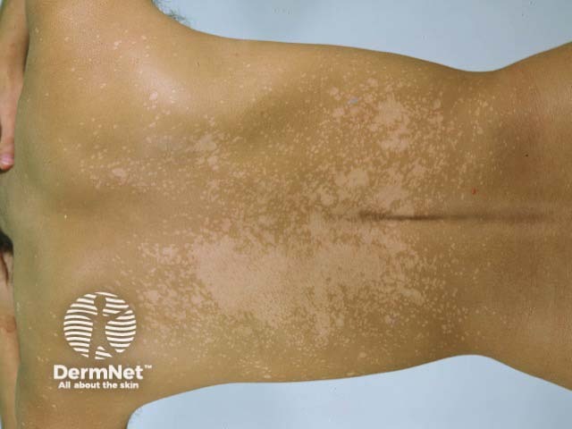
Clinical photography of the back in dorsal/posterior view. The frame shows extensive confluent hypopigmented patches on the upper and mid back with irregular margins and minimal scale. Lesions appear lighter than surrounding skin and coalesce into large hypoesthetic-looking plaques along the scapular region. The overall pattern is diffuse, predominantly on sun-exposed areas of the back, consistent with pityriasis versicolor (tinea versicolor) caused by Malassezia species. There is no erythema, crust, ulceration, or nodularity visible. The skin texture remains intact, and there is no evident lichenification. The color contrast between involved and uninvolved skin is notable, particularly under room lighting; subtle perifollicular fine scaling may be present. This image exemplifies the classic hypopigmented variant rather than hyperpigmented patches. Wood’s lamp examination would be expected to show a coppery yellow-green fluorescence in many cases, aiding diagnosis; KOH preparation may reveal spaghetti and meatballs. Clinically, pityriasis versicolor is common in hot, humid climates and among young adults. Differential considerations include vitiligo, post-inflammatory hypopigmentation, and pityriasis alba. Diagnosis influences management with topical antifungals and, for extensive disease, systemic agents. Educationally, this photo supports recognition, differential diagnosis, and integration of clinical findings with laboratory tests and dermatoscopic correlation.
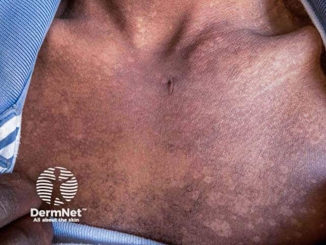
Clinical photography of the anterior trunk in a patient with pityriasis versicolor presenting in the hypopigmented stage. High-resolution, color-balanced digital image captured under standard visible light to document surface skin changes on the chest and upper abdomen with an anterior view. The lesions appear as numerous flat, ill‑defined macules with a pale to slightly hypopigmented hue relative to surrounding skin. The patches are variably coalescent, with fine scale evident at the margins and mild asynchronous tan response in adjacent areas. The distribution favors the central chest and proximal trunk, presenting as asymptomatic patches without erosion or crusting. Dermatoscopic features are not available; the surface is smooth, with subtle textural changes rather than prominent edema or vascular markings. Clinically, pityriasis versicolor is a superficial Malassezia yeast infection that disrupts melanin production, producing hypopigmented or hyperpigmented macules depending on skin type. The image illustrates hypopigmented lesions characteristic of the disease, aiding differential diagnosis from vitiligo, post-inflammatory hypopigmentation, and pityriasis alba. This image supports clinical correlation with Wood’s lamp examination, which often reveals copper-orange fluorescence in affected skin, and with mycological confirmation via KOH preparation showing spaghetti-and-meatballs. Potential clinical applications include education, dermatology training, and research on pigmentary disorders and superficial fungal infections.
Pityriasis (Tinea) Versicolor - Treatment
Pathogen & Background
First-Line: Topical Therapy
| Agent | Regimen | Notes |
|---|---|---|
| Selenium sulfide 2.5% shampoo/lotion | Apply 10 min daily x 3-7 days, rinse | Most cost-effective; sulfur odor may reduce compliance |
| Ketoconazole 2% shampoo | Apply 3-5 min daily x 1 week | Easy to disperse on wet skin |
| Clotrimazole cream | Twice daily x 2-3 weeks | Good efficacy; longer course |
| Zinc pyrithione shampoo | As above | Effective alternative |
| Other azoles | Econazole, oxiconazole, miconazole, bifonazole | All effective |
| Ciclopirox | Topical | Effective; off-label for this indication |
| Terbinafine | Topical (off-label) | Topical effective; oral terbinafine is NOT effective |
Second-Line: Systemic Therapy
- Extensive or widespread disease
- Resistant/recurrent infection
- Cases where topical therapy is impractical
| Drug | Dose | Duration |
|---|---|---|
| Fluconazole (preferred oral agent) | 300 mg once weekly | 2-4 weeks |
| Itraconazole | 200 mg once daily | 1 week |
- Fluconazole and itraconazole are not FDA-approved for pityriasis versicolor (off-label use)
- Oral ketoconazole is strongly discouraged by the FDA due to risk of hepatotoxicity, adrenal insufficiency, and drug interactions (at least one reported fatality) - Red Book 2021
- Topical therapy has appeared equivalent or superior to systemic therapy in several studies
- Monitor for liver toxicity with systemic agents, especially with multiple courses
Treatment Response & Recurrence
- Scale resolution is the primary endpoint; "spaghetti and meatballs" disappears on KOH prep
- Pigmentation normalization may take months - patients must be counseled that skin color does not return immediately after cure
- Recurrence is common - rates approach 60-80% after stopping therapy
- Prophylactic regimens (off-label): Weekly or monthly selenium sulfide or ketoconazole shampoo application reduces recurrence
Special Populations
- Children/infants: Facial involvement is more common; same topical agents used
- Immunocompromised patients: Higher risk of folliculitis; systemic therapy more often required
- Neonates on TPN with lipids: Risk of invasive Malassezia infection
Diagnosis Reminder
- KOH prep: "Spaghetti and meatballs" (short hyphae + yeast clusters)
- Wood's lamp: Yellow-green fluorescence
- Evoked scale sign: Stretching or scraping skin produces visible pale scale