Explain me visual pathway in detail
Reading File
Finding Sources
Finding Sources
Searching Images
visual pathway optic nerve chiasm tract cortex diagram
Reading File
Reading File
Loading Image
Loading Image
Reading File
Finding Sources
Loading Image
Loading Image
Reading File
I now have all the comprehensive information needed to write a detailed answer. Let me compile everything.
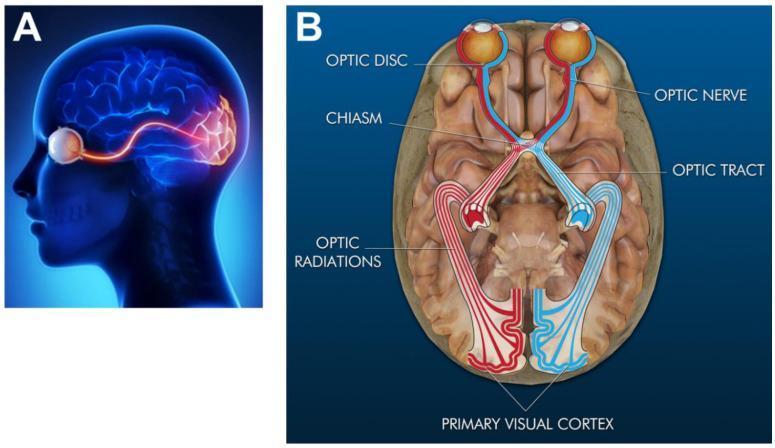
The Visual Pathway — A Detailed Overview
The visual pathway is the series of neural structures that transmit and process visual information from the retina to the primary visual cortex and beyond. It is divided into pre-geniculate, geniculate, and post-geniculate (cortical) segments.
1. The Retina — Origin of the Pathway
The visual journey begins at the retina, a multilayered neural tissue lining the back of the eye. Light is transduced by photoreceptors (rods and cones) into electrical signals.
- Rods: Scotopic vision (dim light, periphery, achromatic)
- Cones: Photopic vision (bright light, color, concentrated at the fovea)
Signals pass through bipolar cells to retinal ganglion cells (RGCs), the projection neurons of the retina. The axons of RGCs form the optic nerve. About 20 distinct types of RGCs extract different features in parallel — local light-dark contrast, color opponent signals (red-green, blue-yellow), motion, etc.
The optic disc (blind spot) is the point where axons converge and exit the eye. It contains no photoreceptors, creating a physiological blind spot.
2. The Optic Nerve (CN II)
- After leaving the optic disc, axons acquire a myelin sheath provided by oligodendrocytes (not Schwann cells — because the optic nerve is a CNS structure, derived embryologically from the diencephalon)
- Covered by all three cranial meninges (dura, arachnoid, pia), with CSF in the subarachnoid space around it
- Travels posteriorly through the orbit, optic canal, and into the cranial cavity
Gray's Anatomy for Students — the optic nerve is defined as a CNS component due to its oligodendrocyte myelination and meningeal coverings.
3. The Optic Chiasm
The two optic nerves meet at the optic chiasm, located anterior to the infundibular stalk and above the pituitary gland.
Partial decussation occurs here:
- Nasal (medial) retinal fibers → cross to the contralateral optic tract
- Temporal (lateral) retinal fibers → remain ipsilateral in the optic tract
Why does this matter?
Because of the optics of the eye (image inversion), the nasal retina sees the temporal visual field, and the temporal retina sees the nasal visual field. After decussation, all fibers representing the left visual hemifield travel in the right optic tract, and vice versa. This ensures the contralateral hemisphere processes each visual hemifield.
Because of the optics of the eye (image inversion), the nasal retina sees the temporal visual field, and the temporal retina sees the nasal visual field. After decussation, all fibers representing the left visual hemifield travel in the right optic tract, and vice versa. This ensures the contralateral hemisphere processes each visual hemifield.
4. The Optic Tract
Beyond the chiasm, fibers from the nasal retina of the contralateral eye and the temporal retina of the ipsilateral eye combine to form the optic tract.
- Each optic tract thus carries information about the contralateral visual hemifield
- The tracts course around the midbrain (cerebral peduncles) posteriorly toward the thalamus
- A small contingent of fibers branch off to:
- Pretectal area: mediates the pupillary light reflex
- Superior colliculus: controls saccadic eye movements (via the pontine reticular formation and extraocular nuclei)
- Suprachiasmatic nucleus (via retinohypothalamic tract): regulates circadian rhythms
5. The Lateral Geniculate Nucleus (LGN) of the Thalamus
The optic tract synapses in the LGN, the primary thalamic relay for vision.
Laminar Organization (6 layers in primates):
| Layers | Cell Type | Input from | Information Carried |
|---|---|---|---|
| 1, 2 | Magnocellular (large cells) | Parasol RGCs (~10%) | Achromatic contrast, motion, depth |
| 3, 4, 5, 6 | Parvocellular (small cells) | Midget RGCs (~70%) | Color (red-green), fine spatial detail |
| Intercalated (thin layers between each) | Koniocellular (dust-like cells) | Bistratified RGCs (~8%) | Blue-yellow color; also other RGC types |
Eye Input Segregation:
- Layers 1, 4, 6 receive input from the contralateral eye
- Layers 2, 3, 5 receive input from the ipsilateral eye
- The layers are precisely aligned so that corresponding visual field points are stacked vertically — forming a retinotopic map
The LGN is not a simple relay — it receives powerful feedback from the visual cortex and modulatory input from brainstem arousal systems (attention, wakefulness strongly modulate LGN responses).
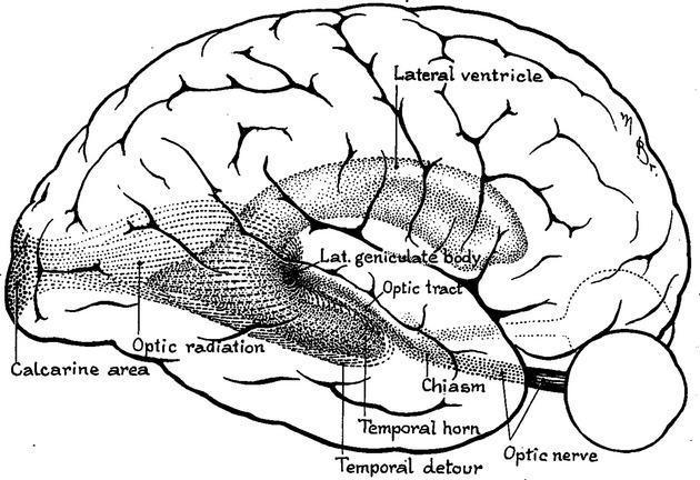
6. The Optic Radiations (Geniculocalcarine Tract)
Axons from LGN neurons form the optic radiations, which fan out through the white matter of the temporal and parietal lobes to reach the primary visual cortex.
Two divisions — critical for clinical localization:
| Division | Path | Visual Field Represented |
|---|---|---|
| Superior (parietal) fibers | Directly posterior through parietal lobe | Lower visual field |
| Inferior (temporal) fibers — Meyer's loop | Arc anteriorly around the temporal horn of the lateral ventricle before curving posteriorly | Upper visual field |
Meyer's loop (temporal detour) is clinically crucial:
- Temporal lobe lesions (e.g., temporal lobectomy for epilepsy, temporal lobe tumors) damage Meyer's loop → "pie in the sky" defect — contralateral superior quadrantanopia
7. Primary Visual Cortex (V1 / Striate Cortex / Brodmann Area 17)
Located in the occipital lobe, specifically in and around the calcarine sulcus. Called "striate cortex" due to the stripe of Gennari — a dense band of myelinated fibers visible in layer IV.
Retinotopic Organization:
- Upper optic radiation fibers → upper bank of calcarine sulcus (lower visual field)
- Lower optic radiation fibers → lower bank of calcarine sulcus (upper visual field)
- The fovea (central vision) is disproportionately represented at the posterior pole — cortical magnification factor
- The peripheral retina maps to more anterior striate cortex
Cortical Columns:
V1 is organized into functional columns perpendicular to the cortical surface:
- Orientation columns: neurons respond to bars/edges at specific angles
- Ocular dominance columns: alternate stripes processing input from each eye
- Blobs (cytochrome oxidase-rich regions): process color information
8. Beyond V1 — Extrastriate Visual Areas
From V1, information diverges into two major processing streams:
The "Two-Stream" Hypothesis:
| Stream | Direction | Function | Key Areas |
|---|---|---|---|
| Ventral ("What") pathway | V1 → V2 → V4 → Inferior temporal cortex | Object identity, color, face recognition, form | V4, IT cortex (areas TE, TEO) |
| Dorsal ("Where/How") pathway | V1 → V2 → V5/MT → Posterior parietal cortex | Motion, spatial location, visuomotor guidance | MT/V5, area MST, PPC |
- V4: Color processing; lesions → achromatopsia
- MT/V5: Motion processing; lesions → akinetopsia (inability to perceive motion)
- Fusiform face area (FFA): Face recognition; lesions → prosopagnosia
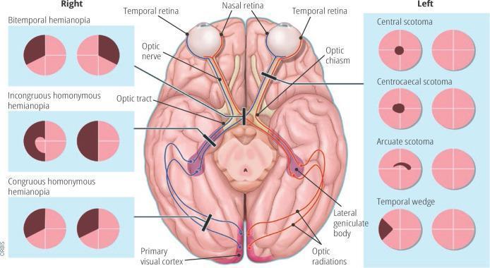
9. Visual Field Deficits — Lesion Localization
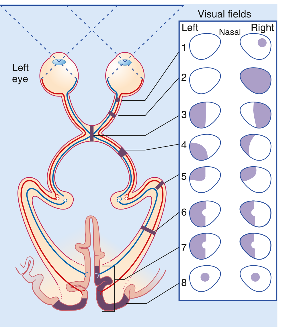
| Site of Lesion | Visual Field Defect |
|---|---|
| Optic nerve (site 1–2) | Ipsilateral monocular scotoma or complete blindness in that eye |
| Optic chiasm — central (site 3) | Bitemporal hemianopia (loss of temporal fields in both eyes — "tunnel vision"); classic with pituitary macroadenoma |
| Optic tract (site 4) | Contralateral homonymous hemianopia (incongruous) |
| Meyer's loop / temporal radiation (site 5) | Contralateral superior homonymous quadrantanopia ("pie in the sky") |
| Parietal optic radiation (site 6) | Contralateral inferior homonymous quadrantanopia ("pie on the floor") |
| Optic radiation (complete) (site 6–7) | Contralateral homonymous hemianopia with macular sparing (posterior cerebral artery infarct) |
| V1 (occipital cortex) (site 8) | Contralateral homonymous hemianopia with macular sparing |
Macular sparing in occipital lesions is due to the dual blood supply of the occipital pole (MCA + PCA) and the large cortical representation of the macula.
Summary: The Complete Visual Pathway at a Glance
Photoreceptors (rods/cones)
        ↓
Bipolar cells
        ↓
Retinal ganglion cells (RGCs)
        ↓ [axons form...]
Optic nerve (CN II) — CNS structure, oligodendrocyte myelination
        ↓
Optic chiasm — nasal fibers decussate
        ↓
Optic tract (carries contralateral hemifield)
        ├── Branches: Superior colliculus (eye movements)
        ├── Pretectal area (pupillary reflex)
        └── Suprachiasmatic nucleus (circadian rhythms)
        ↓
Lateral Geniculate Nucleus (LGN) — 6 laminae (M/P/K channels)
        ↓
Optic radiations
        ├── Superior fibers (parietal) — lower field
        └── Inferior fibers (Meyer's loop, temporal) — upper field
        ↓
Primary Visual Cortex V1 / Striate Cortex (calcarine sulcus, BA 17)
        ↓
Extrastriate cortex (V2, V3, V4, V5/MT)
        ├── Ventral "What" stream → temporal lobe (form, color, face recognition)
        └── Dorsal "Where/How" stream → parietal lobe (motion, spatial awareness)
Sources:
- Gray's Anatomy for Students (Elsevier) — Central Visual Pathway, p. 1346–1347
- Kandel: Principles of Neural Science, 6th Ed. — Ch. 21, Visual Processing and the Geniculostriate Pathway, pp. 544–549
This is a shared conversation. Sign in to Orris to start your own chat.