
angiosarcoma
Reading File
Searching sources...
Finding Sources
Searching PubMed
angiosarcoma
Good results. Now let me read the most comprehensive textbook sections in parallel - Robbins Pathology, Fitzpatrick's Dermatology, Andrews' Dermatology, and Sabiston Surgery.
Reading File
Reading File
Reading File
Reading File
Excellent content retrieved. Let me also get the Robbins histology image and search for molecular/genetic information.
Loading Image
Loading Image
Finding Sources
I now have comprehensive material from multiple authoritative textbooks plus histology images. Let me compile the full answer.
Angiosarcoma
Definition
Angiosarcoma is a malignant neoplasm arising from the endothelial lining of blood (or lymphatic) vessels. It is one of the most aggressive soft-tissue sarcomas (STS), accounting for approximately 2% of all STS. Histology ranges from well-differentiated tumors that mimic hemangiomas to profoundly anaplastic, poorly differentiated lesions.
- Robbins, Cotran & Kumar Pathologic Basis of Disease, p. 493
- Sabiston Textbook of Surgery, 14e
Epidemiology
- Peak incidence in the 7th and 8th decades; older adults predominate
- Male:female ratio ~2:1 for cutaneous head-and-neck form
- Most common sites (decreasing order): skin > soft tissue > breast > liver > spleen
- ~20% of patients present with distant metastasis at diagnosis
- ~40% of all angiosarcomas are radiation-associated
Pathogenesis and Risk Factors
Angiosarcoma arises in four distinct clinical settings:
| Setting | Key Feature |
|---|---|
| Sporadic (de novo) | Head/neck skin in elderly men; most common type |
| Chronic lymphedema | Post-mastectomy upper arm - "Stewart-Treves syndrome"; occurs ~11-12 years post-surgery in ~0.45% of patients; 5-year survival 6-14% |
| Post-irradiation | Latency ~23 years (benign condition) or ~12 years (prior malignancy) |
| Chemical/hepatic | Arsenic, Thorotrast (radioactive contrast agent), polyvinyl chloride - all classic hepatic angiosarcoma carcinogens with long latency |
Molecular Biology
- MYC amplification is the molecular hallmark of secondary (post-radiation and post-lymphedema) angiosarcoma - it is absent in primary cutaneous angiosarcoma and in atypical vascular lesions (AVL), making it a key discriminator
- KDR (VEGFR2) activating mutations have been identified in human angiosarcomas
- High Ki-67 proliferation index is characteristic
Pathology
Gross
- Ill-defined, dark-brown/hemorrhagic mass; often multifocal
- In the heart/right ventricle: appears as a large, dark, infiltrating mass (see image A below)
Histology
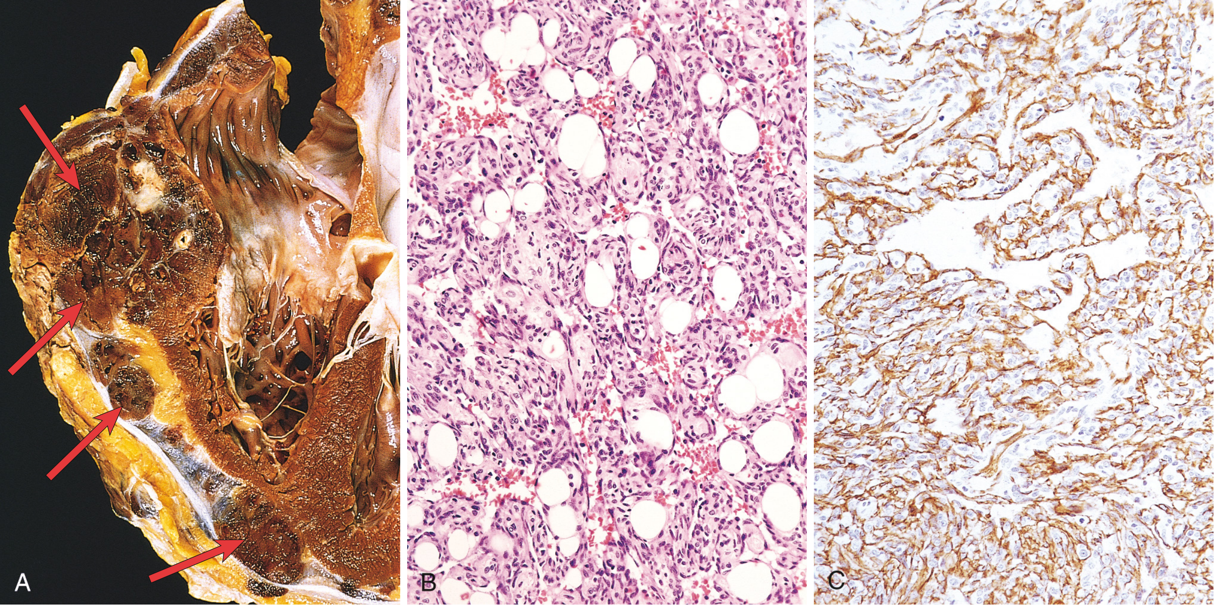
Fig. 11.32 - Robbins, Cotran & Kumar Pathologic Basis of Disease
- Well-differentiated: irregular vascular channels in dermis lined by atypical endothelial cells with enlarged, pleomorphic nuclei
- Poorly differentiated: solid sheets of epithelioid cells, increased cellularity, nuclear atypia, high mitotic activity; subtle infiltration between collagen bundles producing "crack-like" spaces with hyperchromatic nuclei
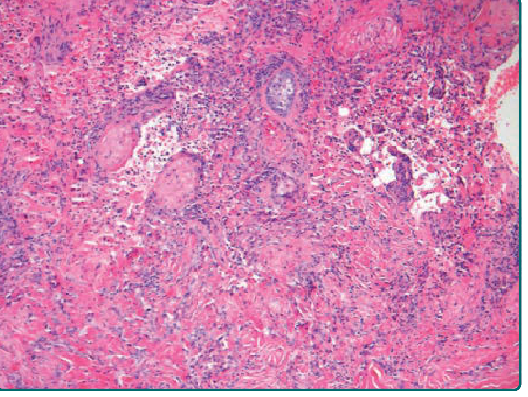
Fitzpatrick's Dermatology
Immunohistochemistry (IHC)
The following markers confirm endothelial origin:
- CD31 - most consistent and specific
- CD34
- FLI-1 (transcription factor)
- ERG (endothelial transcription factor)
- Podoplanin (D2-40) - positive in most malignant vascular tumors
- Ulex europaeus lectin
- c-MYC nuclear staining - helps distinguish radiation-induced angiosarcoma from AVL
Clinical Presentation
Cutaneous (Head/Neck - most common)
- Poorly defined bluish macule on the scalp/face of elderly men - easily mistaken for a bruise
- Distinguishing features: peripheral erythematous ring, satellite nodules, spontaneous bleeding, intratumoral hemorrhage
- Progressive asymmetric enlargement with indurated bluish nodules/plaques
- Sudden thrombocytopenia may herald metastasis or rapid tumor expansion
- Multicentricity and early metastasis are hallmarks; most patients die within 2 years
- Prognosis worse in men >70
Stewart-Treves Syndrome
- Angiosarcoma in chronic lymphedematous upper arm after mastectomy
- Mean survival 19-31 months; 5-year survival ~6-14%
- Lung metastases are the most frequent cause of death
- Early amputation offers the best prognosis
Spread and Prognosis
- Locally invasive with early hematogenous metastasis
- Lung is the most common metastatic site; breast angiosarcoma may spread to liver
- Overall 5-year survival: 10-30% (Robbins)
- 5-year disease-specific survival for resectable disease: ~53% (Sabiston)
- Stage IV median survival: 8-12 months
Management
Surgery
- Wide resection with negative margins (at least 2 cm of normal tissue) is the mainstay
- Tumors microscopically extend well beyond gross margins
- Radiation-associated breast angiosarcoma: mastectomy including all irradiated skin (vs. simple mastectomy - dramatically reduces local recurrence from 76% to 23% and improves 5-year DSS from 46% to 86%)
- Stewart-Treves: amputation
Radiation Therapy
- Adjuvant radiotherapy for head/neck and scalp angiosarcomas (high locoregional recurrence)
- Cannot use for radiation-induced tumors
Systemic Therapy (Palliative/Metastatic)
| Agent | Notes |
|---|---|
| Paclitaxel | First-line palliative for cutaneous angiosarcoma; ~56% objective response rate |
| Doxorubicin ± ifosfamide | Standard chemotherapy; modest response rate |
| Pazopanib | For taxane-resistant disease; controls progression especially in cutaneous angiosarcoma |
| Sirolimus | Shows promise for scalp/facial angiosarcomas |
| Interferon (IFN) | Shows promise for scalp/facial angiosarcomas |
Key Points to Remember
- Malignant endothelial neoplasm - arises from blood/lymphatic vessel endothelium
- Four clinical settings: sporadic head/neck, Stewart-Treves (lymphedema), post-irradiation, chemical (hepatic)
- MYC amplification = molecular hallmark of secondary (radiation/lymphedema-associated) angiosarcoma
- IHC: CD31 + CD34 + FLI-1 + ERG (confirm endothelial origin); c-MYC distinguishes radiation-induced from AVL
- Paclitaxel is first-line systemic therapy; doxorubicin and pazopanib are alternatives
- Overall poor prognosis: 5-year survival 10-30%; stage IV median survival 8-12 months
- Wide surgical margins critical - tumors extend microscopically well beyond gross disease
Recent Literature: A 2025 systematic review (PMID 40666093) in Frontiers in Oncology reviewed biomarkers in angiosarcoma diagnosis, prognosis, and therapeutics - consistent with the molecular data above. No major updates contradict current textbook guidance.
Sources: Robbins, Cotran & Kumar Pathologic Basis of Disease | Sabiston Textbook of Surgery, 14e | Andrews' Diseases of the Skin, 12e | Fitzpatrick's Dermatology, 9e
This is a shared conversation. Sign in to Orris to start your own chat.