
Acid aspiration
I'll look up acid aspiration in the medical library for a thorough answer.
Searching sources...
Reading File
Finding Sources
Finding Sources
Good hits from Fishman's Pulmonary and the Anesthesiology text. Let me read the dedicated sections on Chemical Pneumonitis and the pathophysiology section.
Finding Sources
Reading File
Now let me read the key sections from Fishman's and Murray & Nadel's on aspiration pneumonitis:
Reading File
Reading File
Excellent content. Let me now read the pathophysiology and management sections from Fishman's, and also check Sabiston's surgical perspective:
Reading File
Reading File
Let me get the management section and also search for a relevant image:
Reading File
Reading File
Searching Images
acid aspiration pneumonitis chest X-ray lung infiltrate
I now have comprehensive, authoritative content. Let me compile the full answer.
Acid Aspiration (Aspiration Pneumonitis / Mendelson's Syndrome)
Definition
Acid aspiration - more precisely called aspiration pneumonitis - is acute lung injury following the aspiration of regurgitated gastric contents into the lower respiratory tract. The classic form, Mendelson's syndrome, was first described in 1946 in obstetric patients receiving general anesthesia without endotracheal intubation. - Fishman's Pulmonary Diseases and Disorders, p. 1195
Distinguishing Pneumonitis vs. Pneumonia
These two entities are often confused but have distinct mechanisms and management:
| Feature | Aspiration Pneumonitis | Aspiration Pneumonia |
|---|---|---|
| Mechanism | Aspiration of sterile gastric contents | Aspiration of colonized oropharyngeal material |
| Pathophysiology | Acid/particulate chemical injury | Bacterial inflammatory response |
| Bacteriology | Initially sterile | Gram-negative rods, gram-positive cocci, rarely anaerobes |
| Main predisposing factor | Depressed consciousness | Dysphagia, gastric dysmotility |
| Age group | Any age (often young) | Usually elderly |
| Aspiration event | Often witnessed | Usually not witnessed |
| Onset | Rapid (2-5 h), often self-limiting | Gradual, mimics community-acquired pneumonia |
- Fishman's Pulmonary Diseases and Disorders, Table 69-1
Pathophysiology
Critical Thresholds
The severity of lung injury depends on two key factors:
- pH < 2.5 is required to cause significant pneumonitis
- Volume > 20 mL increases damage severity
- Particulate matter (food particles) independently worsens injury even at pH > 2.5
Two-Phase Injury Model
When acid enters the lungs, a biphasic injury occurs:
- Phase 1 (within 1 hour): Direct caustic chemical injury to pulmonary tissue from the acid itself
- Phase 2 (3-6 hours post-exposure): Neutrophil-mediated injury - recruited neutrophils release reactive oxygen species (ROS) via NADPH oxidase and neutrophil extracellular traps (NETs), activating NF-kB and cytokines TNF-α and IL-8
The resulting inflammation mirrors ARDS - airspace edema, hemorrhage, and hyaline membrane formation from type 2 pneumocyte hyperplasia.
Role of Bacteria
Gastric acid normally keeps the stomach sterile - so bacterial infection does NOT play a significant early role. However:
-
Acid aspiration impairs host defenses, increasing risk of secondary superinfection
-
Gastric colonization occurs when pH is raised by antacids, H2 blockers, or PPIs, or in patients on enteral feeds, gastroparesis, or small bowel obstruction - in these cases, the inflammatory response involves both chemical and bacterial injury
-
Fishman's Pulmonary Diseases and Disorders, pp. 1196-1197; Sabiston Textbook of Surgery, p. 445
Risk Factors
- Anesthesia (especially emergency/unplanned intubation)
- Drug overdose (most common cause of aspiration pneumonitis in hospitalized patients - ~10% of drug overdose admissions)
- Seizures, coma, head trauma, massive CVA
- GLP-1 receptor agonists (semaglutide, liraglutide) - cause delayed gastric emptying; ASA now recommends holding daily GLP-1 agonists 1 day before surgery, and weekly agonists 1 week before surgery
- Small bowel obstruction or ileus
- Emergency surgery
- Older age (pharyngeal dysmotility + GERD + poor oral hygiene)
The risk of aspiration increases with the degree of impaired consciousness (measured by GCS score).
- Sabiston Textbook of Surgery, p. 445; Fishman's Pulmonary Diseases and Disorders, p. 1195
Clinical Features
Symptoms appear 2-5 hours after aspiration and range from:
- Asymptomatic / subclinical
- Nonproductive cough, tachypnea, low-grade fever
- Bronchospasm
- Bloody or frothy sputum
- Hypoxia (earliest and most reliable sign)
- Severe respiratory distress, progressing to ARDS
Radiographic changes begin within 24-36 hours. In uncomplicated chemical pneumonitis, most changes clear within 48 hours. Persistence beyond 48 hours should raise suspicion for superimposed bacterial pneumonia. - Sabiston Textbook of Surgery, p. 445
Management
Immediate
- Suction the airway - clear any aspirated material
- Endotracheal intubation if the patient cannot protect the airway
- Bronchoscopy - consider for retrieval of particulate matter
- Gastric decompression (nasogastric tube)
- Supplemental oxygen; mechanical ventilation with PEEP in severe cases
Antibiotics
- NOT recommended prophylactically in standard aspiration pneumonitis (gastric contents are sterile; early antibiotics may select for resistant organisms)
- ARE indicated if:
- Aspiration in setting of small bowel obstruction or feculent content
- Gastric colonization likely (patient on acid suppression, enteral feeds)
- Pneumonitis fails to resolve within 48 hours
- Empiric regimens: fluoroquinolones, piperacillin/tazobactam, or ceftriaxone
- Anaerobic coverage is not routinely required (recent data challenge classic teaching about anaerobic dominance)
Corticosteroids
-
Used since 1955, but their role as monotherapy is limited - they have minimal effect on neutrophils and ROS
-
Emerging approach: HAT therapy (hydrocortisone + ascorbic acid + thiamine) - vitamin C acts as a potent NADPH oxidase inhibitor and NETs inhibitor; small case series and one RCT in ARDS suggest benefit, but larger studies are still needed
-
Fishman's Pulmonary Diseases and Disorders, pp. 1199-1200; Sabiston Textbook of Surgery, pp. 446-447
Prevention
-
NPO guidelines: nothing by mouth for ≥2 hours before elective procedures requiring sedation/anesthesia
-
Rapid sequence induction (RSI) with cricoid pressure - considered most effective during high-risk intubation, though not 100% reliable
-
Non-particulate antacids (sodium citrate, sodium bicarbonate) before anesthesia - preferred over particulate antacids (aluminum/magnesium hydroxide), which can themselves cause lung injury if aspirated
-
H2 blockers or PPIs in high-risk patients (favorable risk-benefit, but not absolutely reliable)
-
Delay extubation until patient is fully awake with intact protective reflexes
-
Oral hygiene may reduce aspiration pneumonia risk (especially in nursing home patients), though evidence is mixed
-
Morgan and Mikhail's Clinical Anesthesiology, p. [block2]; Sabiston Textbook of Surgery, p. 446
Key Numbers to Remember
| Parameter | Value |
|---|---|
| Critical pH for lung injury | < 2.5 |
| Critical aspirate volume | > 20-25 mL |
| Phase 1 injury onset | Within 1 hour |
| Phase 2 (neutrophil) injury peak | 4-6 hours |
| Symptom onset | 2-5 hours post-aspiration |
| Resolution of simple pneumonitis | Within 48 hours |
| Antibiotic trigger if no resolution | > 48 hours |
Explain how aspiration pneumonia occurs
Searching sources...
Finding Sources
Finding Sources
Good hits from Fishman's, Harrison's, and Robbins. Let me read the dedicated sections in parallel:
Reading File
Reading File
Reading File
Excellent. Now let me get the pathogenesis detail from Fishman's, the oropharyngeal colonization mechanism, and fetch the chest X-ray image found earlier, plus the Goldman-Cecil entry:
Reading File
Reading File
Loading Image
I now have thorough content from multiple authoritative sources. Here is the full explanation:
How Aspiration Pneumonia Occurs
The Starting Point: Everyone Microaspirates
A key concept is that aspiration of small amounts of oropharyngeal secretions during sleep happens in approximately half of all healthy adults. In a healthy person, this causes no harm because:
- The bacterial burden is low and of low virulence
- Coughing forcefully clears the inoculum
- Ciliary transport (mucociliary escalator) moves material upward
- Normal humoral and cellular immune mechanisms eliminate bacteria
Aspiration pneumonia develops when this balance tips - either the amount/virulence of aspirated material is too great, or the host's defenses are too weak. - Fishman's Pulmonary Diseases and Disorders, p. 1199
Step 1: Failure of the Swallowing Mechanism
Normal swallowing is a highly coordinated act involving 5 cranial nerves, more than 50 muscles of the head and neck, and bilateral input from the sensorimotor cortex, insula, basal ganglia, and brainstem medullary swallowing center.
During the oropharyngeal phase, the larynx elevates and moves anteriorly to meet the epiglottis, sealing the airway before the food bolus passes into the esophagus. When this fails - due to neurological or structural disease - food and secretions spill into the airway instead.
Dysphagia is the single most important risk factor for aspiration pneumonia. The more severely swallowing is impaired, the greater the volume aspirated, and in stroke patients there is a direct correlation between aspirate volume and pneumonia development. - Fishman's Pulmonary Diseases and Disorders, p. 1199
Step 2: Oropharyngeal Colonization by Pathogenic Bacteria
The mouth is densely colonized. In healthy people, normal oral flora predominates. In sick or debilitated patients, the oropharynx becomes colonized by pathogenic Gram-negative bacilli (e.g., E. coli, Klebsiella, Pseudomonas) and Gram-positive cocci (e.g., Staphylococcus), along with anaerobes from the gingival crevices (Prevotella, Fusobacterium, Peptostreptococcus, Bacteroides).
Factors that increase this pathogenic colonization:
- Poor oral hygiene and severe dental disease
- Nasogastric tube feeding (reflux of gastric contents colonizes the pharynx)
- Prior antibiotic use (selects resistant organisms)
- Acid suppression therapy (raises gastric pH, allowing bacterial overgrowth that refluxes upward)
- Reduced salivary flow
The lungs are continuously seeded with organisms from the oral microbiota via subclinical microaspiration. Even though the lung is aerobic, culture-independent methods show anaerobes like Prevotella and Veillonella are abundant in the lower respiratory tract at baseline. - Harrison's Principles of Internal Medicine 22E, p. 1419
Step 3: Aspiration of Colonized Material into the Lung
When the colonized oropharyngeal secretions (or gastric contents carrying pathogens) are aspirated past the vocal cords, the outcome depends on:
- Volume of aspirate - larger volumes overwhelm clearance mechanisms
- Bacterial burden and virulence of the aspirated organisms
- Whether gastric fluid co-aspirates - acid damages the airway epithelium, impairs mucociliary clearance, and creates a foothold for bacteria
The combination of increased bacterial burden + impaired airway defenses allows bacteria to establish infection. - Harrison's Principles of Internal Medicine 22E, p. 1419
Step 4: Impaired Host Defense Clears Nothing
In aspiration pneumonia, the host defenses fail for one or more reasons:
- Reduced cough reflex (sedation, stroke, intoxication, anesthesia)
- Dysfunctional mucociliary transport (smoking, chronic illness)
- Impaired cellular immunity (elderly, malnourished, immunocompromised)
- Impaired humoral immunity (hypogammaglobulinemia, splenic dysfunction)
When these are intact, aspiration is cleared. When they fail, bacteria persist, multiply in alveolar spaces, and trigger an acute inflammatory response - i.e., pneumonia.
Step 5: The Inflammatory Response and Progression
Once bacteria establish themselves in the alveoli, a full-blown pneumonic consolidation develops. The process tends to:
- Develop over days to weeks (in contrast to chemical pneumonitis, which declares itself within hours)
- Be polymicrobial - more than one organism is typically recovered
- Be aerobes more common than anaerobes in modern studies (early teachings overemphasized anaerobes; recent data show Gram-negatives and streptococci dominate)
- Pursue a necrotizing course in severe cases
Robbins Basic Pathology notes it is "often necrotizing, pursues a fulminant clinical course, and is a frequent cause of death in individuals predisposed to aspiration." - p. 446
Where in the Lung?
The dependent segments are consistently affected - gravity pulls the aspirate to wherever is lowest at the time:
| Patient position at time of aspiration | Segment most affected |
|---|---|
| Upright/semi-recumbent | Basilar segments of lower lobes |
| Supine | Posterior segment of right upper lobe or superior segment of lower lobe |
The right side is more commonly affected because the right main bronchus is more vertically oriented and wider, offering less resistance to aspirated material. - Harrison's Principles of Internal Medicine 22E, p. 1412
Here is what aspiration pneumonia looks like on a chest X-ray:
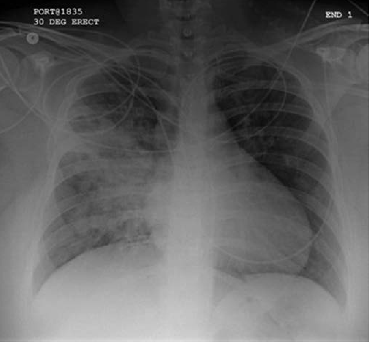
CXR showing opacification of the right perihilar region and superior segment of the right lower and upper lobes - aspiration pneumonia in a 75-year-old post-stroke patient. From Fishman's Pulmonary Diseases and Disorders, p. 1199.
Complications
If the infection is not cleared:
- Lung abscess - localized suppuration forming a cavitary lesion; almost all contain anaerobes (often 3-6 isolates), and ~90% of cultures also show aerobes (S. aureus, S. pneumoniae, Klebsiella)
- Empyema - spread of infection to the pleural space
- Bronchopleural fistula - from necrosis of lung tissue connecting the bronchus to the pleural cavity
Risk Factors at a Glance
| Category | Examples |
|---|---|
| Neurological | Stroke (30%+ develop dysphagia), Parkinson's (52-82%), Alzheimer's (84%), ALS, MS |
| Structural | Head/neck cancer, esophageal malignancy, achalasia, scleroderma |
| Motility disorders | Diabetic gastroparesis, presbyesophagus, GERD |
| Situational | Alcohol intoxication, sedation, anesthesia, coma |
| Institutionalized elderly | Up to 60% of elderly institutionalized patients have dysphagia |
Aspiration pneumonia accounts for 5-15% of community-acquired pneumonia cases overall, but in elderly nursing home patients it is likely the dominant cause of pneumonia - and is considered one of the final common pathways by which most chronically ill patients die. - Fishman's Pulmonary Diseases and Disorders, p. 1199
Which group of people are prone to aspiration pneumonia
Based on the previous discussion, the high-risk groups are already well established. Let me pull the specific risk factor tables from the textbooks for a complete, precise answer.
Searching sources...
Finding Sources
Finding Sources
Reading File
Reading File
Reading File
I now have rich, detailed content from multiple authoritative sources. Let me read the specific substance P / ACE / cough reflex passage and Tintinalli's risk factor table for completeness:
Reading File
Comprehensive data in hand. Here is the complete answer:
Who is Prone to Aspiration Pneumonia?
The core requirement is some combination of impaired swallowing or airway protection AND increased oropharyngeal bacterial colonization. Any condition that disrupts either or both dramatically raises risk. The groups are best organized by the mechanism through which they become vulnerable.
1. People with Neurological Disease (Highest Risk Group)
Swallowing depends on 5 cranial nerves, 50+ muscles, and intact bilateral cortical-brainstem circuitry. Neurological disease dismantles this at multiple levels simultaneously.
| Condition | Prevalence of Dysphagia |
|---|---|
| Acute stroke | 40-70% develop dysphagia |
| Parkinson's disease | 52-82% |
| Alzheimer's dementia | 84% |
| Multi-infarct dementia | High |
| ALS (motor neuron disease) | Progressive, near-universal |
| Multiple sclerosis | Common |
| Subarachnoid haemorrhage | Common |
In stroke patients specifically, silent aspiration (aspiration without cough or sensation) is found in 71% of those who develop community-acquired pneumonia, versus only 10% of age-matched controls. Dysphagia after stroke follows a fluctuating course, with 10-30% having ongoing aspiration long-term. - Fishman's Pulmonary Diseases and Disorders, pp. 1199-1200
A critical additional mechanism in neurological disease is impaired cough reflex. Substance P - released from vagal sensory nerves in the pharynx - mediates the cough reflex. In elderly patients with pneumonia, sputum substance P levels are substantially reduced. Patients with the ACE gene DD allele have faster bradykinin breakdown and a blunted cough, further raising pneumonia risk. This is why ACE inhibitors (which raise bradykinin) are associated with a reduced risk of aspiration pneumonia. - Fishman's Pulmonary Diseases and Disorders, p. 1200
2. The Elderly
Age independently impairs every defense mechanism:
- Swallowing efficiency declines with age due to diminished oropharyngeal sensation, lingual weakness (sarcopenia), silent cerebral infarcts, and slowed synaptic conduction
- Cough reflex weakens - elderly patients need a stronger stimulus to trigger coughing
- Mucociliary clearance slows - cilia beat less effectively
- Cellular and humoral immunity decline - reduced alveolar macrophage activity
- Salivary flow decreases - saliva normally washes bacteria from the mouth; less saliva means more bacterial buildup
- Poor oral hygiene is common in institutionalized elderly - dental plaque is a reservoir for pathogens
The incidence of pneumonia is nearly 6 times higher in those over 75 vs. under 60. Aspiration pneumonia is the leading cause of death in nursing home patients and the leading cause of transfer from nursing home to hospital - the second most common infection in nursing homes after UTI. - Tintinalli's Emergency Medicine, p. 488; Fishman's Pulmonary Diseases and Disorders, p. 1199
3. Patients with Impaired Consciousness
When consciousness is depressed, the gag reflex and cough reflex are suppressed and the epiglottis fails to protect the airway. This group includes:
- Alcohol intoxication - one of the most common causes in younger patients; frequent periods of loss of consciousness with oropharyngeal relaxation
- Illicit drug overdose and sedative overdose
- Seizure disorders - the post-ictal state leaves patients obtunded with pooled secretions
- General anaesthesia - particularly emergency/unplanned intubation
- Traumatic brain injury and coma
- Metabolic encephalopathy
In younger patients, aspiration pneumonia almost always traces back to one of these obtundation-related events. The patient may recall an aspiration incident or have a history of substance misuse. - Tintinalli's Emergency Medicine, p. 488
4. Patients with Head, Neck, and Oesophageal Structural Disease
Any anatomic disruption of the swallowing passage causes food and secretions to misdirect into the airway:
-
Head and neck cancers (oropharyngeal, oral cavity, laryngeal)
-
Oesophageal cancer
-
Post-surgical/post-radiation changes to the pharynx or oesophagus
-
Achalasia - failure of the lower oesophageal sphincter to relax leads to retained food that overflows into the airway, especially at night
-
Zenker's diverticulum - a pharyngeal pouch traps food that is later regurgitated and aspirated
-
Severe GERD / reflux oesophagitis
-
Scleroderma - oesophageal dysmotility and poor LOS tone
-
Fishman's Pulmonary Diseases and Disorders, Table 69-3
5. Patients with Poor Oral Health
The mouth is the direct source of the aspirated bacteria. Poor oral hygiene leads to large reservoirs of pathogens in dental plaque and periodontal pockets.
Independent predictors of aspiration pneumonia include:
- 4 or more decayed teeth
- Gingivitis or periodontitis
- Dental plaque colonized with periodontal pathogens
- Never or only occasionally brushing teeth
- Having 10 or more periodontal pockets (associated with increased risk of death from pneumonia)
Medications that reduce salivary flow compound this: diuretics, anticholinergics, antipsychotics, anxiolytics, and levodopa all reduce saliva production, allowing bacterial counts in the mouth to rise. - Tintinalli's Emergency Medicine, p. 488; Fishman's Pulmonary Diseases and Disorders, p. 1200
6. Patients on Enteral Feeding or with Tubes
- Nasogastric tubes bypass the upper oesophageal sphincter and provide a conduit for gastric contents to reflux back into the pharynx
- Percutaneous gastric (PEG) or jejunal tubes in debilitated patients
- Prolonged supine positioning (e.g., ICU patients) favours reflux and passive flow of secretions into the airway
7. Patients with Gastric Motility Disorders
Delayed gastric emptying causes accumulation of large gastric volumes that can reflux and be aspirated:
- Diabetic gastroparesis
- GLP-1 receptor agonist use (semaglutide, liraglutide) - now specifically flagged by ASA guidelines as a perioperative aspiration risk
- Small bowel obstruction - and importantly, in this setting the gastric contents are bacterially colonized, making the resulting pneumonia more severe
8. Patients on Acid-Suppressive Therapy
Proton pump inhibitors and H2 blockers raise gastric pH, which removes the bactericidal effect of gastric acid. This leads to colonization of the stomach with Gram-negative pathogens. When gastric contents reflux, they carry a much higher bacterial burden into the oropharynx. Studies show concurrent PPI use in patients over 60 significantly increases risk of community-acquired aspiration pneumonia. - Fishman's Pulmonary Diseases and Disorders, p. 1200
Summary Table
| Population | Key Mechanism |
|---|---|
| Stroke / neurological disease | Dysphagia + impaired cough reflex |
| Elderly (especially institutionalised) | All defenses weakened; swallowing, cough, immunity, oral hygiene |
| Alcohol/drug intoxication | Suppressed gag and cough reflex |
| Head/neck/oesophageal structural disease | Anatomic misdirection of food bolus |
| Poor oral hygiene / dental disease | High bacterial burden in aspirated material |
| Nasogastric/PEG tube patients | Reflux of colonized material into pharynx |
| Gastroparesis / GLP-1 agonist users | Large retained gastric volumes |
| PPI / H2 blocker users | Gastric bacterial colonization |
| ICU patients (supine, ventilated) | Passive reflux + impaired mucociliary clearance |
The unifying theme: aspiration alone does not cause pneumonia - about half of all healthy adults aspirate during sleep without consequences. It is the failure of normal clearance mechanisms, combined with a sufficiently large or virulent bacterial inoculum, that allows infection to take hold. - Tintinalli's Emergency Medicine, p. 484; Frameworks for Internal Medicine, p. 425
This is a shared conversation. Sign in to Orris to start your own chat.