
Radial artery full laq for 15 marks
I'll look up the radial artery in the medical library for a comprehensive 15-mark answer.
Searching sources...
Reading File
Finding Sources
Finding Sources
Gray's Anatomy for Students has a dedicated "Radial artery" section. Let me read it along with the imaging anatomy text.
Reading File
Reading File
Good - I have solid content from Gray's. Let me also read the hand section of the radial artery (branches in the hand) and the imaging anatomy text for the forearm arteries overview.
Reading File
Reading File
Now let me get the radial artery in the hand section from Gray's, and also check the imaging anatomy text for the hand branches.
Finding Sources
Finding Sources
Excellent! Now let me read the Thieme Atlas branches table and the Color Atlas hand section for a clean branch list:
Reading File
Reading File
I now have comprehensive information from multiple authoritative sources. Let me also fetch the key anatomical diagram from Gray's to include in the answer.
Loading Image
I now have all the information needed for a thorough 15-mark answer. Here is the complete LAQ:
Radial Artery - Long Answer Question (15 Marks)
1. Introduction / Origin
The radial artery is one of the two terminal branches of the brachial artery, arising at the level of the neck of the radius (apex of the cubital fossa). It is the smaller of the two terminal branches - the ulnar artery is typically larger and primarily supplies the proximal forearm, while the radial artery typically supplies the distal forearm and hand.
2. Course
The radial artery has a continuous course that can be divided into three segments:
A. In the Forearm (Anterior Compartment)
The artery passes along the lateral aspect of the forearm, running from the cubital fossa to the wrist.
- Proximal half: lies just deep to the brachioradialis muscle
- Middle third: the superficial branch of the radial nerve lies on its lateral side (an important anatomical relation)
- Distal forearm: lies between the tendon of brachioradialis laterally and the tendon of flexor carpi radialis medially, covered only by deep fascia, superficial fascia, and skin - this is where the radial pulse is palpated. It lies directly anterior to the pronator quadratus muscle and the distal end of the radius.
B. At the Wrist and Dorsum of the Hand
The artery leaves the forearm by passing around the lateral side of the wrist (lateral to the scaphoid and trapezium). It travels across the floor of the anatomical snuffbox (between the tendons of abductor pollicis longus/extensor pollicis brevis laterally and extensor pollicis longus medially). It then penetrates the posterolateral aspect of the hand by passing between the bases of the 1st and 2nd metacarpals, entering the palm.
C. In the Palm (Deep)
Once in the palm, the radial artery passes deep between the two heads of the first dorsal interosseous muscle and then curves medially across the palm as the deep palmar arch, lying on the bases of the metacarpal bones, deep to the long flexor tendons.
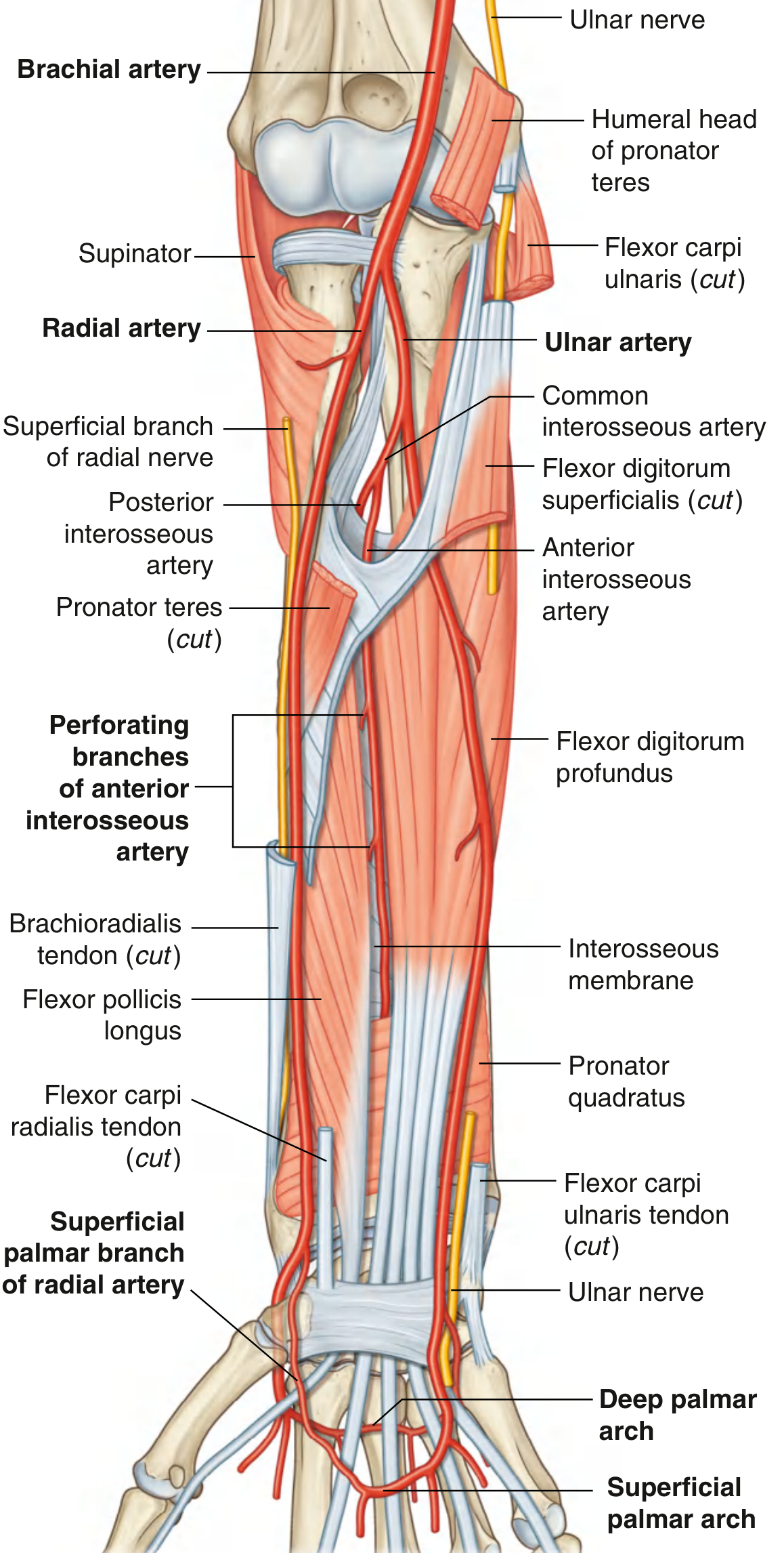
Fig. 7.88 - Arteries of the Anterior Compartment of the Forearm (Gray's Anatomy for Students)
3. Relations
| Region | Medial | Lateral | Anterior | Posterior |
|---|---|---|---|---|
| Proximal forearm | Pronator teres, FDS | Brachioradialis (overlying) | - | Supinator, FDS |
| Middle forearm | - | Superficial branch of radial nerve | - | FPL, pronator quadratus |
| Distal forearm | FCR tendon | Brachioradialis tendon | Skin, fascia | Pronator quadratus, distal radius |
4. Branches
In the Forearm:
- Radial recurrent artery - first branch, arises just distal to the bifurcation; passes superiorly to anastomose with the radial collateral branch of the profunda brachii artery, contributing to the periarticular anastomosis around the elbow joint. It also supplies brachioradialis, extensor carpi radialis longus and brevis, and supinator.
- Muscular branches - multiple unnamed branches supply the muscles on the lateral (radial) side of the forearm.
- Palmar carpal branch - a small branch near the wrist; contributes to the palmar carpal arch/network on the palmar surface of the carpus (supplies carpal bones and joints).
- Superficial palmar branch - arises just before the artery leaves the forearm; passes through or superficial to the thenar muscles at the base of the thumb, entering the hand to anastomose with the superficial palmar arch formed mainly by the ulnar artery.
At the Wrist / Dorsum:
- Dorsal carpal branch - contributes to the dorsal carpal network/arch; from this network arise the dorsal metacarpal arteries → dorsal digital arteries to the fingers.
In the Palm:
- First dorsal metacarpal artery - arises as the radial artery crosses the first dorsal interosseous muscle; supplies the dorsal surface of the thumb and the lateral side of the index finger.
- Princeps pollicis artery - arises as the radial artery turns into the palm; runs on the palmar (volar) surface of the 1st metacarpal and divides into two proper digital arteries supplying both sides of the thumb.
- Radialis indicis artery - arises from the radial artery (or from the princeps pollicis); supplies the radial side of the index finger.
- Deep palmar arch - the terminal continuation of the radial artery; curves medially across the bases of metacarpals 2-5, lying deep to the long flexor tendons. It anastomoses with the deep palmar branch of the ulnar artery to complete the arch. From the deep palmar arch arise:
- Palmar metacarpal arteries (3 arteries) - pass distally and join the common palmar digital arteries of the superficial arch
- Perforating branches - pass dorsally through interosseous spaces to anastomose with the dorsal metacarpal arteries
5. Summary Table of All Branches
| Segment | Branch | Destination / Anastomosis |
|---|---|---|
| Forearm | Radial recurrent a. | Elbow anastomosis (with profunda brachii) |
| Forearm | Muscular branches | Radial-side forearm muscles |
| Forearm | Palmar carpal branch | Palmar carpal arch |
| Forearm | Superficial palmar branch | Superficial palmar arch |
| Wrist | Dorsal carpal branch | Dorsal carpal arch → dorsal digitals |
| Palm | First dorsal metacarpal a. | Dorsum of thumb + index finger |
| Palm | Princeps pollicis a. | Both sides of thumb |
| Palm | Radialis indicis a. | Radial side of index finger |
| Palm | Deep palmar arch + palmar metacarpal a. | Completes deep arch; finger supply |
6. Clinical Importance
A. Radial Pulse
The pulse is palpated in the distal forearm between the brachioradialis tendon (laterally) and flexor carpi radialis tendon (medially), with the artery lying directly on the pronator quadratus and the distal radius.
B. Allen's Test
Before radial artery cannulation or harvest, Allen's test is performed to confirm the patency of the ulnar artery and the palmar arches. The test ensures collateral flow to the hand will be adequate if the radial artery is occluded.
C. Arterial Cannulation (Monitoring)
The radial artery at the wrist is the most common site for arterial line placement in ICU and anesthesia because it is:
- Superficial and easily accessible
- Has excellent collateral supply via the ulnar artery and palmar arches
- Complications (ischemia, thrombosis) are rare
D. Coronary Artery Bypass Surgery (CABG)
The radial artery is harvested as a conduit for coronary artery bypass grafting due to its muscular wall and good long-term patency. The left radial artery is commonly used; the surgeon verifies intact ulnar collateral circulation before harvest.
E. Radial Forearm Flap (Chinese Flap)
A fasciocutaneous flap based on the radial artery is used in reconstructive surgery for coverage of defects of the head, neck, and hand. The largest such flaps can be up to 16 cm x 9 cm (Foucher et al.).
F. Anatomical Snuffbox
The radial artery is palpable in the floor of the anatomical snuffbox. This region is also important because tenderness here after a fall on an outstretched hand raises suspicion for a scaphoid fracture.
G. Variations
High origin of the radial artery (from the axillary or brachial artery proximal to the cubital fossa) occurs in approximately 15% of individuals - an important consideration before arterial catheterization or harvesting.
7. Anastomoses
The radial artery participates in several important anastomotic networks:
| Network | Vessels Involved |
|---|---|
| Elbow anastomosis | Radial recurrent + radial collateral (from profunda brachii) |
| Palmar carpal arch | Palmar carpal branches of radial + ulnar arteries |
| Dorsal carpal arch | Dorsal carpal branches of radial + ulnar + anterior interosseous arteries |
| Superficial palmar arch | Superficial palmar branch of radial + ulnar artery (mainly) |
| Deep palmar arch | Radial artery (mainly) + deep branch of ulnar artery |
Sources: Gray's Anatomy for Students, p. 903 | Color Atlas of Human Anatomy Vol. 2, p. 109-110 | Imaging Anatomy Vol. 3 (Bones, Joints, Vessels and Nerves), p. 160-161 | Thieme Atlas of Anatomy: General Anatomy and Musculoskeletal System, p. 375
This is a shared conversation. Sign in to Orris to start your own chat.