
Explain cardiac cycle as a20 marks answer
Reading File
Now let me search the medical library for cardiac cycle content in parallel:
Finding Sources
Finding Sources
Excellent sources found. Let me read the most comprehensive ones in parallel - Guyton & Hall, Costanzo Physiology, and Ganong's:
Reading File
Reading File
Reading File
Great content. Let me read more from Ganong's and also fetch the Wiggers diagram image:
Reading File
Reading File
Loading Image
Excellent content gathered. Let me also fetch the Costanzo Wiggers diagram and read the remaining phases:
Loading Image
Reading File
I now have comprehensive content from multiple authoritative textbooks. Let me compile the full 20-mark answer.
THE CARDIAC CYCLE
Definition
The cardiac cycle refers to the complete sequence of electrical and mechanical events that occur from the beginning of one heartbeat to the beginning of the next. At a normal resting heart rate of 72 beats/min, one complete cycle lasts approximately 0.833 seconds (duration = 1/heart rate). The cycle is identical in both the left and right sides of the heart, though slight timing differences exist between them.
- Guyton and Hall Textbook of Medical Physiology
- Ganong's Review of Medical Physiology, 26th Edition
Initiation of the Cardiac Cycle
Each cycle begins with spontaneous generation of an action potential in the sinoatrial (SA) node, located in the superior lateral wall of the right atrium near the opening of the superior vena cava. This impulse travels:
- Rapidly through both atria via internodal pathways
- Through the AV node (where there is a delay of >0.1 second)
- Down the Bundle of His - right and left bundle branches - Purkinje fibers to ventricular myocardium
The AV nodal delay is physiologically important: it allows atria to contract first and "prime" the ventricles before ventricular contraction begins. The atria therefore act as primer pumps, and the ventricles provide the major power for systemic and pulmonary circulation.
Phases of the Cardiac Cycle (Wiggers Diagram)
The cardiac cycle is best understood using the Wiggers diagram, which plots simultaneous changes in left ventricular pressure, aortic pressure, left atrial pressure, ventricular volume, ECG, and heart sounds over time.
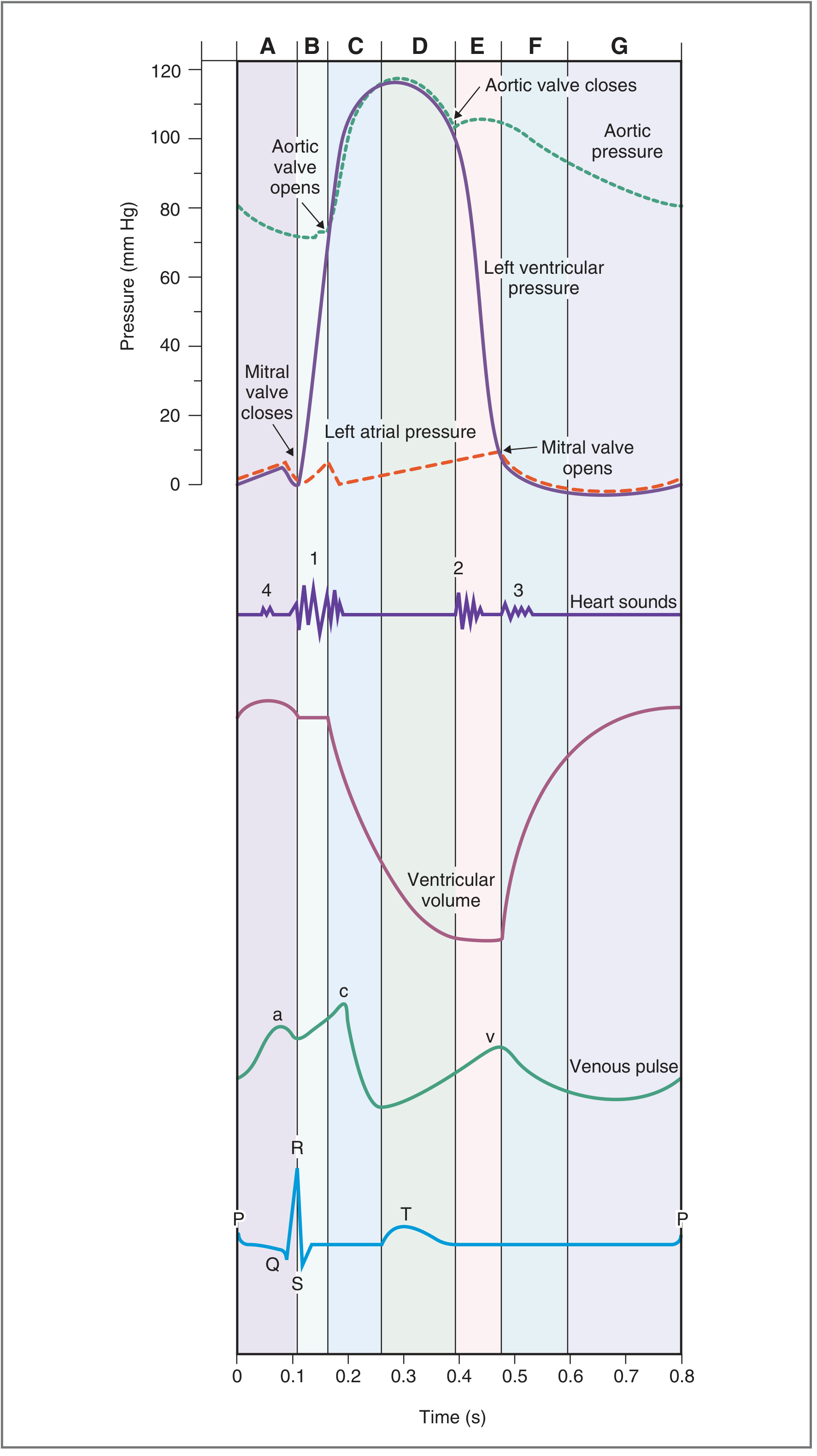
The cycle is classically divided into 7 phases (labeled A through G in Costanzo's framework):
DIASTOLE
Phase A - Atrial Systole (0 - 0.1 sec)
- ECG event: P wave (atrial depolarization)
- Mechanical event: Both atria contract simultaneously
- Atrial contraction raises left atrial pressure, pushing blood through the open mitral valve into the left ventricle
- About 70% of ventricular filling occurs passively during early diastole; atrial contraction contributes the final 30% (atrial kick)
- Left ventricular pressure rises slightly due to the additional volume
- The venous pulse shows the "a" wave (due to atrial contraction)
- Heart Sound: S4 (not normally audible; heard in ventricular hypertrophy where compliance is reduced)
- Duration: ~0.1 sec
Phase G - Reduced Ventricular Filling / Diastasis (late diastole)
- Passive slow filling of the ventricles as the filling rate decreases
- No valve events or heart sounds
- ECG: baseline (between T wave of previous cycle and next P wave)
SYSTOLE
Phase B - Isovolumetric Ventricular Contraction (0.1 - 0.16 sec)
- ECG event: QRS complex (ventricular depolarization)
- The ventricles contract; left ventricular pressure rises sharply
- As soon as LV pressure exceeds left atrial pressure (~5-10 mmHg), the mitral valve closes (tricuspid closes on right side)
- The aortic valve has not yet opened (LV pressure has not exceeded aortic pressure of ~80 mmHg)
- Therefore, all valves are closed - no blood enters or leaves the ventricle
- Ventricular volume remains constant (isovolumetric) while pressure rises dramatically
- Heart Sound: S1 ("lub") - due to mitral + tricuspid valve closure (mitral closes slightly before tricuspid, causing possible physiological splitting)
- Duration: ~0.05 sec
Phase C - Rapid Ventricular Ejection (0.16 - 0.30 sec)
- LV pressure exceeds aortic diastolic pressure (~80 mmHg) → aortic valve opens
- Blood is rapidly ejected from LV into the aorta
- Most of the stroke volume (~70%) is ejected during this phase
- LV pressure and aortic pressure both rise to their peak (~120 mmHg)
- Ventricular volume decreases dramatically
- ECG: ST segment
- Left atrial pressure begins to rise as pulmonary venous return continues to fill the atria
- The venous pulse shows the "c" wave (bulging of tricuspid valve toward atrium)
Phase D - Reduced Ventricular Ejection (0.30 - 0.43 sec)
- Ventricular repolarization begins (T wave on ECG)
- Ventricular pressure begins to fall as myocardium relaxes
- The aortic valve is still open - blood continues to be ejected at a reduced rate
- Ventricular volume continues to fall (reaches minimum = End-Systolic Volume, ESV ≈ 50 mL)
- Blood "runs off" into the arterial tree faster than ejection, so aortic pressure begins to fall
- Left atrial pressure continues to rise (the "v" wave on venous pulse - due to venous return during closed AV valves)
- ECG: T wave (ventricular repolarization)
EARLY DIASTOLE
Phase E - Isovolumetric Ventricular Relaxation (0.43 - 0.50 sec)
- After complete ventricular repolarization, LV pressure drops rapidly
- When LV pressure falls below aortic pressure, the aortic valve closes
- Aortic valve closure produces the dicrotic notch (incisura) on the aortic pressure tracing
- All valves are again closed - ventricular volume is constant (isovolumetric) while pressure falls
- Heart Sound: S2 ("dub") - due to aortic + pulmonic valve closure (aortic closes slightly before pulmonic)
- During inspiration, pulmonic valve closure is delayed (increased venous return to right heart prolongs RV ejection) → physiological splitting of S2
- Duration: ~0.07 sec
Phase F - Rapid Ventricular Filling (0.50 - 0.65 sec)
- When LV pressure falls below left atrial pressure (~5-8 mmHg), the mitral valve opens
- Blood rushes rapidly from the left atrium into the left ventricle down the pressure gradient
- Ventricular volume increases rapidly
- Left atrial pressure falls sharply as the "v" wave descends (the "y" descent)
- Heart Sound: S3 - heard in rapid filling phase; normal in children and young adults; pathological in adults (indicates ventricular volume overload, e.g., heart failure)
- Duration: ~0.15 sec
Key Quantitative Parameters
| Parameter | Normal Value |
|---|---|
| Duration of cardiac cycle (at 72 bpm) | 0.833 sec |
| Systole | ~0.3 sec |
| Diastole | ~0.5 sec |
| End-Diastolic Volume (EDV) | ~130 mL |
| End-Systolic Volume (ESV) | ~50 mL |
| Stroke Volume (SV = EDV - ESV) | ~70-90 mL |
| Ejection Fraction (EF = SV/EDV) | ~65% (normal >55%) |
| Peak LV systolic pressure | ~120 mmHg |
| Peak RV systolic pressure | ~25 mmHg |
| Aortic diastolic pressure | ~80 mmHg |
Pressure-Volume Loop
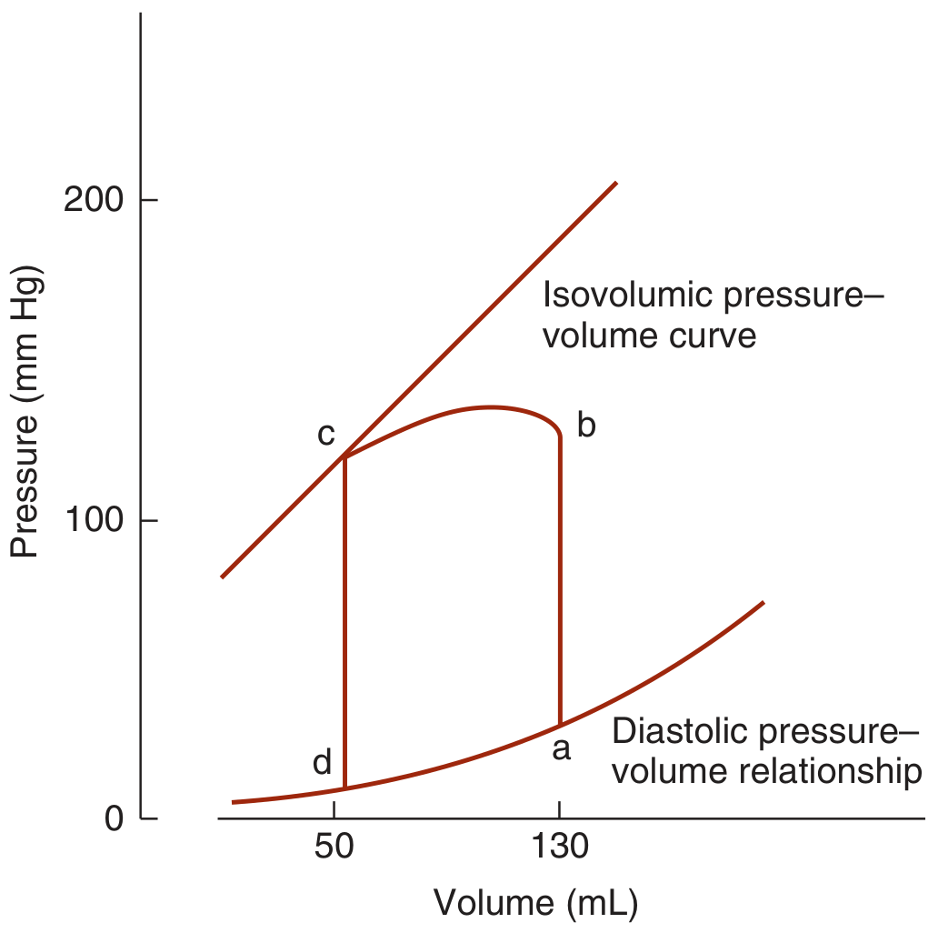
The P-V loop is a graphical representation of one complete cardiac cycle:
- d → a: Passive ventricular filling (diastole) - volume increases, pressure rises slightly
- a → b: Isovolumetric contraction - pressure rises sharply, volume constant
- b → c: Ventricular ejection - volume decreases as blood enters aorta
- c → d: Isovolumetric relaxation - pressure falls rapidly, volume constant
The width of the loop = stroke volume; the area of the loop = stroke work.
Heart Sounds Summary
| Sound | Cause | Phase | Clinical Note |
|---|---|---|---|
| S1 (lub) | Mitral + tricuspid closure | Start of ventricular systole | Best heard at apex/LLSB |
| S2 (dub) | Aortic + pulmonic closure | End of ventricular systole | Physiologically split on inspiration |
| S3 | Rapid ventricular filling | Early diastole | Normal in children; pathological in adults (LV failure) |
| S4 | Atrial contraction against stiff LV | Late diastole (atrial systole) | Pathological; heard in hypertrophy/ischemia |
Venous Pulse Waves
The jugular venous pulse reflects events in the right atrium and has three positive deflections:
- "a" wave - right atrial contraction (atrial systole)
- "c" wave - tricuspid valve bulging toward atrium at onset of ventricular systole
- "v" wave - venous return filling the atrium while tricuspid valve is closed (during ventricular systole)
Corresponding descents: x descent (atrial relaxation + tricuspid being pulled down during ejection) and y descent (tricuspid valve opening, atrial emptying).
Effect of Heart Rate on Cardiac Cycle
When heart rate increases, the duration of the cardiac cycle decreases. Diastole is shortened proportionally more than systole. At very high heart rates:
- Incomplete ventricular filling occurs (reduced EDV → reduced SV)
- At 3× normal heart rate, systole comprises ~65% of the cycle vs ~40% at rest
This is why tachycardia can reduce cardiac output despite a higher rate - insufficient filling time.
Right vs. Left Heart Timing
Although both sides go through identical phases, there are slight asynchronies:
- Right atrial systole precedes left atrial systole
- Right ventricular contraction starts after left ventricular contraction
- However, right ventricular ejection begins before left because pulmonary artery pressure (25 mmHg) is much lower than aortic pressure (80 mmHg)
- The pulmonic valve closes after the aortic valve - basis of physiological S2 splitting
Clinical Correlations
| Condition | Alteration in Cardiac Cycle |
|---|---|
| Aortic stenosis | Prolonged isovolumetric contraction; delayed aortic valve opening |
| Mitral stenosis | Prolonged rapid filling phase; loud S1; opening snap |
| Heart failure | Reduced ejection fraction; S3 gallop; elevated EDV |
| Aortic regurgitation | Wide pulse pressure; increased EDV; dicrotic notch absent |
| Atrial fibrillation | Loss of "a" wave; no S4; variable stroke volumes |
Summary
The cardiac cycle is a precisely coordinated sequence of electrical and mechanical events divided into systole (isovolumetric contraction + ejection) and diastole (isovolumetric relaxation + ventricular filling). Coordinated valve opening and closure, regulated by pressure gradients, ensures unidirectional blood flow. The Wiggers diagram and P-V loop together provide a complete picture of cardiac mechanics. Understanding the cardiac cycle is fundamental to interpreting ECGs, heart sounds, venous pulse waveforms, and the pathophysiology of cardiac diseases.
Sources: Guyton and Hall Textbook of Medical Physiology | Costanzo Physiology, 7th Ed. | Ganong's Review of Medical Physiology, 26th Ed. | Morgan & Mikhail's Clinical Anesthesiology, 7th Ed.
This is a shared conversation. Sign in to Orris to start your own chat.