
Sudden sensorineural hearing loss
"sudden sensorineural hearing loss" treatment
sudden sensorineural hearing loss audiogram
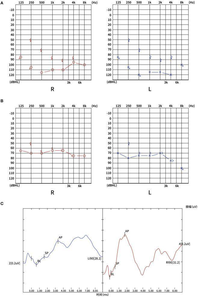
This composite diagnostic image illustrates the audiological evaluation of a patient with bilateral sudden sensorineural hearing loss (SSNHL). Panel A displays the admission pure-tone audiograms (PTA) for the right (R) and left (L) ears, showing total hearing loss with thresholds primarily between 80-120 dBHL across frequencies from 125 Hz to 8 kHz. Panel B shows the follow-up audiogram after three months, demonstrating a partial recovery to severe hearing loss, with thresholds improved to the 60-80 dBHL range. Panel C presents electrocochleogram (ECochG) waveforms for both ears at 90 dBnHL. The waveforms clearly identify the summating potential (SP) and action potential (AP) peaks. The SP/AP ratios (0.25 for the right ear and 0.33 for the left ear) are within normal clinical limits, helping to localize the pathology and assess cochlear function. The images provide a longitudinal view of disease progression and recovery, useful for medical education on otolaryngology and neuro-otology cases involving vascular or idiopathic hearing loss.
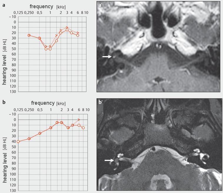
Educational medical composite featuring clinical audiograms and diagnostic MRI images focusing on intralabyrinthine schwannomas. Panel (a) shows a pure-tone audiogram demonstrating a mid-frequency 'cookie-bite' sensorineural hearing loss. Corresponding axial T1-weighted contrast-enhanced MRI (a') reveals a focal, hyperintense enhancing mass within the right cochlea (arrow), consistent with an intracochlear schwannoma. Panel (b) shows an audiogram with low-frequency sensorineural hearing loss. The associated axial T2-weighted MRI (b') identifies a small, hypointense filling defect in the high-signal fluid of the right vestibule (arrow), diagnostic of an intravestibular schwannoma. These images illustrate the correlation between specific inner ear tumor locations and their characteristic audiological presentations. The radiological views highlight the importance of thin-section temporal bone imaging in detecting small intralabyrinthine lesions that may present with symptoms mimicking idiopathic sudden sensorineural hearing loss or Meniere's disease.
This diagnostic image displays two comparative pure-tone audiograms representing initial audiology testing for the right (a) and left (b) ears. Each graph plots frequency in Hertz (Hz) on the x-axis, ranging from 125 Hz to 8000 Hz, against hearing level in decibels (dB) on the y-axis, ranging from -10 dB to 120 dB. The audiogram for the right ear (a) demonstrates a severe-to-profound high-frequency sensorineural hearing loss (SNHL), characterized by a sharp downward slope with thresholds exceeding 80-100 dB at higher frequencies. In contrast, the audiogram for the left ear (b) shows hearing thresholds within the normal to mild loss range across most frequencies, illustrating a significant asymmetrical hearing impairment. These clinical visuals are used in Otolaryngology (ENT) to diagnose and monitor auditory dysfunction, specifically identifying the severity and configuration of sensorineural hearing loss often associated with pathology or sudden onset syndromes.
Sudden Sensorineural Hearing Loss (SSNHL)
Definition
- Cummings Otolaryngology Head and Neck Surgery
Epidemiology
| Feature | Detail |
|---|---|
| Incidence | 5-20 per 100,000 persons/year |
| Peak age | 6th decade |
| Sex | M = F |
| Laterality | Almost always unilateral; simultaneous bilateral is very rare |
| Share of ENT outpatient visits | ~2-3% |
Etiology
Infectious
- Viral (most commonly implicated): mumps, measles, herpes zoster oticus, HSV, VZV, CMV, HIV, EBV, influenza, parainfluenza, Lassa fever, Zika, West Nile virus
- 28% of patients report a viral URTI within 1 month before onset
- Mumps virus has been directly isolated from perilymph of SSNHL patients
- Bacterial: syphilis (important and treatable), Lyme disease, mycoplasma, meningitis (Streptococcal, cryptococcal)
Neoplastic
- Vestibular schwannoma (acoustic neuroma): ~1% of SSNHL presentations - this is the key "must not miss" diagnosis
- Meningioma, epidermoid, hemangioma, temporal bone metastases, lymphoma, leukemia
Traumatic
- Temporal bone fracture, acoustic trauma, inner ear concussion, perilymphatic fistula, barotrauma
Ototoxic
- Aminoglycosides, loop diuretics, cisplatin, quinine, aspirin (high dose)
Immunologic/Autoimmune
- Autoimmune inner ear disease (AIED), Cogan syndrome, polyarteritis nodosa, SLE, Wegener granulomatosis
Vascular
- Labyrinthine artery occlusion, hypercoagulable states, polycythemia, leukemia, macroglobulinemia
Other / Idiopathic
- The majority of cases remain idiopathic after thorough evaluation
- Principal pathogenetic theories for idiopathic cases:
- Viral cochleitis/neuritis
- Vascular occlusion of the labyrinthine artery
- Intracochlear membrane breaks
- Autoimmune damage
Clinical Presentation
- Most common: unilateral hearing loss noticed on awakening (patient woke up deaf in one ear)
- Progressive loss over hours to days
- Aural fullness - very common, sometimes the only complaint
- Tinnitus - variable; may precede hearing loss
- Vertigo/dysequilibrium - present in ~40% of patients; a poor prognostic sign
Diagnosis
Pure-Tone Audiogram
Investigations
| Investigation | Purpose |
|---|---|
| MRI internal auditory canals with gadolinium | Exclude acoustic neuroma / other retrocochlear lesion (mandatory - 1% of acoustic neuromas present as SSNHL) |
| Pure-tone audiometry + speech audiometry | Confirm and characterize loss |
| Tympanometry | Rule out middle ear pathology |
| Blood tests | CBC, ESR, CRP, ANA, FTA-ABS (syphilis serology), lipids, coagulation studies, blood glucose |
| FTA-ABS / VDRL | Syphilis - treatable cause |
| Lyme serology | In endemic areas |
Prognosis
- Severity of loss - more severe = worse prognosis; profound losses have exceptionally poor prognosis
- Audiogram shape - upsloping and mid-frequency losses recover better; downsloping and flat losses do worse
- Presence of vertigo - poor prognostic indicator, especially with downsloping loss
- Age - older age associated with worse outcomes
Management
First-Line: Systemic (Oral) Corticosteroids
- Cummings Otolaryngology
Intratympanic (IT) Steroids
- Primary treatment: randomized controlled data shows no significant difference vs. oral steroids - reinforces oral route as first-line
- Salvage therapy: for patients who fail oral steroids - multiple retrospective and prospective studies show audiometric improvement. Key principle: IT salvage should be used as soon as it is clear oral steroids are not working, preferably within the first 2 weeks of the original insult. The longer the delay, the worse the salvage outcome.
- Dexamethasone: 1-25 mg/mL; or methylprednisolone 62.5 mg/mL
- Volume: 0.3-0.5 mL to fill the middle ear space
- Route: direct IT injection or through tympanostomy tube
Other Treatments (Limited/Investigational)
- Hyperbaric oxygen therapy (HBO): used as adjunct in some centers; a 2026 systematic review (PMID 40747804) examined the evidence - benefit remains debated, especially when added to steroids
- Vasodilators / rheological agents (carbogen, histamine, dextran, pentoxifylline): used in some European centers; evidence quality is low
- Antiviral therapy: acyclovir - no robust evidence supporting routine use for idiopathic cases
- Anticoagulants: no proven benefit in idiopathic SSNHL
Treat Identified Causes
- Syphilis → IV penicillin
- Lyme disease → doxycycline or IV ceftriaxone
- Autoimmune inner ear disease → prolonged immunosuppression
Rehabilitation
- Patients with residual loss: hearing aids, or cochlear implant evaluation for profound losses
- Contralateral CROS/BiCROS hearing aids for single-sided deafness
Summary Flowchart
Sudden hearing loss (≥30 dB, 3 frequencies, ≤72 hrs)
        ↓
Confirm SNHL on audiogram (rule out CHL with tuning forks / tympanometry)
        ↓
Treat as otologic emergency — start oral prednisone ASAP
        ↓
Urgent MRI IAC + gadolinium (exclude acoustic neuroma)
        ↓
Targeted bloodwork (FTA-ABS, CBC, ESR, ANA, glucose, lipids)
        ↓
No improvement after 2 weeks?
        ↓
Intratympanic steroid salvage (within 2 weeks of onset)
        ↓
Audiology follow-up; consider HBO, hearing rehabilitation
Recent Evidence (2024-2026)
- SSNHL is an otologic emergency - treat promptly with oral steroids (prednisone 1 mg/kg/day)
- MRI IAC with gadolinium is mandatory to exclude retrocochlear pathology (acoustic neuroma)
- Intratympanic steroids are valuable salvage if oral steroids fail - use within 2 weeks
- Spontaneous recovery occurs in 30-65% without treatment, so all recovery rates must be interpreted against this background rate
Invasive Fungal sinusitis classification
"invasive fungal sinusitis" classification diagnosis
invasive fungal sinusitis mucormycosis nasal black eschar necrosis
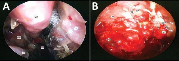
This composite of two endoscopic images (Panels A and B) illustrates advanced rhino-orbital mucormycosis. Panel A (Right Nasal Cavity) demonstrates characteristic signs of invasive fungal infection, including a thick blackish eschar containing fungal elements situated between the middle turbinate (MT) and the nasal septum (S). Localized necrosis is visible on the septum inferior to the eschar. The middle meatus (MM) exhibits significant crusting and mucopurulent discharge trickling toward the nasopharynx (NP), suggestive of secondary sinusitis. The inferior turbinate (IT) appears hypertrophic. Panel B (Left Nasal Cavity) shows more extensive tissue destruction and complications. Key findings include an extradural abscess (EDA) undergoing transnasal drainage and multiple polyps near the ethmoidal fovea (FP). There is notable destruction of the crista ethmoidalis (CE), the middle turbinate, and the orbital roof (OR) due to direct fungal invasion. These images highlight the angioinvasive nature of Mucorales, leading to hallmark features of tissue infarction, black eschar formation, and rapid destruction of cartilaginous and bony sinonasal structures in immunocompromised patients, such as those following COVID-19 treatment.
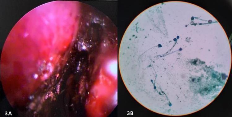
The image consists of two panels demonstrating clinical and laboratory findings associated with invasive fungal sinusitis. Panel A is a diagnostic nasal endoscopy (DNE) photograph showing the right nasal cavity. The mucosa is significantly erythematous and inflamed, featuring extensive dark-brown to black devitalized tissue (eschar) along the turbinates and lateral wall, characteristic of angio-invasive fungal necrosis. Panel B is a bright-field microscopy image of a potassium hydroxide (KOH) mount prepared from a nasal biopsy. It demonstrates fungal elements consistent with Mucorales, characterized by broad, pauciseptate (ribbon-like) hyphae and rounded sporangia/spores. These findings are pedagogically relevant for identifying rhino-orbital-cerebral mucormycosis (ROCM), particularly in immunocompromised or diabetic patients. The combination of visual necrotic tissue (black turbinate sign) and specific fungal morphology provides a comprehensive diagnostic overview of acute invasive fungal infection.
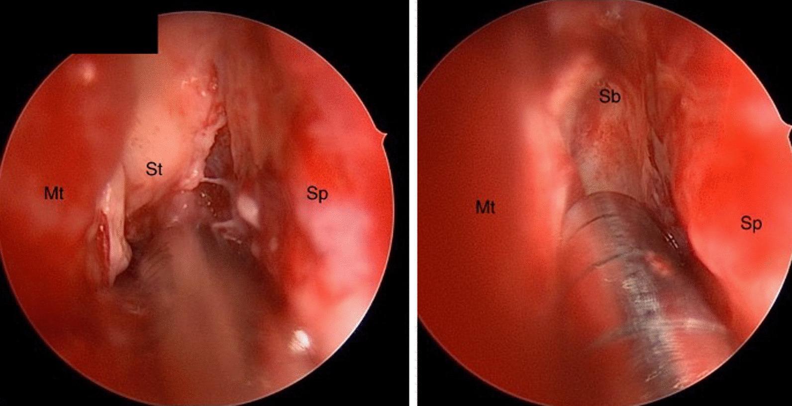
Clinical endoscopic images of the right nasal cavity demonstrating key anatomical landmarks and pathological changes associated with invasive fungal sinusitis. The view identifies the nasal septum (Sp) medially and the middle turbinate (Mt) laterally. Centrally, the superior turbinate (St) exhibits avascular fibrinous conversion, characterized by a pale, blanched tissue appearance lacking normal vascularity. The pathology extends superiorly to the level of the anterior skull base (Sb). Notably, there is an absence of the classic black necrotic eschar frequently observed in advanced invasive fungal infections. The images provide a comparison of the spatial relationships between these structures, with labels highlighting the attachment points at the skull base and the relative positioning of the turbinates. These findings are clinically significant for the early identification of rhinocerebral mucormycosis in high-risk patients, such as those with diabetic ketoacidosis, where tissue devitalization may precede overt necrosis.
Invasive Fungal Sinusitis (IFS) - Classification
Overview Classification Scheme
Fungal Rhinosinusitis (FRS)
│
├── NON-INVASIVE
│   ├── Fungal Ball (Mycetoma)
│   ├── Allergic Fungal Sinusitis (AFS)
│   └── Eosinophilic Fungal Rhinosinusitis (EFRS)
│
└── INVASIVE (IFS) ← histologic tissue invasion
    ├── 1. Acute Invasive Fungal Sinusitis (AIFS)
    ├── 2. Chronic Invasive Fungal Sinusitis (CIFS)
    └── 3. Granulomatous Invasive Fungal Sinusitis (GIFS)
Defining hallmark of all invasive forms: Perineural invasion or angioinvasion by fungal elements on histopathology - KJ Lee's Essential Otolaryngology
Comparative Summary Table
| Feature | Acute Invasive (AIFS) | Chronic Invasive (CIFS) | Granulomatous Invasive (GIFS) |
|---|---|---|---|
| Time course | <4 weeks (days) | >4 weeks (months) | Months to years |
| Immune status | Immunocompromised | Immunocompetent / mildly immunocompromised | Immunocompetent |
| Key pathogens | Mucorales (Rhizopus, Mucor) > Aspergillus | Aspergillus spp., dematiaceous molds > Mucorales | A. flavus, A. fumigatus |
| Geography | Worldwide | Worldwide | Middle East, South Asia, Africa (Sudan, India, Pakistan, Saudi Arabia) |
| Mortality | ~50% (systematic review data) | Lower; depends on extent | Low if treated |
| Histology | Angioinvasion, perineural invasion, necrosis | Dense hyphae with tissue invasion & necrosis | Noncaseating granulomas, giant cells, fibrosis, vasculitis |
| Hallmark presentation | Rapid deterioration in immunocompromised patient | Chronic CRS with proptosis / cranial neuropathy | Unilateral proptosis - often mistaken for tumor |
1. Acute Invasive Fungal Sinusitis (AIFS)
Risk Factors / Predisposing Conditions
- Hematologic malignancies (leukemia, lymphoma) and bone marrow transplantation
- Chemotherapy-induced neutropenia (<500 neutrophils/μL = severe risk)
- Solid organ transplantation (chronic immunosuppression)
- Diabetic ketoacidosis (DKA) - acidic environment activates Mucorales enzyme ketone reductase
- Advanced HIV/AIDS with neutrophil dysfunction
- Dialysis patients receiving deferoxamine (acts as a siderophore for Rhizopus species, increasing iron availability and fungal proliferation)
Causative Fungi
- Rhizopus (most common), Mucor, Rhizomucor, Absidia, Cunninghamella
- Morphology: fast-growing, non-septate (coenocytic) hyphae branching at wide angles (~90°)
- "Mucormycosis" refers specifically to Mucorales infection - do NOT use interchangeably with AIFS unless confirmed by culture
- A. fumigatus (most common Aspergillus species in AIFS), A. flavus, A. niger, A. terreus
- Morphology: regular septate hyphae branching at acute angles (45°)
Clinical Features
- Nasal obstruction, mucous drainage, facial pressure, smell loss, fever, epistaxis
- Pale / ischemic nasal mucosa on endoscopy
- Anesthesia of mucosa to manipulation, minimal bleeding on biopsy (early ischemia)
- Black eschar on turbinates / septum (classic mucormycosis finding)
- Orbital: proptosis, ecchymosis, ophthalmoplegia, blindness
- Palatal necrosis / ulceration
- Infraorbital nerve numbness (premaxillary anesthesia - frequently present)
- Cranial nerve deficits, altered mental status
Diagnosis
- Antero-inferior portion of middle turbinate is the preferred biopsy site (85% sensitivity, 100% specificity for frozen section)
- Frozen section intraoperatively is key for rapid diagnosis
- Pathognomonic finding: angioinvasion or perineural invasion by fungal elements
- Histologic stains: Silver stain (GMS) - most sensitive (silver deposits into fungal cell wall); Calcofluor white; H&E has high false negatives
- Culture: slow - do not await results; absence of growth does not rule out disease
- Early: unilateral soft tissue mucosal thickening (turbinates, septum, nasal walls)
- Late: soft tissue emphysema, bony erosion (late-phase change)
- Extrasinus fat stranding - evaluate for periantral, orbital, pterygopalatine fossa involvement
- T1/T2: low-to-intermediate signal intensity in affected regions
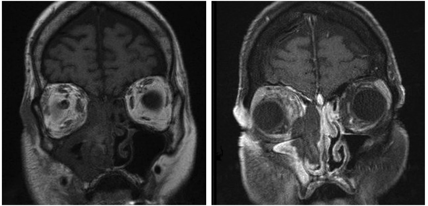
- "Black turbinate sign" - non-enhancement of sinonasal mucosa on post-contrast T1/T2 = mucosal necrosis from angioinvasion (highly suggestive)
- Extrasinus invasion enhances with gadolinium (contrast to the non-enhancing necrotic mucosa)
- Look for: orbital invasion, cavernous sinus thrombosis, leptomeningeal involvement, intracranial extension
Prognosis
- Overall mortality ~50% (largest systematic review)
- Negative prognosticators: advanced age, palatal/facial involvement, severe neutropenia (<500/μL), CRP ≥5.50 mg/dL, altered mental status, renal/liver failure, intracranial extension
- Positive prognosticators: reversible immunosuppression (e.g., DKA), surgical resection, treatment with liposomal amphotericin B
Treatment - Three Pillars
- Endoscopic surgery preferred for disease limited to sinonasal cavity
- Open approaches (maxillectomy, orbital exenteration) for intraorbital/palatal/intracerebral involvement
- Note: orbital exenteration has NOT been shown to improve survival outcomes
- Endoscopic resection showed 64% survival vs. open resection 54% (systematic review)
- Debride until normal bleeding mucosa or non-eroded bone is reached
- "Second-look" endoscopy at regular intervals until mucosal stabilization
- Endocrinology: control DKA / hyperglycemia (reversal improves survival significantly)
- Reduce/stop immunosuppressive drugs where possible
- Adjuncts: HBO (limited evidence), GM-CSF / G-CSF cytokine therapy, granulocyte transfusion (investigational)
- Amphotericin B (liposomal) - antifungal of choice empirically when culture unavailable or zygomycosis confirmed
- Voriconazole - drug of choice when Aspergillus is confirmed
- Treatment duration: prolonged (weeks to months)
2. Chronic Invasive Fungal Sinusitis (CIFS)
- Duration >4 weeks (months of symptoms)
- Considered a less fulminant form of AIFS
- Affects immunocompetent or mildly immunocompromised patients (e.g., poorly controlled diabetes mellitus, low-dose glucocorticoids)
- Pathogens: Aspergillus spp. and dematiaceous (black) molds more common than Mucorales
- Months of chronic rhinosinusitis symptoms (maxillary pressure, congestion) before complications develop
- Proptosis or visual changes (orbital invasion)
- Neurologic changes / cranial neuropathies
- Pre-antral cellulitis
- Tissue biopsy: dense hyphae similar to fungal ball + tissue invasion with necrosis
- CT: mass lesion with mucosal thickening; bony erosion and extrasinus involvement are common
- MRI: iso- or hypointense T1/T2 signals in affected regions
- Time course and immune status distinguish CIFS from AIFS
3. Granulomatous Invasive Fungal Sinusitis (GIFS)
- Rare form of chronic invasive FRS with characteristic non-caseating granuloma formation
- Geography: Primarily Middle East, South Asia, and Sub-Saharan Africa (Sudan, India, Pakistan, Saudi Arabia); rarely reported in USA/Europe
- Affects healthy, immunocompetent patients
- Pathogens: Aspergillus flavus and Aspergillus fumigatus (most common)
- Most common symptom: unilateral proptosis (not typical CRS symptoms)
- Often misdiagnosed as suspected tumor until intraoperative biopsy confirms
- Disease is often advanced at presentation due to its indolent nature
- Non-caseating granuloma with foreign body reaction
- Multinucleated giant cells
- Dense background fibrosis and vasculitis
- CT: large expansive, homogeneously hyperdense mass with contrast enhancement; lacks dual-density secretions seen in fungal balls; bony erosion and extrasinus extension common
- MRI: T1/T2 hypointensity; intense homogenous enhancement; cerebritis (foci of parenchymal enhancement adjacent to the solid lesion)
- Surgical resection + prolonged antifungal therapy
- Conservative, orbit-sparing surgery supported by case reports given good responsiveness to therapy
- Note: GIFS is considered by some authorities to be a subset of CIFS rather than a separate entity - Scott-Brown's Otorhinolaryngology
Key Distinguishing Points at a Glance
| AIFS | CIFS | GIFS | |
|---|---|---|---|
| Hallmark path finding | Angioinvasion / perineural invasion | Dense hyphae + necrosis | Non-caseating granuloma + giant cells |
| Urgency | Surgical emergency | Weeks to months | Months to years |
| Fungus shape | Mucorales: broad non-septate 90° branching; Aspergillus: septate 45° branching | Septate hyaline/dematiaceous molds | Aspergillus (septate 45°) |
| Imaging hallmark | "Black turbinate sign" on MRI | Bony erosion, extrasinus mass | Expansive homogenous mass; no dual density |
| Presentation clue | Immunocompromised + rapid deterioration | Chronic CRS symptoms + orbital/neuro signs | Unilateral proptosis in immunocompetent |
Name of fungal classification
Which fungal classification are you asking about?
De shazo classification
deShazo classification fungal rhinosinusitis 1997 categories
deShazo Classification of Fungal Rhinosinusitis (1997)
- Arch Otolaryngol Head Neck Surg. 1997;123:1181-8 (invasive types)
- J Allergy Clin Immunol. 1995;96:24-35 (allergic fungal sinusitis criteria)
CATEGORY 1: NON-INVASIVE (No mucosal invasion)
| Type | Key Features |
|---|---|
| 1. Saprophytic Fungal Infestation | Fungi colonizing crusts/mucus; no tissue reaction; incidental finding |
| 2. Fungal Ball (Mycetoma) | Dense conglomeration of hyphae in sinus cavity; separate from mucosa; no invasion; usually maxillary sinus |
| 3. Allergic Fungal Rhinosinusitis (AFRS) | Hypersensitivity (Type I + III) to fungal antigens; atopic patients; eosinophilic allergic mucin with Charcot-Leyden crystals; no tissue invasion |
CATEGORY 2: INVASIVE (Histologic tissue invasion present)
| Type | Time Course | Immune Status | Pathology |
|---|---|---|---|
| 1. Acute Invasive (Fulminant) | <4 weeks | Immunocompromised (neutropenia, DKA, transplant) | Angioinvasion, perineural invasion, necrosis |
| 2. Chronic Invasive | >4 weeks (months) | Immunocompetent / mildly immunocompromised | Dense hyphae + tissue necrosis |
| 3. Granulomatous Invasive | Months to years | Immunocompetent | Non-caseating granulomas, giant cells, fibrosis |
The Schema in One Line
deShazo FRS Classification
├── NON-INVASIVE
│   ├── Saprophytic infestation
│   ├── Fungal ball (Mycetoma)
│   └── Allergic Fungal Rhinosinusitis (AFRS)
│
└── INVASIVE
    ├── Acute Invasive (Fulminant)
    ├── Chronic Invasive
    └── Granulomatous Invasive
Key Points to Remember
- The unifying criterion for invasive disease is histopathologic evidence of fungal invasion of mucosa, submucosa, blood vessels, or bone
- AFRS (non-invasive) is the most common form overall
- Acute invasive is the most dangerous form - 50% mortality even with treatment
- Granulomatous invasive is geographically restricted (Sudan, India, Pakistan, Saudi Arabia) and occurs in immunocompetent hosts - almost exclusively Aspergillus flavus
- The deShazo classification was later refined by Chakrabarti et al. (2009) in Laryngoscope to address controversies, but deShazo's framework remains the most widely taught and referenced schema
Pathogenesis of allergic rhinitis
allergic rhinitis IgE mast cell sensitization pathogenesis diagram
allergic rhinitis early late phase response eosinophil Th2
Pathogenesis of Allergic Rhinitis
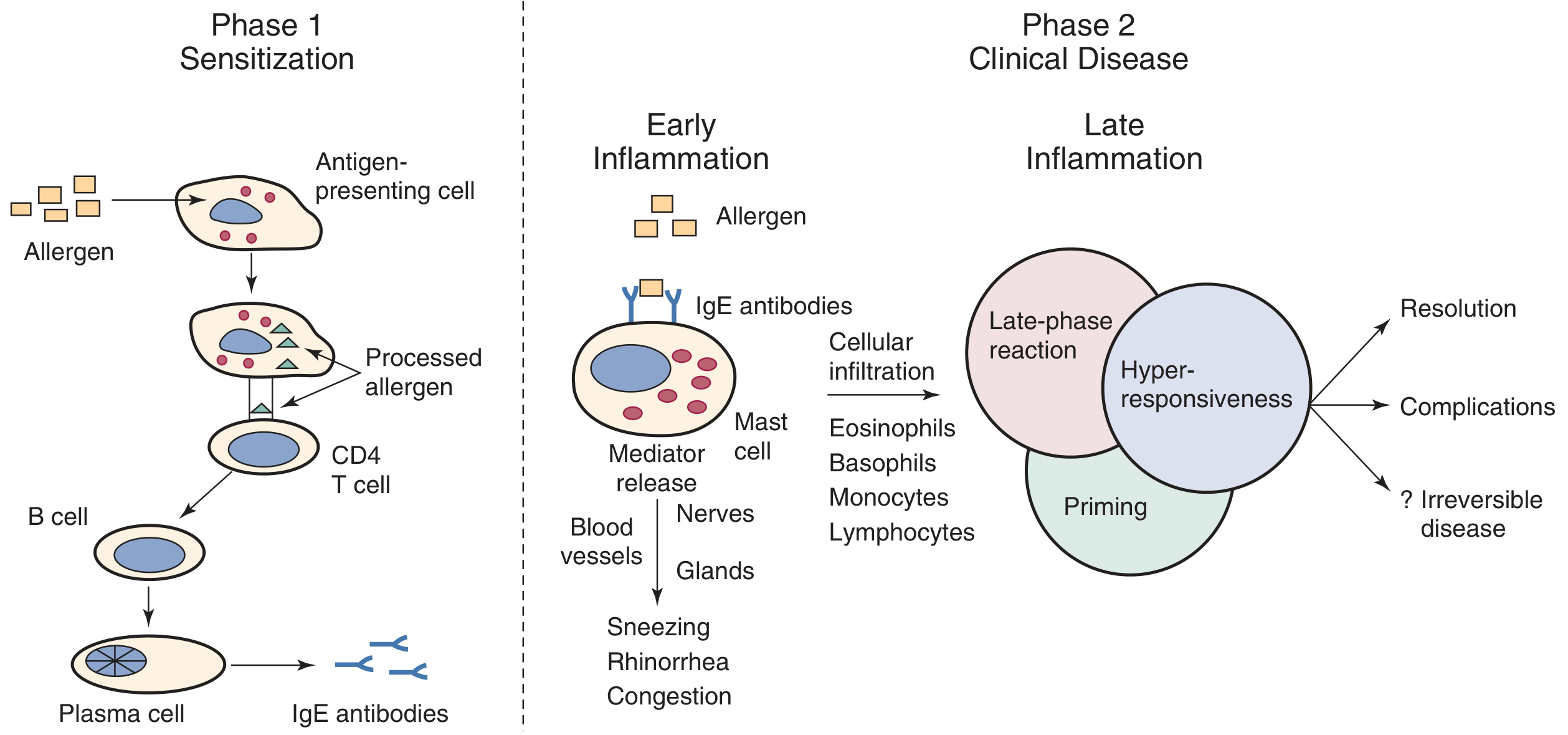
PHASE 1: SENSITIZATION (Initial Allergen Exposure)
Step 1 - Allergen Deposition and Epithelial Signaling
- Inhaled allergen (pollen, dust mite, pet dander) deposits on the nasal mucosal surface
- Mucosal epithelial cells are the first responders - they secrete key cytokines including:
- TSLP (thymic stromal lymphopoietin)
- IL-25 and IL-33
- These cytokines drive maturation of dendritic cells into TH2-promoting subtypes
Step 2 - Antigen Processing by APCs
- Allergen is engulfed by antigen-presenting cells (APCs): macrophages, dendritic cells, Langerhans cells
- Allergen is partially degraded and peptide fragments are presented on MHC class II molecules
- APCs also secrete cytokines that attract and activate TH2 lymphocytes
Step 3 - TH2 Lymphocyte Polarization
- APCs present antigen to naive CD4+ T cells, which polarize to the TH2 phenotype
- TH2 cells secrete a characteristic cytokine profile:
- IL-4 → drives IgE class switching in B cells
- IL-5 → eosinophil recruitment, activation, and survival
- IL-13 → mucus hypersecretion, goblet cell hyperplasia
- IL-3 → mast cell and basophil proliferation
Step 4 - IgE Production and Mast Cell Sensitization
- TH2 cytokines (primarily IL-4 and IL-13) instruct B cells to undergo class switching → differentiate into plasma cells secreting allergen-specific IgE
- IgE antibodies bind with high affinity to FcεRI receptors on:
- Mast cells (in nasal mucosa)
- Basophils (circulating)
- The patient is now sensitized - future allergen exposure will trigger immediate symptoms
At this stage: no symptoms. The patient only develops symptoms on re-exposure.
PHASE 2: CLINICAL DISEASE (Re-exposure to Allergen)
EARLY PHASE RESPONSE (Immediate - within minutes)
Mechanism:
- Inhaled allergen cross-links adjacent allergen-specific IgE molecules on mast cell surface
- IgE cross-linking activates FcεRI signaling cascade → mast cell degranulation
- Release of preformed mediators (stored in granules) and newly synthesized mediators
Mediators Released:
| Mediator | Source | Effect |
|---|---|---|
| Histamine | Mast cell granules | Vasodilation, increased vascular permeability, stimulates nerve endings → sneezing, itch, rhinorrhea |
| Tryptase | Mast cell granules | Marker of mast cell activation; activates matrix metalloproteinases, recruits more inflammatory cells |
| Prostaglandin D2 (PGD2) | Newly synthesized (arachidonic acid via COX) | Vasodilation, bronchoconstriction |
| Leukotriene C4 (LTC4) | Newly synthesized (arachidonic acid via 5-LOX) | Mucus secretion, vasodilatation, increased permeability |
| Leukotriene B4 (LTB4) | Newly synthesized | Neutrophil chemoattractant |
| Platelet-activating factor (PAF) | Mast cells | Vasodilation, increased permeability |
| Bradykinin / Kinins | Generated in nasal secretions | Glandular secretion, pain, nerve stimulation |
Symptoms (onset within minutes):
- Pruritus and tingling (first sensation)
- Sneezing (reflex arc stimulated by histamine on sensory nerve endings)
- Watery rhinorrhea (glandular hypersecretion + transudation)
- Nasal congestion (vasodilation + vascular engorgement)
Neuronal Amplification:
- Sensory C-fibers in nasal mucosa release neuropeptides (substance P, CGRP, neurokinin A) → neurogenic inflammation
- Axon reflex causes bilateral response even with unilateral challenge
- Parasympathetic reflex (cholinergic): amplifies glandular secretion - blocked by atropine
- Nasal-ocular reflex: explains conjunctivitis accompanying nasal challenge
LATE PHASE RESPONSE (4-10 hours after exposure, peak ~6 hours)
Mechanism:
- Mediators from the early phase act as chemoattractants and upregulate adhesion molecules (ICAM-1, VCAM-1, E-selectin) on vascular endothelium
- Massive cellular infiltration into nasal mucosa:
- Eosinophils (dominant cell - recruited by IL-5, eotaxin)
- Basophils
- CD4+ TH2 lymphocytes
- Monocytes/macrophages
- Neutrophils (early)
Eosinophil-Mediated Damage:
- Activated eosinophils release:
- Major basic protein (MBP) - toxic to epithelium
- Eosinophil cationic protein (ECP) - toxic to nerves and epithelium
- Eosinophil peroxidase
- Leukotrienes (LTC4, LTD4)
- Results in epithelial damage, goblet cell hyperplasia, subepithelial fibrosis
Dominant Symptom:
- Nasal congestion (most prominent symptom of late phase)
- Persists 4-24 hours after exposure
PHASE 3: CHRONIC CHANGES - PRIMING AND NASAL HYPERRESPONSIVENESS
1. Priming Effect
- Repeated allergen exposures lower the threshold for allergic response
- The same dose of allergen causes progressively larger responses
- Explains why symptoms worsen through pollen season even with constant allergen levels
- Mechanism: inflammatory cell infiltration → increased density of IgE-bearing cells → amplified mediator release
2. Nasal Hyperresponsiveness
- The inflamed nasal mucosa becomes hyperreactive to non-specific stimuli (cold air, cigarette smoke, strong odors, pollution, exercise)
- Resembles bronchial hyperreactivity in asthma
- Caused by:
- Increased density of mast cells and eosinophils
- Sensory nerve sensitization (lowered threshold)
- Altered mucosal barrier
3. Systemic Allergic Inflammation
- An allergic reaction at one site generates a systemic inflammatory response
- Can reach the systemic circulation → involvement of other predisposed end organs (eyes, bronchi)
- Explains the strong association of AR with allergic asthma, allergic conjunctivitis, and atopic dermatitis ("united airway" concept)
Summary Flowchart
SENSITIZATION PHASE
Allergen → Epithelial cells (TSLP, IL-25, IL-33)
    ↓
APCs (dendritic cells) → process & present allergen
    ↓
TH2 polarization → IL-4, IL-5, IL-13 secretion
    ↓
B cells → Plasma cells → Allergen-specific IgE
    ↓
IgE binds to FcεRI on mast cells/basophils in nasal mucosa
    ↓ [Patient now sensitized - no symptoms yet]

RE-EXPOSURE PHASE
Allergen → IgE cross-linking → Mast cell degranulation
    ↓
EARLY (0-30 min): Histamine, PGD2, LTC4 →
    Sneezing, Rhinorrhea, Pruritus, Congestion
    ↓
LATE (4-10 hrs): Eosinophil/basophil infiltration →
    Persistent congestion, chronic inflammation
    ↓
CHRONIC: Priming + Nasal hyperresponsiveness →
    Lower allergen threshold, non-specific reactivity