
Gradinego syndrome
Gradenigo syndrome petrous apex
Gradenigo syndrome petrous apex anatomy diagram
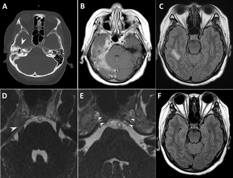
This composite figure displays diagnostic imaging of a patient with Gradenigo’s syndrome, emphasizing complications of chronic otitis media. Panel A is an axial non-contrast CT scan of the head showing decreased pneumatization of the right mastoid air cells and opacification of the petrous apex (arrowhead), indicative of petrous apicitis. Panels B and C are axial MRI slices (Post-contrast T1-weighted and FLAIR, respectively) demonstrating right-sided pachymeningitis, leptomeningitis, and hyperintense signal in the temporal lobe representing cerebritis. Panel D focuses on the right Meckel’s cave, showing inflammatory enhancement involving the trigeminal nerve (arrowhead). Panel E provides a detailed view of the Dorello canal, highlighting inflammatory swelling (arrowheads) which correlates with abducens nerve palsy. Panel F shows a follow-up axial MRI demonstrating complete resolution of the right temporal lobe cerebritis. This series illustrates the classic clinical triad of Gradenigo’s syndrome: otorrhoea, retro-orbital pain (trigeminal nerve involvement), and abducens nerve palsy, linked to apical petrositis.
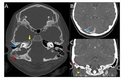
This composite diagnostic image features head computed tomography (CT) scans illustrating complications of Gradenigo's syndrome. Image A is an axial CT scan in a bone window demonstrating complete opacification of the right tympanic cavity (blue arrow) and mastoid air cells (red arrow) with fluid, consistent with acute otomastoiditis. Additionally, there is inflammatory involvement and morphological alteration of the right petrous apex (yellow arrow), indicating petrous apicitis. Images B (axial) and C (coronal) are contrast-enhanced CT scans showing a central filling defect within the right dural venous sinuses. Specifically, there is evidence of acute thrombosis involving the right transverse sinus (blue arrowhead), sigmoid sinus (red arrowhead), and the jugular bulb (yellow arrowhead). This constellation of findings—otomastoiditis, petrous apicitis, and subsequent dural venous sinus thrombosis—highlights the progression of severe middle ear infections into intracranial vascular complications and Gradenigo's syndrome.
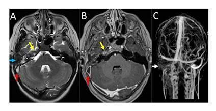
This composite diagnostic image displays brain MRI findings in a patient with Gradenigo syndrome and associated complications. Panel A is an axial T2-weighted sequence showing hyperintense inflammatory changes in the right petrous apex (yellow arrow) and the right tympanic cavity (blue arrow), consistent with petrous apicitis and otomastoiditis. A red arrow indicates signal abnormality in the right sigmoid sinus. Panel B is a post-contrast T1-weighted axial image demonstrating peripheral enhancement in the right petrous apex (yellow arrow) and a filling defect in the sigmoid sinus (red arrow). Panel C presents a Time-of-Flight (TOF) venous MR angiography (MRV), which reveals a complete absence of flow signal in the right transverse sinus, sigmoid sinus, and internal jugular vein (white arrows), confirming extensive dural venous sinus thrombosis. The left-sided venous structures appear patent. These findings illustrate the infectious spread from the middle ear to the petrous apex and the subsequent development of intracranial venous thrombosis.
Gradenigo Syndrome (Petrous Apicitis)
Definition
The Classic Triad
| Feature | Mechanism |
|---|---|
| Otorrhea (suppurative otitis media) | Infection spreading from middle ear/mastoid to petrous apex |
| Retro-orbital / deep facial pain | Irritation of CN V (trigeminal nerve) at Meckel's cave |
| Diplopia | CN VI (abducens) palsy - nerve compressed in Dorello's canal |
- KJ Lee's Essential Otolaryngology defines it as: otorrhea + retro-orbital pain (CN V irritation) + diplopia due to CN VI palsy.
- Cummings Otolaryngology: "The classic symptoms of petrous apicitis include deep facial pain, otitis media, and ipsilateral abducens nerve paralysis."
Pathophysiology
- Only ~30% of temporal bones have a pneumatized petrous apex; infection can only spread here when pneumatization is present.
- Infection tracks from the middle ear and mastoid through perilabyrinthine air cell tracts into the petrous apex.
- Results in a spectrum from asymptomatic effusion to frank abscess formation (petrous apicitis / extradural abscess).
- The abducens nerve (CN VI) is vulnerable because it runs a long intracranial course through Dorello's canal, directly adjacent to the petrous apex.
- The trigeminal nerve (CN V) is irritated where it lies in Meckel's cave at the petrous tip, causing deep retro-orbital pain.
- Now rare in the antibiotic era, but still encountered.
Additional Cranial Nerve Involvement
- CN VII (facial nerve) - facial palsy
- CN VIII (vestibulocochlear) - sensorineural hearing loss, vertigo
- CN IX, X, XI (jugular foramen syndrome)
- CN XII (hypoglossal)
Causative Organisms
- Historically: Streptococcus pneumoniae, Haemophilus influenzae
- Recent case reports highlight anaerobes as well, including Fusobacterium necrophorum - a cause that may be underrecognized (Ibrahim et al., 2024)
Diagnosis
- HRCT temporal bones (high-resolution CT) - first-line imaging:
- Dissolution of bony septa with coalescence
- Opacification of petrous apex air cells
- Bone erosion patterns
- MRI with contrast - to evaluate for intracranial complications (meningitis, cerebritis, venous sinus thrombosis, epidural abscess)
- Shows inflammatory enhancement at Meckel's cave, Dorello's canal, and petrous apex
- MR venography if venous sinus thrombosis is suspected
Complications
- Dural venous sinus thrombosis (transverse sinus, sigmoid sinus, internal jugular vein)
- Meningitis / leptomeningitis
- Cerebritis / brain abscess
- Pachymeningitis
- As noted in Harrison's Principles of Internal Medicine 22E: transverse sinus thrombosis may itself present with the Gradenigo picture.
Treatment
| Approach | Indication |
|---|---|
| Prolonged IV antibiotics | First-line; broad spectrum targeting otogenic pathogens |
| Petrosectomy / partial apicectomy | Abscess formation, necrotic bone, or failure of medical therapy |
- Retrolabyrinthine
- Infraochlear
- Infralabyrinthine
- Subarcuate ("hole in the doughnut")
- Middle fossa craniotomy
- Translabyrinthine or transcochlear (non-hearing ear)
Historical Note
- First described as a triad by Giuseppe Gradenigo (Italian otologist) in the early 1900s.
- A patient with petrous apicitis and the Gradenigo triad was initially described by Goris.
- In Gradenigo's own review of 57 patients with petrositis, only 24 had the pure triad - others had multiple complications.
- The classic petrous apex exenteration operation was described by Ramandier (1933) and Lempert shortly after.
Key Images
Memory Aid
- The petrous apex sits between CN V (Meckel's cave) and CN VI (Dorello's canal)
- Pus here = pain (V) + diplopia (VI) + ear discharge