
Apoptosis
Reading File
Now let me search the medical library for apoptosis content across multiple angles simultaneously.
Finding Sources
Searching PubMed
apoptosis mechanisms caspase pathways
Robbins Pathologic Basis of Disease and Robbins Basic Pathology have the most authoritative coverage. Let me read those in parallel with the immunology textbook content.
Reading File
Reading File
Reading File
Good - Robbins Pathologic Basis of Disease is the most detailed source. Let me read more of it - including the mechanisms section (intrinsic/extrinsic pathways) and also check for images.
Reading File
Reading File
Excellent content. Let me fetch the key diagram and read more of both texts for morphology, execution, and diseases.
Loading Image
Reading File
Reading File
I now have comprehensive content. Let me also get the morphology and the Robbins PBD detailed pathway sections.
Reading File
I have all the key information needed. Let me fetch the morphology image for visual completeness.
Loading Image
I now have all the material needed for a comprehensive response. Let me compile this into a thorough, well-structured answer.
Apoptosis
Apoptosis is a form of programmed cell death in which a cell activates internal enzymes to systematically dismantle itself - degrading its own DNA and proteins - and packages its contents into membrane-bound fragments (apoptotic bodies) that are quietly removed by phagocytes. Unlike necrosis, the plasma membrane remains intact throughout, cellular contents are never released into the extracellular space, and no inflammatory reaction is triggered.
The name comes from the Greek for "falling off" (like leaves from a tree) and was first described morphologically in 1972.
Definition
Apoptosis is a type of cell death induced by a tightly regulated suicide program in which cells destined to die activate intrinsic enzymes that degrade cellular DNA and nuclear and cytoplasmic proteins.
- Robbins, Cotran & Kumar: Pathologic Basis of Disease
Causes of Apoptosis
Physiologic (Normal) Situations
Apoptosis is the mechanism by which ~1 million cells are turned over per second in the human body. It is essential for:
| Situation | Example |
|---|---|
| Embryonic development (removal of supernumerary cells) | Webbing between fingers eliminated by apoptosis |
| Hormone-dependent tissue involution | Endometrial breakdown during menstruation; ovarian follicular atresia in menopause; lactating breast regression after weaning |
| Tissue homeostasis | Turnover of intestinal crypt epithelial cells |
| Lymphocyte selection | Elimination of immature lymphocytes failing to produce functional antigen receptors in bone marrow and thymus |
| Elimination of self-reactive lymphocytes | Preventing autoimmunity |
| Post-response cell death | Neutrophils after acute inflammation; lymphocytes after immune responses |
Pathologic Situations
Apoptosis also eliminates irreparably damaged cells to minimize tissue harm:
- DNA damage - Radiation, cytotoxic drugs, and free radicals. If repair fails, the cell triggers apoptosis to prevent survival of potentially malignant, mutated cells.
- Misfolded protein accumulation - ER stress activates proapoptotic BH3-only proteins.
- Viral infections - The virus itself (adenovirus, HIV) or the host CTL response (viral hepatitis) induces apoptosis of infected cells. The same CTL mechanism kills tumor cells, mediates transplant rejection, and underlies graft-versus-host disease.
- Duct obstruction - Pathologic atrophy in the pancreas, parotid gland, and kidney.
Mechanisms of Apoptosis
Two major pathways both converge on caspase activation. Caspases are cysteine proteases that cleave after aspartic acid residues - the name is a portmanteau of "cysteine-aspartic proteases."
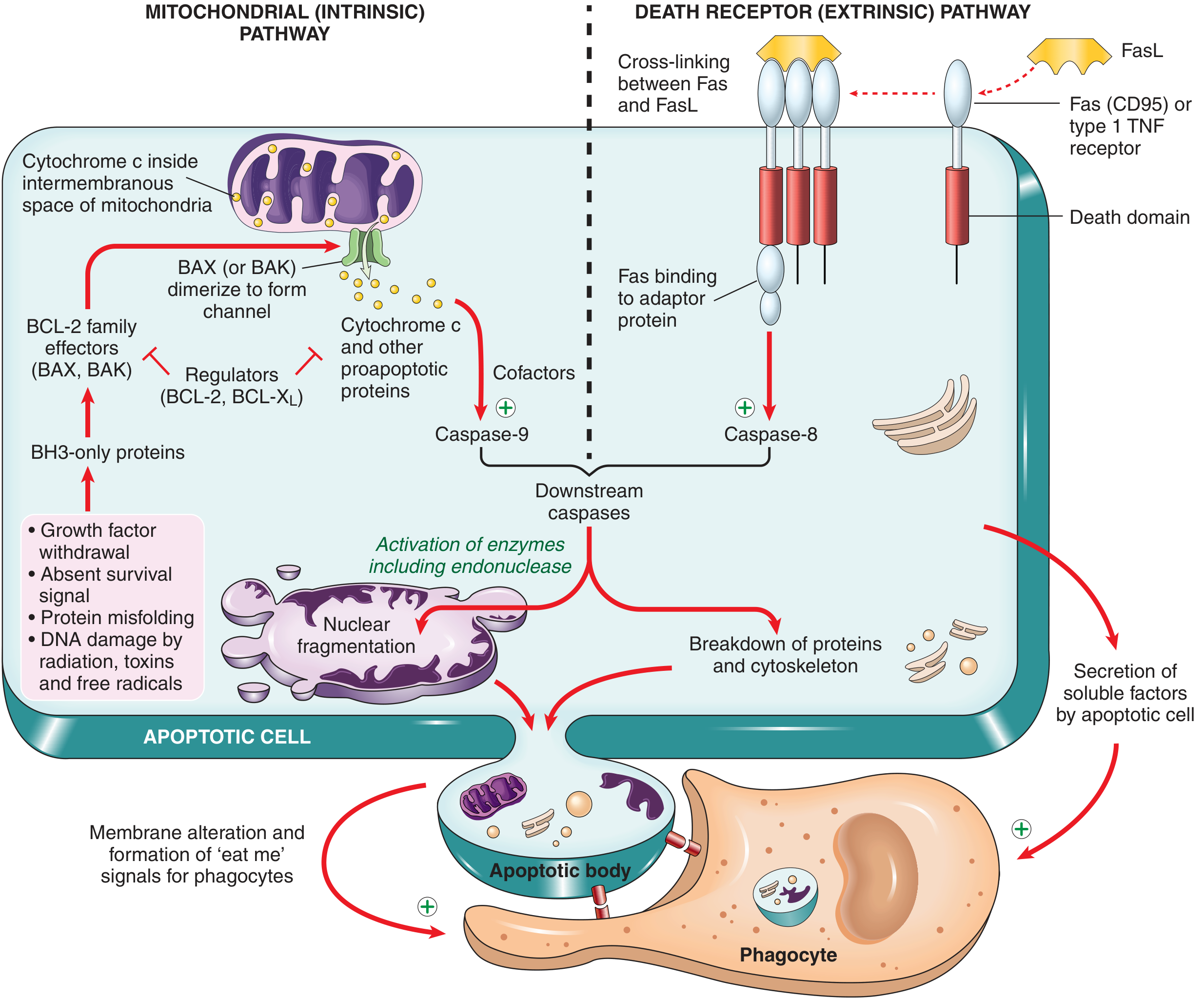
1. Mitochondrial (Intrinsic) Pathway
This is the dominant pathway in most physiologic and pathologic situations.
Key players - the BCL-2 family (>20 members):
| Category | Members | Role |
|---|---|---|
| Anti-apoptotic | BCL-2, BCL-X | Maintain mitochondrial membrane integrity; produced in response to growth factors |
| Pro-apoptotic effectors | BAX, BAK | Dimerize, insert into outer mitochondrial membrane, form channels |
| Pro-apoptotic sensors | BH3-only proteins (e.g., BID, BAD, PUMA, NOXA) | Shift the balance toward BAX/BAK; activated by cellular stress |
Sequence of events:
- A cellular stress signal (growth factor withdrawal, DNA damage, protein misfolding) activates BH3-only proteins
- BH3-only proteins inhibit BCL-2/BCL-X
Land activate BAX/BAK - BAX/BAK dimerize and insert into the outer mitochondrial membrane, forming pores
- Cytochrome c (normally in the intermembranous space) leaks into the cytosol
- Cytochrome c + cofactors (Apaf-1) activate caspase-9 (initiator caspase)
- Caspase-9 triggers a cascade of executioner caspases (caspase-3, caspase-6)
2. Death Receptor (Extrinsic) Pathway
This pathway is especially important for:
- Elimination of self-reactive lymphocytes
- CTL-mediated killing of virus-infected and tumor cells
Key players:
- Death receptors: Fas (CD95) and Type 1 TNF receptor - transmembrane proteins with a cytoplasmic "death domain"
- Ligands: FasL (expressed on CTLs and other cells), TNF
Sequence of events:
- FasL or TNF cross-links death receptors on the cell surface
- Receptor clustering recruits adaptor proteins via the death domain
- Adaptor proteins assemble a DISC (Death-Inducing Signaling Complex)
- DISC activates caspase-8 and caspase-10 (initiator caspases)
- These activate downstream executioner caspases (caspase-3, caspase-6)
Note: Caspase-8 can also cleave the BH3-only protein BID, creating a link between the extrinsic and intrinsic pathways.
3. Execution Phase (Common Final Pathway)
Once initiator caspases (caspase-8 or caspase-9) are activated:
- They rapidly and sequentially activate executioner caspases (caspase-3, caspase-6)
- Executioner caspases cleave an inhibitor of DNase → the freed DNase degrades nuclear DNA
- Caspases also proteolyze structural components of the nuclear matrix → nuclear fragmentation
- The cytoskeleton and cellular proteins are dismantled
- Membrane blebs form and pinch off as apoptotic bodies
Morphologic Features
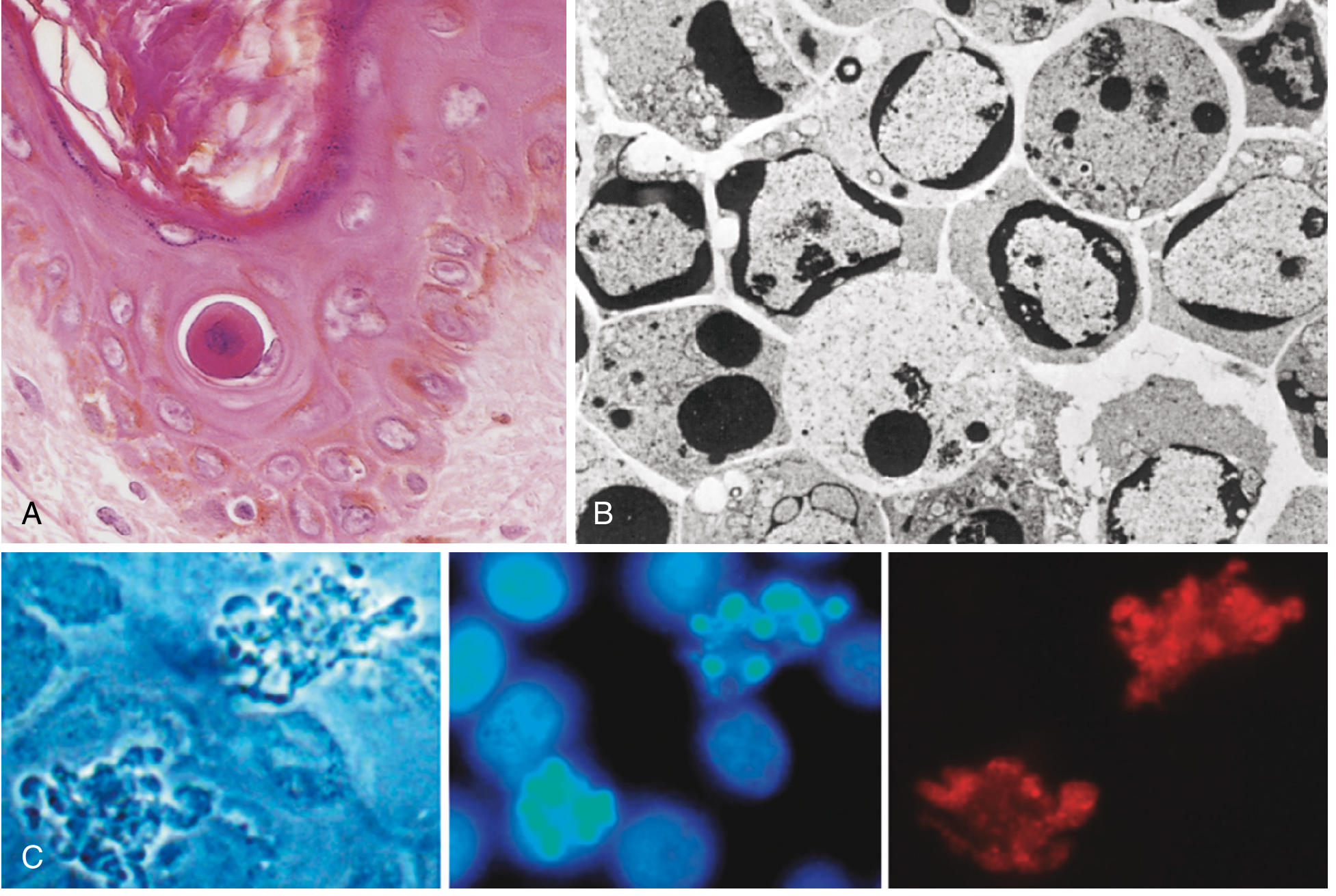
| Feature | Description |
|---|---|
| Cell shrinkage | Reduced cell volume; dense, eosinophilic cytoplasm (contrast: necrosis = cell swelling) |
| Chromatin condensation | Most characteristic feature. Chromatin aggregates peripherally under the nuclear membrane into dense crescents; nucleus may fragment |
| Membrane blebbing | Surface membrane forms blebs |
| Apoptotic bodies | Cell fragments into membrane-bound apoptotic bodies containing cytoplasm + tightly packed organelles ± nuclear fragments |
| Phagocytosis | Rapidly ingested by macrophages and degraded by lysosomal enzymes |
On H&E: round/oval mass of intensely eosinophilic cytoplasm with fragments of dense nuclear chromatin.
Removal of Apoptotic Cells (Efferocytosis)
This is a critical step that distinguishes apoptosis from necrosis:
- In healthy cells, phosphatidylserine resides on the inner leaflet of the plasma membrane
- In apoptotic cells, it flips to the outer leaflet - recognized by macrophage receptors as an "eat me" signal
- Apoptotic cells also secrete "find me" signals (soluble factors) to recruit phagocytes
- Apoptotic bodies become coated with natural antibodies and complement C1q, further facilitating recognition
- Phagocytes that engulf apoptotic cells reduce production of proinflammatory cytokines, maintaining immune quiescence
This process is called efferocytosis and is so efficient that dead cells disappear within minutes - leaving no trace and no inflammation.
Apoptosis vs. Necrosis - Key Distinctions
| Feature | Apoptosis | Necrosis |
|---|---|---|
| Cell volume | Shrinks | Swells |
| Membrane integrity | Maintained until fragmentation | Lost early |
| DNA degradation | Orderly, laddering pattern | Random, diffuse |
| Inflammation | None (no leakage) | Yes (contents released) |
| Trigger | Programmed (physiologic or controlled pathologic) | Uncontrolled injury |
| Morphology | Eosinophilic bodies, chromatin condensation | Cell lysis, ghost outlines |
Clinical Relevance - Dysregulation of Apoptosis
Insufficient apoptosis (cells that should die survive):
- Cancer - overexpression of BCL-2 is the hallmark of follicular lymphoma (t(14;18) translocation)
- Autoimmune diseases - failure to eliminate self-reactive lymphocytes
- Viral infections - viruses may encode BCL-2 homologues to prevent apoptosis of infected cells
Excessive apoptosis (cells die when they should survive):
- Neurodegenerative diseases (Alzheimer, Parkinson, ALS) - pathologic neuronal loss
- Ischemia-reperfusion injury - post-infarct cardiac and renal cell death
- AIDS - CTL-mediated apoptosis of CD4+ T cells
- Graft-versus-host disease
Key Concepts Summary
- Apoptosis = regulated cell death; intrinsic enzymes degrade DNA and proteins; no inflammation
- Two pathways: Mitochondrial (intrinsic) - regulated by BCL-2 family, caspase-9; Death receptor (extrinsic) - FasL/TNF, caspase-8
- Both converge on executioner caspases (3, 6) → DNA fragmentation, nuclear and cytoplasmic breakdown
- Dead cells are cleared by efferocytosis via phosphatidylserine "eat me" signals
- Dysregulation: too little → cancer/autoimmunity; too much → neurodegeneration/ischemia
Sources: Robbins, Cotran & Kumar: Pathologic Basis of Disease | Robbins & Kumar: Basic Pathology
This is a shared conversation. Sign in to Orris to start your own chat.