
Ckd
What would you like to know about Chronic Kidney Disease (CKD)?
- Overview - definition, causes, stages
- Diagnosis & staging criteria
- Management/treatment
- Complications
- Something else (just tell me)
Ckd review for pg medical student
"chronic kidney disease" AND guideline
CKD staging KDIGO GFR albuminuria heat map chart
kidney biopsy glomerulosclerosis chronic kidney disease histology
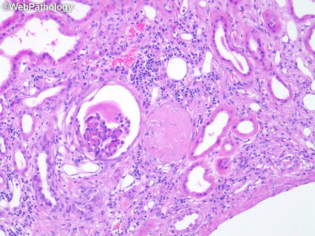
High-power brightfield histopathology of a renal cortical biopsy stained with Hematoxylin and Eosin (H&E) evaluated at approximately 400x magnification. The cortical nephron structures reveal changes typical of Autosomal Dominant Polycystic Kidney Disease (ADPKD) including a sclerotic, enlarged glomerulus with saccular expansion, indicating global or segmental sclerosis. Adjacent nephrons show distortion and dilatation of tubules with irregular luminal profiles and flattened, attenuated lining epithelium. The interstitial compartment displays mild inflammatory cells and periglomerular fibrotic changes, consistent with chronic injury and nephron loss. The overall architectural disruption reflects progressive cystic disease with reduced functional nephrons and compensatory remodeling, commonly seen in ADPKD. The image illustrates well the renal parenchymal consequences of polycystic disease—glomerulosclerosis, tubulomegaly, and tubulointerstitial fibrosis—contributing to decreased glomerular filtration rate. Clinically, these histologic features correlate with imaging-detected bilateral renal cysts and polycystic kidney disease progression, abnormal renal function tests, hypertension, and potential renal failure in affected adults. This slide is valuable for education, differential diagnosis against other glomerulopathies and cystic kidney diseases, and research into cystogenesis, nephron loss, and remodeling in polycystic kidney disease. Keywords for search include: ADPKD histology, glomerulosclerosis, tubulomegaly, polycystic kidney disease pathology, kidney cortex, renal biopsy, brightfield, H&E, renal cysts. educational, clinical, research, nephrology, nephron remodeling.
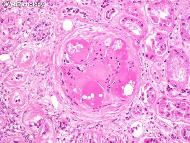
This is a renal biopsy histology image captured under bright-field light microscopy after Hematoxylin and Eosin staining. The focal structure is a glomerulus in the renal cortex showing near-complete global glomerulosclerosis with a markedly hyalinized tuft. Several hyaline caps outline the sclerotic core, appearing as pink, glassy deposits along capillary loops. Within these hyaline rims, lipid droplets and lipid-laden macrophages are evident, reflecting lipid accumulation associated with chronic diabetic microangiopathy. The surrounding tubulointerstitium displays mild interstitial fibrosis and preserved tubules in the periglomerular region. Collectively, the pattern is typical of advanced diabetic nephropathy with extensive glomerulosclerosis and hyaline arteriolosclerosis. The image highlights pathognomonic features such as noncellular sclerosis and lipid-laden inflammatory cells, which have prognostic implications for progressive renal insufficiency. Clinically, these findings correlate with long-standing diabetes mellitus, hypertension, proteinuria, and reduced GFR, indicating high risk for progression to end-stage kidney disease if glycemic control is poor. This specimen serves educational purposes in renal pathology, nephrology, and medical training to illustrate diabetic glomerulosclerosis, hyaline change, and lipid-related macrophage infiltration; it supports differential diagnosis with hypertensive nephrosclerosis and focal segmental glomerulosclerosis.
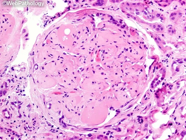
Imaging modality: brightfield histopathology. This slide shows a renal glomerulus from a biopsy specimen of diabetic nephropathy. The mesangial matrix is markedly expanded, producing round to nodular consolidations within the tuft. Several Kimmelstiel‑Wilson nodules are evident as acellular, hyaline nodules that distend the mesangial stalks and encroach on capillary loops. The glomerular basement membranes appear thickened on corresponding PAS‑positive and silver‑stained sections (referenced in adjacent images), consistent with nodular diabetic glomerulosclerosis. The surrounding cortex exhibits mild chronic interstitial changes and arteriolar hyalinosis in keeping with chronic diabetic kidney disease. The glomerulus displays relatively extensive mesangial deposition with focally patent capillary lumina, creating a characteristic nodular pattern. Overall, the morphology is diagnostic for diabetic microangiopathy with progressive nodular sclerosis. Clinically, these findings correlate with long‑standing hyperglycemia, proteinuria, and reduced GFR. Pathological significance: KW nodules indicate advanced disease and higher risk of progression to end‑stage renal disease. Differential considerations include non‑diabetic nodular glomerulosclerosis and other glomerulopathies, but the classic KW nodules and mesangial expansion strongly support diabetic nephropathy. This image is valuable for education, pathology review, and correlating histology with clinical diabetes management. It highlights key features for learners: mesangial expansion, KW nodules, PAS positivity, and altered capillary luminal flow in practice.
Chronic Kidney Disease (CKD) - PG Review
1. Definition
-
GFR < 60 mL/min/1.73 m², OR
-
A marker of kidney damage: albuminuria (>30 mg/day or ACR >30 mg/g), urine sediment abnormalities, electrolyte/tubular disorders, structural abnormalities on imaging, or history of transplantation
-
Goldman-Cecil Medicine, p. 2459 (Ch. 116)
2. Epidemiology
- Goldman-Cecil Medicine, p. 2467; Brenner and Rector's The Kidney, p. 2988
3. Staging - KDIGO 2012 "Heat Map"
| Stage | GFR | Description |
|---|---|---|
| G1 | ≥90 | Normal or high |
| G2 | 60-89 | Mildly decreased |
| G3a | 45-59 | Mildly to moderately decreased |
| G3b | 30-44 | Moderately to severely decreased |
| G4 | 15-29 | Severely decreased |
| G5 | <15 | Kidney failure (add "D" if on dialysis) |
| Category | mg/g | mg/mmol | Description |
|---|---|---|---|
| A1 | <30 | <3 | Normal to mildly increased |
| A2 | 30-300 | 3-30 | Moderately increased |
| A3 | >300 | >30 | Severely increased |
- The Washington Manual of Medical Therapeutics, p. 497-498; Comprehensive Clinical Nephrology, 7e, Ch. 80
4. Etiology
-
Diabetes mellitus (most common cause globally and in incident dialysis populations, ~37%)
-
Hypertension / nephrosclerosis
-
Glomerulonephritis (primary or secondary, e.g., lupus nephritis)
-
Polycystic kidney disease and other congenital/hereditary disorders
-
Chronic pyelonephritis/reflux nephropathy, obstructive uropathy, nephrotoxins (NSAIDs, heavy metals, analgesics), amyloidosis, atherosclerotic renovascular disease
-
Guyton and Hall Textbook of Medical Physiology, Table 32.4, p. 429; Goldman-Cecil Medicine, p. 2459
5. Pathophysiology - The "Vicious Cycle"
- Loss of nephron mass → compensatory hyperfiltration in surviving nephrons (increased glomerular capillary pressure and flow)
- Hyperfiltration → glomerular capillary injury → progressive glomerulosclerosis
- Further nephron loss → greater hyperfiltration burden on remaining nephrons → accelerating decline
- Guyton and Hall Textbook of Medical Physiology, p. 429-430
6. Complications
| System | Complication | Key mechanism |
|---|---|---|
| Hematologic | Anemia of CKD | Reduced erythropoietin production plus relative iron deficiency/inflammation; typically normocytic, normochromic |
| Bone/mineral | CKD-MBD | Phosphate retention, reduced 1,25(OH)2D, secondary hyperparathyroidism, vascular calcification |
| Metabolic | Hyperkalemia, metabolic acidosis | Reduced excretory capacity as GFR falls |
| Cardiovascular | Accelerated atherosclerosis, LVH, heart failure | Volume overload, hypertension, vascular calcification, uremic toxins |
| Nutritional | Protein-energy wasting | Anorexia, metabolic acidosis-driven catabolism, dietary restrictions |
| Immune | Increased infection risk | Uremia-related immune dysfunction |
- Brenner and Rector's The Kidney, Ch. 100 (Anemia of CKD)
-
Dietary phosphate restriction (limit dairy, meat, dark cola, processed foods)
-
Phosphate binders: calcium-based (calcium carbonate/acetate - cheap, effective, but risk vascular calcification) vs non-calcium-based (sevelamer, or iron-based binders like ferric citrate/sucroferric oxyhydroxide, which also help anemia)
-
Calcium and active vitamin D sterols (calcitriol) to manage hypocalcemia and suppress PTH
-
Avoid overcorrection - target near-normal-for-age levels, not supraphysiologic, especially in children who need positive phosphate balance for growth
-
Rheumatology, 2-Vol Set, p. 5364-5365 (Strategies for Treatment of CKD-MBD)
7. Management Principles
- Treat the underlying cause where possible (glycemic control in diabetes, BP control, immunosuppression for glomerulonephritis, relief of obstruction)
- Slow progression:
- RAAS blockade (ACEi/ARB) - reduces intraglomerular pressure, first-line for proteinuric CKD
- SGLT2 inhibitors - now guideline-directed across most CKD (with or without diabetes) for kidney and cardiovascular protection; efficacy for glucose-lowering falls as eGFR declines, but kidney/CV benefit is preserved even at low eGFR (dapagliflozin approved down to very low eGFR ranges)
- Blood pressure control (<130/80 mmHg typically), statins for cardiovascular risk reduction
- Dietary protein moderation (~0.8 g/kg/day in diabetic CKD per KDIGO) and sodium restriction
- Manage complications as above (anemia, CKD-MBD, acidosis, hyperkalemia)
- Avoid nephrotoxins (NSAIDs, contrast when avoidable, dose-adjust renally cleared drugs)
- Vaccinate (influenza annually; pneumococcal and hepatitis B per KDIGO, especially once eGFR <30)
- Plan for renal replacement therapy early when progressing toward G4-G5 (vascular access planning, transplant referral, dialysis modality education)
- Comprehensive Clinical Nephrology, 7e, Ch. 82 (SGLT2 inhibitors, vaccination); The Washington Manual of Medical Therapeutics, p. 497-498
8. Renal Replacement Therapy
- Hemodialysis (facility-based or home)
- Peritoneal dialysis
- Kidney transplantation - preferred when feasible, offers best long-term survival and quality of life
- Brenner and Rector's The Kidney, Ch. 81, p. 2988-2995
Recent Evidence Note
- CKD definition requires persistence >3 months
- Staging = GFR (G1-G5) x Albuminuria (A1-A3) heat map, not GFR alone
- Diabetes > hypertension > glomerulonephritis as global causes
- Anemia of CKD is typically normocytic normochromic
- Hyperfiltration injury explains progression independent of the original insult
- SGLT2 inhibitors are now foundational CKD therapy, not just diabetes drugs