
Familial adenomatous polyposis pathology
"familial adenomatous polyposis"[MeSH Terms] AND pathology
Familial Adenomatous Polyposis (FAP) - Pathology
Definition and Genetics
- Robbins, Cotran & Kumar Pathologic Basis of Disease - Robbins & Kumar Basic Pathology, p. 584
Molecular Pathogenesis: The APC/Wnt Pathway
- In normal cells, APC forms a destruction complex with axin, conductin, and GSK3-beta kinase
- This complex binds and phosphorylates beta-catenin, marking it for ubiquitin-mediated degradation in the cytoplasm
- When APC is mutated/truncated, beta-catenin is no longer degraded and accumulates in the cytoplasm
- Beta-catenin translocates to the nucleus and interacts with transcription factor TCF-4, upregulating genes that drive adenoma formation
- Truncated APC also disrupts normal chromosomal segregation, generating chromosomal instability (CIN)
- Sleisenger and Fordtran's Gastrointestinal and Liver Disease, p. 2578
Gross and Histologic Pathology
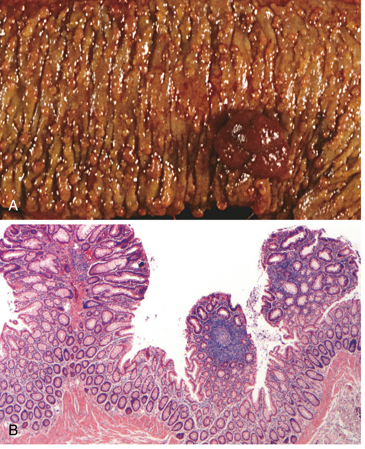
- FAP-associated polyps are morphologically indistinguishable from sporadic adenomas - tubular or villous architecture, dysplastic epithelium
- Flat or depressed adenomas are prevalent in FAP
- Microscopic adenomas (1-2 dysplastic crypts only) occur in otherwise normal-appearing mucosa
- Polyposis involves the entire colon and rectum with no predilection for a single region
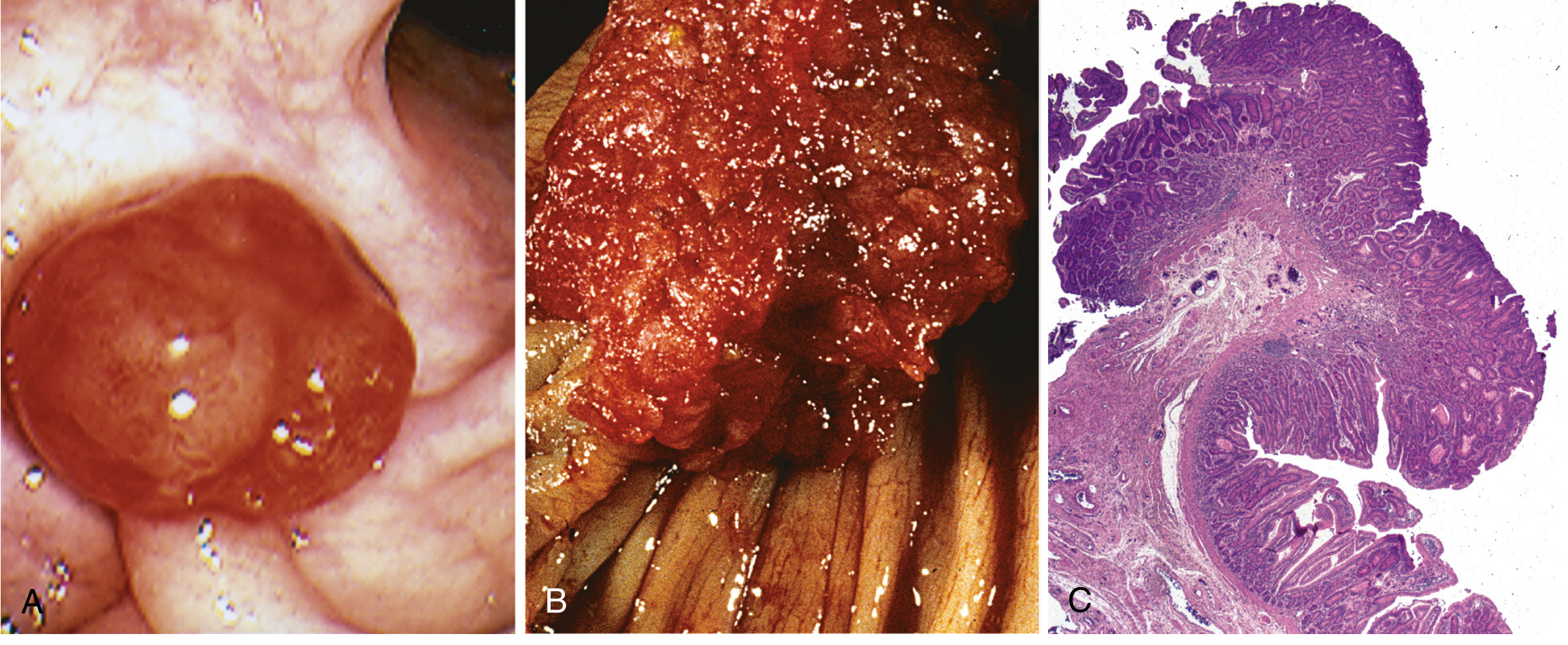
Malignant Potential
- Tubular or villous adenoma architecture
- Typical (not mucinous) adenocarcinoma histology - in contrast to HNPCC/Lynch syndrome
Upper GI and Small Bowel Pathology
| Site | Findings |
|---|---|
| Stomach | Fundic gland polyps (most common); dysplastic change occasionally; rarely adenomas |
| Duodenum | Adenomas in 60-90% of FAP patients; ampulla of Vater involved in 50-85% |
| Periampullary | 4-12% lifetime risk of duodenal/ampullary adenocarcinoma - leading cause of cancer death after prophylactic colectomy |
| Jejunum | Adenomas in ~40% |
| Ileum | Adenomas in ~20% |
FAP Variants
Gardner Syndrome
- Osteomas of the mandible, skull, and long bones
- Epidermoid/sebaceous cysts
- Desmoid tumors (intra-abdominal fibromatosis) - a leading cause of morbidity
- Thyroid tumors (papillary thyroid carcinoma)
- Dental abnormalities (unerupted and supernumerary teeth)
- Congenital hypertrophy of the retinal pigment epithelium (CHRPE) - present from birth, useful as a screening marker
Turcot Syndrome
- FAP + CNS tumors
- Two-thirds have APC mutations - develop medulloblastomas
- One-third have DNA mismatch repair mutations - develop glioblastomas
Attenuated FAP (AFAP)
- Caused by mutations in the 5' and 3' regions of APC
- Fewer than 100 adenomas (typically 10-99), predominantly in the proximal colon
- Later age of polyp onset and cancer development
- Duodenal/periampullary adenomas still occur; gastric fundic gland polyps present; mandibular osteomas rare
Summary table of adenomatous polyposis syndromes
| Syndrome | Gene | Inheritance | Polyp Count | Key Extraintestinal Features |
|---|---|---|---|---|
| Classic FAP | APC | Autosomal dominant | Thousands | CHRPE, osteomas, desmoids, epidermoid cysts, thyroid tumors, medulloblastoma |
| Attenuated FAP | APC (5'/3' ends) | Autosomal dominant | <100, proximal | Rare mandibular osteomas |
| MUTYH-associated polyposis (MAP) | MUTYH (MYH) | Autosomal recessive | 5-100s, later onset | CHRPE, osteomas; sessile serrated polyps, KRAS mutations |
| Turcot syndrome (APC type) | APC | Autosomal dominant | Hundreds | Medulloblastoma |
| Turcot syndrome (MMR type) | MSH2/MLH1 | Autosomal dominant | Few | Glioblastoma |
| Polymerase proofreading-associated polyposis (PPAP) | POLD1, POLE | Autosomal dominant | Adenomas + duodenal | Endometrial cancer |
- Sleisenger and Fordtran's Gastrointestinal and Liver Disease, Table 126.9
Desmoid Tumors in FAP
- Locally invasive, non-metastasizing fibrous tumors arising from musculoaponeurotic structures
- Predominantly intra-abdominal (mesenteric) or abdominal wall in FAP
- A major cause of morbidity and post-colectomy mortality due to bowel obstruction and vascular encasement
- Strongly associated with APC mutations downstream of codon 1310
- Can be triggered by surgical trauma (post-colectomy)
Comparison with HNPCC/Lynch Syndrome (Pathology Key Points)
| Feature | FAP | HNPCC (Lynch) |
|---|---|---|
| Polyp number | Hundreds to thousands | Few (1-3) |
| Molecular pathway | APC/Wnt (CIN) | DNA mismatch repair (MSI) |
| Polyp histology | Tubular/villous adenoma | Sessile serrated adenoma |
| Carcinoma histology | Typical adenocarcinoma | Mucinous adenocarcinoma (often) |
| Carcinoma site | No predilection | Right colon predominant |
| Key gene | APC | MSH2, MLH1 |
Key Points Summary
- FAP = APC tumor suppressor gene mutation, autosomal dominant, 5q21-q22
- >100 adenomas required for classic diagnosis; can number in thousands
- Polyps histologically identical to sporadic adenomas (tubular/villous architecture)
- 100% lifetime risk of colorectal carcinoma if untreated
- Mechanism: loss of APC → beta-catenin accumulation → Wnt pathway activation → adenoma formation
- Prophylactic colectomy is standard care
- Leading extracolonic cancer risk post-colectomy: periampullary/duodenal adenocarcinoma
- Variants: Gardner syndrome (osteomas + desmoids), Turcot syndrome (medulloblastoma), AFAP (<100 polyps, proximal colon)
- MUTYH-associated polyposis (MAP) is the autosomal recessive phenocopy
Carcinoma
What aspect of carcinoma are you interested in?
squamous cell carcinoma histology keratinization pearls
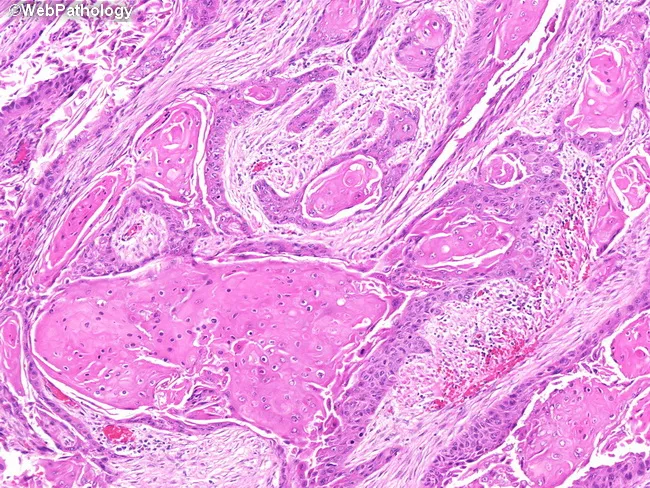
High-magnification histopathology image of oral mucosa showing keratinizing, well-differentiated squamous cell carcinoma with classic keratin pearl formation. The neoplasm displays nests and cords of malignant squamous cells invading the fibrous lamina propria, with abundant eosinophilic cytoplasm and intercellular bridges. Keratinization is evident both centrally within tumor nests and at the periphery, producing concentric keratin pearls of pale eosinophilic keratin. Tumor cells are relatively well differentiated, with recognizable squamous maturation and occasional mitotic figures. The adjacent mucosa demonstrates diverse epithelial alterations, including squamous metaplasia (keratinizing and nonkeratinizing variants), dysplasia, and areas consistent with squamous cell carcinoma in situ, reflecting field cancerization around the invasive focus. Inflammatory infiltrate and edema are present in the superficial lamina propria, while deeper stroma shows variable desmoplastic response. The surface epithelium may show thinning or ulceration over the lesion. No clear vascular invasion is evident at this magnification, though deeper planes could reveal invasion into connective tissue and perineural spread. This image provides valuable diagnostic features for distinguishing keratinizing SCC from nonkeratinizing variants and other mucosal neoplasms, and highlights the relevance of adjacent dysplasia/CIS as indicators of tumor field effect and oncologic margins. It serves as an educational reference for pathology, histology, and clinical tumor biology.
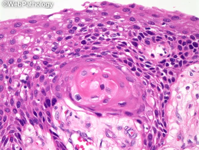
Imaging modality: Light microscopy of a hematoxylin and eosin stained tissue section, often obtained by biopsy. The slide demonstrates conventional histology of a keratinizing squamous cell carcinoma with well-differentiated epithelial nests infiltrating the surrounding stroma. Tumor cells are polygonal with abundant eosinophilic cytoplasm and intercellular bridges; keratinization is evident as keratin pearls within tumor nests. The basal layer shows increased mitotic activity and nuclear atypia, with occasional keratinizing halos surrounding nests. In focal areas, intracellular mucin may be present in scattered tumor cells, adding diagnostic nuance. The overall architecture includes cohesive nests of malignant squamous cells, keratin formation, and a fibrous stromal response. Immunohistochemistry would support squamous lineage if keratinization is less prominent or ambiguous, with p40, p63, CK5/6 positivity favoring squamous differentiation and TTF-1 negativity helping exclude pulmonary adenocarcinoma or other TTF-1 expressing tumors. Clinically, this pattern correlates with a well-differentiated squamous carcinoma that tends to show better differentiation than non-keratinizing variants but still requires complete excision or staged therapy based on invasion depth and anatomic site. This image is representative for pathology education, tumor grading, and diagnostic pathology workflow, illustrating classic keratinization, keratin pearls, and intercellular bridges characteristic of well-differentiated keratinizing squamous cell carcinoma.
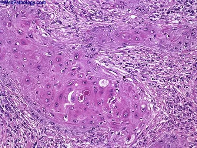
This histopathology image depicts a cutaneous squamous cell carcinoma on routine hematoxylin and eosin stained section. The biopsy shows nests of malignant squamous cells with keratinization and abundant keratin pearls, indicating squamous differentiation. The tumor exhibits moderate differentiation, evidenced by intercellular bridges, eosinophilic cytoplasm, nuclear pleomorphism, hyperchromasia, and mitotic activity. Invasion into the surrounding dermal stroma is evident, with desmoplastic-type fibrous response in some areas. The epidermis overlying the lesion shows dysplastic changes, and keratin pearl formation within tumor nests is a hallmark of keratinizing SCC. The architecture includes irregular cords and nests of polygonal cells, occasional central keratinization, and keratinized pearls of varying sizes. Clinically, these features suggest a malignant cutaneous neoplasm with potential for local invasion; staging would consider depth of invasion and perineural or lymphovascular involvement. This image is relevant for diagnostic pathology, histology education, and tumor biology research, illustrating characteristic squamous differentiation, keratin production, and invasion patterns. Differential considerations include well-differentiated keratinizing SCC versus moderately-to-poorly differentiated forms, verrucous carcinoma, and basal cell carcinoma with squamous features. The visual cues—keratin pearls, keratinization, and cohesive epidermal-derived tumor islands—assist in confirming diagnosis and guiding surgical management. Correlation with clinical data will inform prognosis and adjuvant therapy decisions and multidisciplinary care planning.
Carcinoma - General Pathology
Definition
- Sarcomas - malignant tumors of mesenchymal/connective tissue origin
- Leukemias/Lymphomas - malignant tumors of blood-forming and lymphoid tissues
- Gliomas - malignant tumors of glial cells
- Robbins, Cotran & Kumar Pathologic Basis of Disease, p. 255
Classification by Cell/Tissue of Origin
1. Squamous Cell Carcinoma (SCC)
- Arises from stratified squamous epithelium or epithelia that undergo squamous metaplasia
- Tumor cells resemble stratified squamous epithelium
- Key histologic features:
- Keratin pearl formation (concentric whorls of keratinizing cells)
- Intercellular bridges (desmosomes)
- Polygonal cells with eosinophilic cytoplasm and nuclear pleomorphism
- Sites: skin, oral cavity, esophagus, cervix, lung, larynx, penis, anus
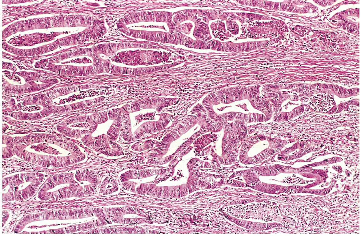
2. Adenocarcinoma
- Arises from glandular epithelium (ductal or acinar) or from epithelia capable of mucus secretion
- Tumor cells grow in a glandular/acinar pattern
- Key histologic features:
- Irregular, back-to-back glands lined by dysplastic columnar or cuboidal cells
- Loss of normal glandular architecture
- Mucin production (variable)
- Desmoplastic stromal reaction with invasion
- Sites: colon, stomach, lung, breast, prostate, endometrium, pancreas
3. Transitional Cell Carcinoma (Urothelial Carcinoma)
- Arises from transitional (urothelial) epithelium lining the urinary tract
- Graded as low or high grade
- Sites: bladder, ureter, renal pelvis
4. Basal Cell Carcinoma
- Arises from basal cells of the skin and adnexal structures
- Locally invasive but rarely metastasizes
- Histology: nests of basaloid cells with peripheral palisading
5. Undifferentiated (Anaplastic) Carcinoma
- ~2% of carcinomas are composed of cells of unknown or unrecognizable origin
- No clear line of epithelial differentiation
- Designated undifferentiated malignant neoplasm
6. Other Descriptive Subtypes
| Subtype | Definition |
|---|---|
| Papillary carcinoma | Finger-like fronds lined by malignant epithelium (e.g., thyroid, bladder) |
| Cystadenocarcinoma | Malignant cystic glandular tumor (e.g., ovary) |
| Mucinous (colloid) carcinoma | Abundant extracellular mucin pools (e.g., colon, breast) |
| Signet ring carcinoma | Intracellular mucin displaces nucleus peripherally (e.g., stomach) |
| Medullary carcinoma | Sheets of cells with little stroma (e.g., breast, thyroid) |
| Scirrhous (desmoplastic) carcinoma | Dense fibrous stroma (e.g., breast, pancreas) |
| Adenosquamous carcinoma | Mixed glandular and squamous differentiation |
| Small cell carcinoma | Neuroendocrine differentiation; extremely aggressive (e.g., lung) |
| Tissue of Origin | Benign | Malignant |
|---|---|---|
| Squamous cells | Squamous cell papilloma | Squamous cell carcinoma |
| Basal cells of skin | - | Basal cell carcinoma |
| Epithelial lining of glands/ducts | Adenoma | Adenocarcinoma |
| Epithelial lining (papillary) | Papilloma | Papillary carcinoma |
| Epithelial lining (cystic) | Cystadenoma | Cystadenocarcinoma |
| Renal tubular epithelium | Renal tubular adenoma | Renal cell carcinoma |
| Liver cells | Hepatic adenoma | Hepatocellular carcinoma |
| Bile ducts | Bile duct adenoma | Cholangiocarcinoma |
| Transitional epithelium | Transitional cell papilloma | Urothelial carcinoma |
Histologic Features of Malignancy (Carcinoma)
Cellular Features
- Pleomorphism - variation in cell and nuclear size and shape; cells may range from small with scant cytoplasm to bizarre tumor giant cells
- Nuclear abnormalities:
- Nuclei disproportionately large (nuclear:cytoplasm ratio approaches 1:1, vs. normal 1:4 to 1:6)
- Irregular nuclear shape
- Hyperchromasia - coarsely clumped, darkly stained chromatin
- Prominent, large nucleoli
- Mitoses:
- Increased mitotic count
- Atypical/bizarre mitotic figures (tripolar, quadripolar) - the most diagnostically significant feature
- Anaplasia - loss of structural and functional differentiation; most pronounced in high-grade tumors
Architectural Features
- Loss of normal tissue organization and polarity
- Invasion through the basement membrane (key distinction from carcinoma in situ)
- Desmoplastic stromal reaction - fibroblast proliferation in response to invasive tumor
- Lymphovascular invasion (lymphatic or blood vessel invasion)
- Perineural invasion
In Situ vs. Invasive Carcinoma
- Carcinoma in situ (CIS): Malignant cytologic changes confined above the basement membrane; does not invade stroma - pre-invasive
- Invasive carcinoma: Malignant cells have breached the basement membrane and entered the stroma - capable of metastasis
Grading of Carcinoma
- Degree of differentiation (resemblance to normal tissue)
- Number of mitoses per high-power field
- Extent of tumor necrosis
- Architectural features (e.g., loss of gland formation, replacement by solid sheets)
| Grade | Term | Differentiation | Features |
|---|---|---|---|
| GX | Cannot assess | - | |
| G1 | Well differentiated | High | Closely resembles normal tissue; few mitoses |
| G2 | Moderately differentiated | Intermediate | Intermediate features |
| G3 | Poorly differentiated | Low | Little resemblance to normal tissue; many mitoses, necrosis |
| G4 | Undifferentiated/Anaplastic | Very low | No recognizable differentiation |
Some tumors use a 2-tier system (low grade vs. high grade), e.g., urothelial carcinoma, glioma.
- Robbins & Kumar Basic Pathology, p. (Grading and Staging section)
Staging of Carcinoma (TNM System)
| Component | Meaning |
|---|---|
| T (Tumor) | Size/extent of primary tumor |
| N (Nodes) | Regional lymph node involvement |
| M (Metastasis) | Presence of distant metastases |
- T0 - In situ lesion (bounded by basement membrane)
- T1-T4 - Increasing size and/or invasion of adjacent structures
- N0 - No regional lymph node involvement
- N1-N3 - Increasing number and range of involved nodes
- M0 - No distant metastases
- M1 (or M2) - Presence of distant metastases
Routes of Spread
1. Direct (Local) Invasion
- Infiltration of adjacent tissues by tumor cells breaching basement membrane
- Often preceded by upregulation of proteases (MMPs) degrading extracellular matrix
2. Lymphatic Spread
- Most common route for carcinomas (vs. sarcomas which prefer hematogenous spread)
- Follows regional lymphatic channels to sentinel and regional lymph nodes
- Sentinel node biopsy assesses the first draining node
- Examples: breast cancer to axillary nodes; colorectal cancer to mesenteric nodes
3. Hematogenous Spread
- Enters blood vessels (veins more than arteries due to thinner walls)
- Leads to distant organ metastases
- Common metastatic sites: liver, lungs, bone, brain (mnemonic: "Lead Better Be Bright")
- Portal vein drainage explains liver metastases from GI carcinomas
- Vertebral (Batson's) plexus explains vertebral metastases from prostate/breast
Common Carcinomas by Organ - Summary
| Organ | Most Common Type | Key Feature |
|---|---|---|
| Lung | Adenocarcinoma (most common overall) | EGFR, ALK mutations; peripheral location |
| Lung | Squamous cell carcinoma | Central/hilar; cavitates; PTHrP hypercalcemia |
| Lung | Small cell carcinoma | Neuroendocrine; SCLC; very aggressive |
| Breast | Invasive ductal carcinoma (NST) | Most common; scirrhous |
| Colon | Adenocarcinoma | APC/Wnt pathway; left > right |
| Prostate | Adenocarcinoma | Gleason grading; PSA |
| Cervix | Squamous cell (exocervix); Adenocarcinoma (endocervix) | HPV-driven |
| Stomach | Adenocarcinoma | Intestinal (Lauren) vs. diffuse (signet ring) |
| Pancreas | Ductal adenocarcinoma | Dense desmoplasia; KRAS mutation |
| Thyroid | Papillary carcinoma (most common) | Orphan Annie nuclei; psammoma bodies |
| Liver | Hepatocellular carcinoma | HBV/HCV/cirrhosis; AFP elevated |
| Bladder | Urothelial (transitional cell) carcinoma | Aniline dye/smoking exposure |
| Kidney | Renal cell carcinoma (clear cell) | VHL mutation; "golden yellow" gross appearance |
| Skin | Basal cell carcinoma (most common skin cancer) | Rarely metastasizes |
| Skin | Squamous cell carcinoma | UV/HPV; can metastasize |
Key Points Summary
- Carcinoma = malignant epithelial tumor; the most common category of human cancer
- Classified by differentiation pattern: squamous cell, adenocarcinoma, transitional cell, basal cell, undifferentiated
- Histologic malignancy features: pleomorphism, hyperchromasia, atypical mitoses, invasion through basement membrane
- Grading (G1-G4) reflects degree of differentiation - higher grade = worse prognosis
- Staging (TNM) reflects extent of spread - more clinically significant than grade
- Carcinomas preferentially spread via lymphatics (vs. sarcomas via blood vessels)
- Common distant metastatic sites: liver, lung, bone, brain