
Enumerate swellings of jaw differential diagnosis and treatment
Reading File
Now let me search the medical library and web in parallel for jaw swellings.
Finding Sources
Finding Sources
Searching the Web
swellings of jaw differential diagnosis classification treatment oral surgery
Finding Sources
Finding Sources
Finding Sources
Reading Web Page
https://www1.racgp.org.au/ajgp/2020/september/common-causes-…
Now let me read relevant textbook sections on odontogenic cysts and tumors from Scott-Brown's and Cummings:
Reading File
Reading File
Finding Sources
Reading File
Finding Sources
I now have comprehensive information to compile a thorough answer. Let me also fetch a relevant textbook image:
Loading Image
I now have sufficient information from the textbooks and web sources to compile a comprehensive answer. Here is the full response:
Swellings of the Jaw - Differential Diagnosis and Treatment
Jaw swellings arise from a wide range of pathologies spanning inflammatory, cystic, odontogenic, non-odontogenic, fibro-osseous, and neoplastic categories. The structured approach below follows the classification used in Scott-Brown's Otorhinolaryngology Head & Neck Surgery and Cummings Otolaryngology.
Classification Overview
Swellings of the Jaw
├── 1. Inflammatory / Infective
├── 2. Odontogenic Cysts
│   ├── Developmental
│   └── Inflammatory
├── 3. Non-Odontogenic Cysts
├── 4. Non-Epithelial (Pseudocysts)
├── 5. Odontogenic Tumors (Benign)
├── 6. Fibro-Osseous Lesions
├── 7. Giant Cell Lesions
├── 8. Malignant Tumors (Primary & Secondary)
└── 9. Soft Tissue / Mucosal Swellings
1. Inflammatory / Infective Swellings
Dental Abscess / Periapical Abscess
- Pathology: Bacterial infection from a non-vital (pulp-necrotic) tooth spreading through the apical foramen into the alveolar bone
- Features: Acute onset, tender, fluctuant swelling; tooth non-vital to electric pulp test; fever, malaise
- Radiology: Periapical radiolucency on OPG/periapical X-ray
- Treatment:
- Drainage (incision and drainage if fluctuant)
- Root canal treatment (RCT) or tooth extraction
- Antibiotics (amoxicillin 500 mg TDS or metronidazole) if systemic signs present
Osteomyelitis
- Acute: Severe throbbing pain, swelling, high fever, trismus, pus discharge; associated non-vital teeth
- Chronic / Primary Chronic Osteomyelitis: Insidious jaw swelling, normal overlying mucosa, no fever, no leukocytosis; "onion-skin" periosteal reaction on imaging; peaks in childhood/adolescence and after age 50
- Treatment: Acute - IV antibiotics (penicillin-based), surgical debridement; Chronic - long-course antibiotics, sequestrectomy, hyperbaric oxygen in refractory cases
Cellulitis / Fascial Space Infections
- Ludwig's angina (bilateral submandibular/sublingual space infection) - life-threatening
- Treatment: Airway protection, IV antibiotics, surgical drainage
2. Odontogenic Cysts
(Arise from remnants of the tooth-forming apparatus - Scott-Brown's, p. 443)
A. Developmental Odontogenic Cysts
Dentigerous (Follicular) Cyst
- Most common developmental odontogenic cyst
- Arises from reduced enamel epithelium; envelops the crown of an unerupted tooth, attached at the cemento-enamel junction
- Most commonly involves mandibular third molars and maxillary canines
- Radiology: Well-defined radiolucency around the crown of an impacted tooth
- Treatment: Enucleation + extraction of involved tooth; marsupialization for large cysts in children to allow eruption
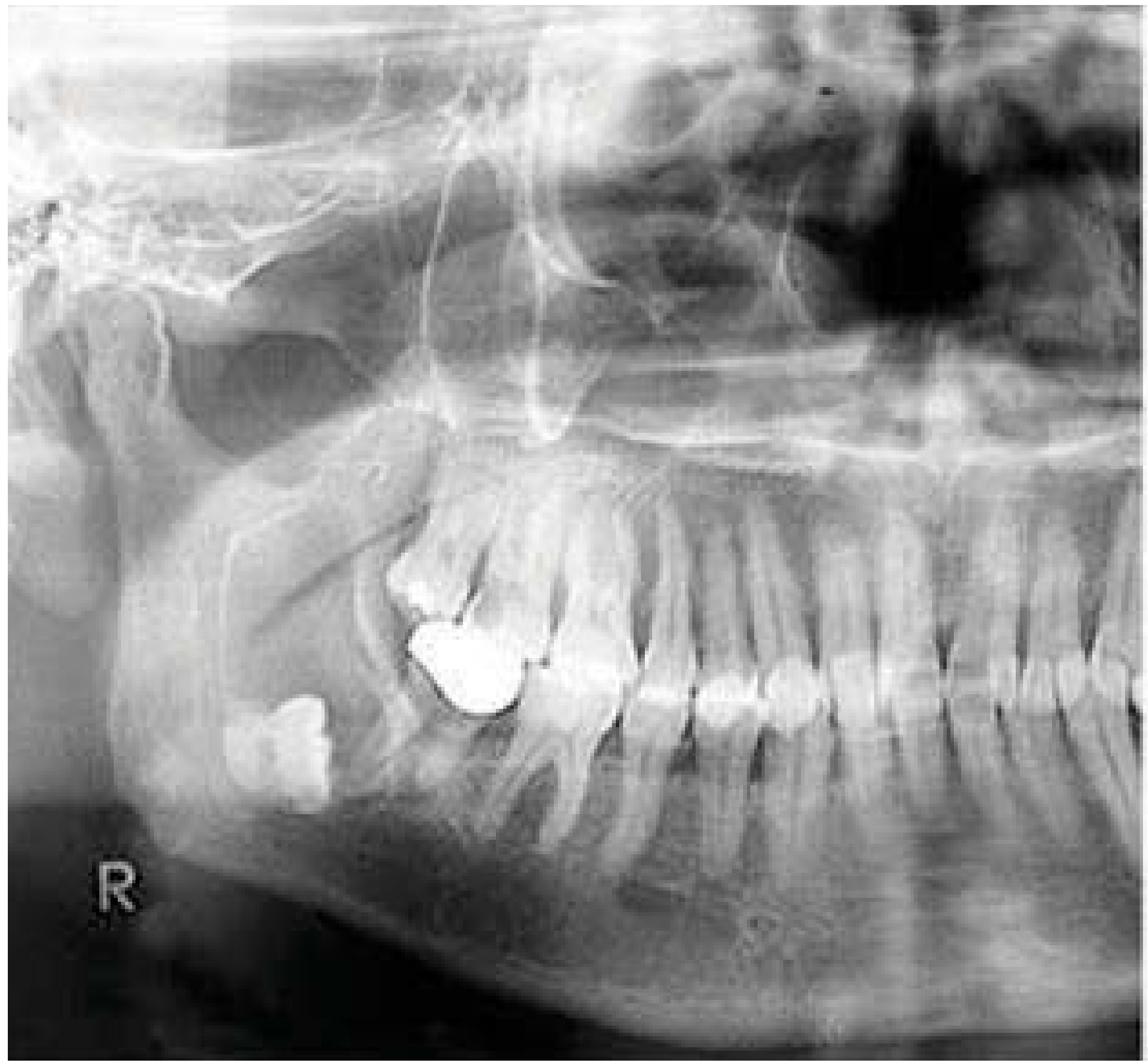
Odontogenic Keratocyst (OKC) / Keratocystic Odontogenic Tumour (KCOT)
- Peak incidence 20-30 years; mandible:maxilla ratio 2:1
- Arises from rests of Serres (dental lamina)
- Features: Usually asymptomatic jaw enlargement; may be detected incidentally on OPG
- Radiology: Unilocular or multilocular radiolucency with well-defined sclerotic margins; tendency to grow anteroposteriorly within the medullary cavity without bony expansion
- Treatment: Surgical excision; adjunctive methods to reduce recurrence - peripheral ostectomy, Carnoy's solution application, cryotherapy; enucleation alone has ~30% recurrence; associated with Gorlin-Goltz syndrome (multiple OKCs + basal cell carcinomas + calcified falx cerebri)
Lateral Periodontal Cyst
- Arises from rests of Serres lateral to the root of a vital tooth
- Treatment: Enucleation; botryoid variant (multilocular) has higher recurrence
Glandular Odontogenic Cyst (Sialo-Odontogenic Cyst)
- Rare; lined by epithelium containing mucous cells resembling salivary gland tissue
- More aggressive behavior; higher recurrence rate
- Treatment: Wide surgical excision with margins
B. Inflammatory Odontogenic Cysts
Radicular (Periapical / Dental) Cyst
- Most common cyst of the jaws overall
- Arises from rests of Malassez stimulated by infection at the root apex of a non-vital tooth
- Radiology: Periapical radiolucency, usually <2 cm, well-defined
- Treatment: Root canal treatment often leads to resolution; enucleation if persistent; tooth extraction + curettage
Residual Cyst
- Radicular cyst that persists after tooth extraction
- Treatment: Surgical enucleation
Paradental Cyst
- Lateral to the crown of a partially erupted mandibular third molar
- Treatment: Enucleation + extraction of the associated tooth
3. Non-Odontogenic Cysts
Nasopalatine Duct Cyst (Incisive Canal Cyst)
- Most common non-odontogenic cyst
- Arises from embryological remnants of the nasopalatine duct in the incisive canal
- Features: Asymptomatic swelling of the anterior hard palate/nasopalatine papilla; adjacent teeth remain vital
- Radiology: Heart-shaped or oval radiolucency in the midline of the anterior maxilla
- Treatment: Surgical enucleation
Nasolabial (Nasoalveolar) Cyst
- Soft tissue cyst; lateral to midline; arises from remnants of nasolacrimal duct
- Treatment: Surgical excision
Median Palatal / Median Mandibular Cyst
- Rare; arise along lines of fusion
- Treatment: Enucleation
Dermoid / Epidermoid Cyst
- Entrapped epithelium at embryonic lines of closure
- Presents as sublingual/submental swelling in the floor of mouth
- Treatment: Surgical excision
4. Non-Epithelial Bone Cysts (Pseudocysts)
Solitary (Simple / Traumatic) Bone Cyst
- Not a true cyst - no epithelial lining; empty or serosanguinous fluid-filled cavity
- Young patients; mandible; often asymptomatic; vital teeth
- Radiology: Radiolucency with scalloped margins between tooth roots
- Treatment: Surgical exploration - opening the cavity to promote bleeding and healing is often curative
Aneurysmal Bone Cyst
- Not a true cyst; presents in mandible of children/young adults; rapid growth
- Radiology: Unilocular/multilocular radiolucency; scalloped margins; blood-filled spaces on MRI
- Treatment: Curettage or excision; may heal spontaneously after biopsy
Stafne's Idiopathic Bone Cavity
- An asymptomatic, below-the-inferior-alveolar-canal radiolucency in the posterior mandible; actually a lingual cortical defect containing salivary gland tissue
- Treatment: None required; biopsy/follow-up to confirm
5. Odontogenic Tumors (Benign)
Ameloblastoma
- Most common clinically significant odontogenic tumor
- Locally aggressive, infiltrative into bone marrow; multicystic or unicystic
- Features: Asymptomatic early; later - extensive jaw expansion and facial deformity; can cause tooth displacement/loosening
- Radiology: "Soap bubble" multilocular radiolucency; can be unilocular; mandible >> maxilla (80%)
- Treatment:
- Unicystic: Enucleation + curettage (with risk of recurrence ~15-30%)
- Multicystic/solid: Resection with 1-1.5 cm bony margins; immediate reconstruction with bone grafts/implants
- Maxillary ameloblastoma: Often needs more radical resection due to proximity to skull base
Adenomatoid Odontogenic Tumor (AOT)
- Also called "adenoameloblastoma"; predominantly in young females; follicular (73%), extra-follicular, peripheral variants
- Associated with unerupted tooth (usually canine)
- Features: Painless, slow-growing swelling
- Treatment: Enucleation - excellent prognosis, rarely recurs
Odontoma
- Most common odontogenic tumor; hamartomatous rather than true neoplasm
- Complex odontoma: Disorganized mass of dental hard tissue; posterior jaws
- Compound odontoma: Multiple small tooth-like structures; anterior maxilla
- Treatment: Surgical excision (conservative); excellent prognosis
Cementoma / Cementifying Fibroma / Cemento-Ossifying Fibroma
- Benign; mesenchymal origin; slow-growing, painless
- Treatment: Surgical excision; recurrence rate low for cemento-ossifying fibroma
Myxoma (Odontogenic Myxoma)
- Locally aggressive; myxoid tissue in jaw bone; gelatinous appearance
- Radiology: "Tennis racket" or multilocular radiolucency
- Treatment: Resection with margins (high recurrence with curettage alone)
Calcifying Epithelial Odontogenic Tumor (CEOT / Pindborg Tumor)
- Rare; locally aggressive; mandible; calcified Liesegang ring deposits on histology
- Radiology: "Driven snow" calcifications mixed with radiolucency
- Treatment: Resection with margins
6. Fibro-Osseous Lesions
Fibrous Dysplasia
- Most common fibro-osseous lesion of the jaw; usually under age 20
- Monostotic (most common) - localized; affects maxilla more often
- Polyostotic - rare; Albright's syndrome (café-au-lait spots + precocious puberty)
- Features: Painless, smooth, rounded swelling; may cause malocclusion by displacing teeth; growth typically ceases at skeletal maturity
- Radiology: "Ground glass" appearance; poorly defined border blending into normal bone
- Treatment: Observation until skeletal maturity; surgical recontouring for cosmetic/functional deformity; no excision needed as margins poorly defined
Ossifying Fibroma
- Well-defined margins (distinguishes from fibrous dysplasia); mandible more common
- Juvenile aggressive ossifying fibroma - seen in pediatric patients; high recurrence
- Treatment: Surgical excision (conservative); juvenile variant - wide excision due to recurrence risk
Paget's Disease of Bone (Osteitis Deformans)
- Older patients; maxilla more commonly affected (jaw enlargement causing denture problems)
- Elevated ALP; "cotton wool" radiological appearance
- Treatment: Bisphosphonates (zoledronate, alendronate); calcitonin for pain
Cemento-Osseous Dysplasia
- Primarily periapical; teeth remain vital; rarely presents as jaw swelling
- Treatment: Usually none required; observe
7. Giant Cell Lesions
Central Giant Cell Granuloma (CGCG)
- Benign but locally aggressive; mandible of young females; anterior to first molars
- Radiology: Multilocular radiolucency; may cause root resorption
- Treatment: Curettage; intralesional corticosteroids; calcitonin spray; subcutaneous interferon alfa-2a; resection for aggressive variants
- Note: Must be distinguished from giant cell tumor of bone, hyperparathyroidism (brown tumor), Cherubism, and aneurysmal bone cyst
Cherubism
- Autosomal dominant; bilateral giant cell lesions of the mandible (and sometimes maxilla)
- Angel/cherub-like facies due to bilateral jaw expansion
- Treatment: Observation (often self-limiting at puberty); surgical recontouring if severe
8. Malignant Tumors
Primary Malignant Tumors of the Jaw
| Tumor | Features | Radiology | Treatment |
|---|---|---|---|
| Osteosarcoma | Most common primary malignant bone tumor; M>F; ages 10-20 (jaw: 10 yrs later than limb); rapidly growing hard swelling | "Sunburst" periosteal reaction; ill-defined lytic/mixed lesion; "Codman's triangle" | Radical resection + chemotherapy ± radiation |
| Chondrosarcoma | Jaw is rare site; midface/mandibular condyle; adults | Radiolucent with irregular calcifications | Wide resection; limited radiosensitivity |
| Ewing's Sarcoma | Rare in jaw; children/young adults; pain; fever | Permeative lytic lesion; "onion-skin" periosteum | Chemotherapy + radiation ± surgery |
| Multiple Myeloma | Punched-out radiolucencies; older adults; back pain; elevated protein | Multiple radiolucencies; no sclerotic margin | Systemic chemotherapy, stem cell transplant, bisphosphonates |
| Primary Lymphoma of Bone | Non-Hodgkin's; diffuse ill-defined lytic lesion | Permeative lysis | Chemotherapy ± radiotherapy |
| Ameloblastic Carcinoma | Malignant transformation; aggressive; rapid growth | Ill-defined radiolucency | Radical resection + adjuvant therapy |
Secondary (Metastatic) Tumors to the Jaw
- Jaw metastases most commonly from: breast, lung, prostate, kidney, thyroid, colon
- Mandible > maxilla (80% posterior mandible)
- Features: Pain, paresthesia of lip/chin (Vincent's symptom), tooth mobility
- Treatment: Palliative; primary tumor management; local radiotherapy; bisphosphonates
Squamous Cell Carcinoma (SCC) of the Jaw
- Usually arises from overlying gingival/alveolar mucosa and invades bone secondarily
- Treatment: Hemimandibulectomy/maxillectomy + neck dissection + post-operative radiotherapy
9. Soft Tissue / Mucosal Swellings
| Condition | Features | Treatment |
|---|---|---|
| Epulis (Fibrous/Vascular/Giant cell) | Pedunculated gingival swelling | Excision + scaling of causative tooth |
| Peripheral Giant Cell Granuloma | Purple-red gingival mass; mandible; anterior to first molars | Excision + curettage to bone |
| Pyogenic Granuloma | Vascular; bleeds easily; pregnancy granuloma common | Excision; address local irritant |
| Gingival Hyperplasia | Drug-induced (phenytoin, cyclosporin, nifedipine) | Withdraw drug if possible; gingivectomy |
| Torus Mandibularis / Palatinus | Bony hard, bilateral lingual mandible or palatal midline; normal mucosa | No treatment unless causing prosthetic problems |
| Mucocele / Ranula | Floor of mouth; bluish fluctuant swelling from minor salivary gland | Excision/marsupialization (ranula: marsupialize ± sublingual gland removal) |
| Salivary Gland Tumors | Pleomorphic adenoma, Warthin's tumor, mucoepidermoid carcinoma | Surgical excision ± neck dissection |
| Lymphoma (NHL) | Painless, rubbery, rapidly enlarging; associated systemic features | Chemotherapy ± radiotherapy |
| Haemangioma | Blanches on pressure; blue-purple discoloration | Sclerotherapy, laser, surgical excision |
Investigations for Any Jaw Swelling
- OPG (Orthopantomogram) - first-line imaging for all jaw pathology
- CT scan (with contrast) - extent, bony destruction, relationship to structures
- MRI - soft tissue involvement, marrow infiltration
- Biopsy - mandatory for all tumors/cysts; incisional biopsy first for suspected malignancy
- Blood tests - FBC, ESR, CRP, ALP (Paget's), serum calcium/PTH (brown tumor), serum protein electrophoresis (myeloma)
- Aspiration - yellow keratin content = OKC; straw-colored fluid = other cyst; blood = vascular lesion/aneurysmal bone cyst; negative = traumatic bone cyst
Key Diagnostic Mnemonics
- "VINDICATE" - Vascular, Inflammatory, Neoplastic, Degenerative, Idiopathic, Congenital, Autoimmune, Traumatic, Endocrine
- Periapical radiolucency with non-vital tooth → radicular cyst or periapical abscess
- Radiolucency with impacted tooth crown → dentigerous cyst
- Multilocular "soap bubble" → ameloblastoma, central giant cell granuloma, OKC
- Ground glass appearance → fibrous dysplasia
- "Sunburst" periosteal reaction → osteosarcoma
- Multiple punched-out lesions → multiple myeloma, metastases, Langerhans cell histiocytosis
Sources:
- Scott-Brown's Otorhinolaryngology Head & Neck Surgery, Chapter 25 - Cysts of the Jaws (p. 443-445)
- Cummings Otolaryngology Head and Neck Surgery - Odontogenic Cysts and Tumors (Block 18-19)
- RACGP AJGP - Common causes of swelling in the oral cavity
- Mayo Clinic - Jaw tumors and cysts: Diagnosis and treatment
This is a shared conversation. Sign in to Orris to start your own chat.