
dysapnea
dyspnea causes diagnostic algorithm approach
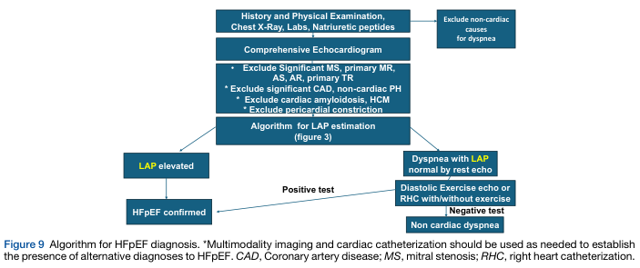
Summary : This flowchart presents an algorithm for diagnosing Heart Failure with preserved Ejection Fraction (HFpEF), detailing the stepwise evaluation of patients with dyspnea, including exclusion of non-cardiac causes, echocardiographic assessment, and further testing for left atrial pressure (LAP) elevation. flowchart: # Nodes : • History and Physical Examination, Chest X-Ray, Labs, Natriuretic peptides (rectangle) • Exclude non-cardiac causes for dyspnea (rectangle, side branch) • Comprehensive Echocardiogram (rectangle) • Exclude Significant MS, primary MR, AS, AR, primary TR (rectangle, bullet points) • Exclude significant CAD, non-cardiac PH (rectangle, bullet point) • Exclude cardiac amyloidosis, HCM (rectangle, bullet point) • Exclude pericardial constriction (rectangle, bullet point) • Algorithm for LAP estimation (figure 3) (rectangle) • LAP elevated (rectangle, yellow highlight) • HFpEF confirmed (rectangle) • Dyspnea with LAP normal by rest echo (rectangle, yellow highlight) • Diastolic Exercise echo or RHC with/without exercise (rectangle) • Positive test (arrow label) • Negative test (arrow label) • Non cardiac dyspnea (rectangle) # Connectors : • Top-down arrows connect each step in the main diagnostic pathway. • Side branch from "History and Physical Examination" to "Exclude non-cardiac causes for dyspnea". • After "Algorithm for LAP estimation", two branches: one to "LAP elevated" and one to "Dyspnea with LAP normal by rest echo". • "LAP elevated" leads directly to "HFpEF confirmed". • "Dyspnea with LAP normal by rest echo" leads to "Diastolic Exercise echo or RHC with/without exercise". • "Diastolic Exercise echo or RHC with/without exercise" splits into "Positive test" (leading to "HFpEF confirmed") and "Negative test" (leading to "Non cardiac dyspnea"). # Layout : • Vertical flow from initial assessment to final diagnosis. • Side branch for exclusion of non-cardiac causes. • Decision points create two main diagnostic branches after LAP estimation. • Use of color (yellow) to highlight LAP-related nodes. # Analysis : • The algorithm prioritizes exclusion of non-cardiac causes and alternative cardiac diagnoses before confirming HFpEF. • LAP elevation is a key diagnostic criterion; if not elevated, further exercise or invasive testing is recommended. • The flowchart ensures systematic, stepwise evaluation, minimizing misdiagnosis by requiring multiple exclusion steps and confirmatory tests.
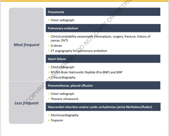
Summary : This figure presents a categorized list of diagnostic approaches for common causes of acute dyspnea, divided into "Most frequent" and "Less frequent" etiologies, with associated recommended tests for each condition. list diagram: # Most frequent : • Pneumonia – Chest radiograph • Pulmonary embolism – Clinical probability assessment (Hemoptysis, surgery, fracture, history of cancer, DVT) – D-dimer – CT angiography for pulmonary embolism • Heart failure – Chest radiograph – NT-Pro-Brain Natriuretic Peptide (Pro-BNP) and BNP – Echocardiography # Less frequent : • Pneumothorax, pleural effusion – Chest radiograph – Thoracic ultrasound • Myocardial infarction and/or cardiac arrhythmias (atrial fibrillation/flutter) – Electrocardiography – Troponin # Layout : • Two main sections: "Most frequent" and "Less frequent" causes of acute dyspnea. • Each section lists conditions in bold, followed by bullet points of recommended diagnostic tests. • The design uses dark blue headers for each condition and yellow bullet points for tests. # Analysis : • The figure emphasizes that pneumonia, pulmonary embolism, and heart failure are the most frequent causes of acute dyspnea, with specific diagnostic tests recommended for each. • Less frequent causes include pneumothorax, pleural effusion, myocardial infarction, and cardiac arrhythmias, with their own diagnostic approaches. • Imaging (chest radiograph, CT angiography, ultrasound) and laboratory tests (D-dimer, BNP, troponin) are central to the diagnostic process. • The structure aids rapid clinical decision-making by grouping conditions and tests by frequency.
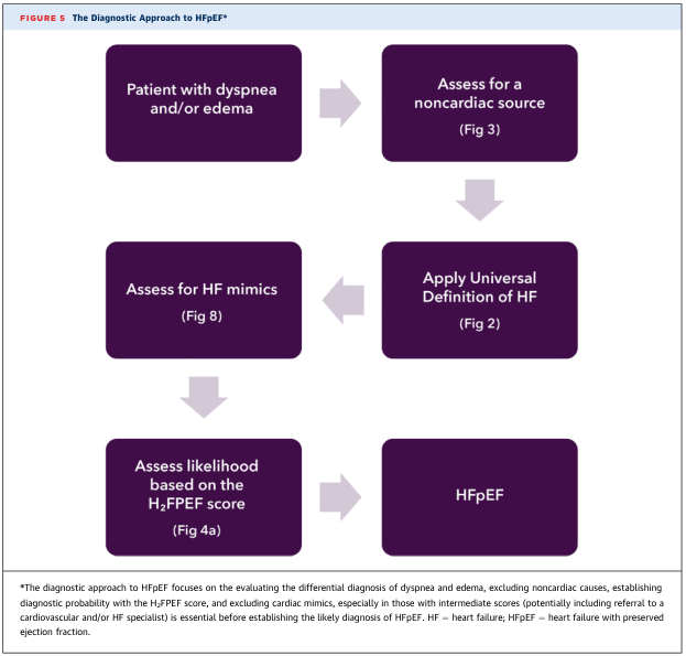
Summary : This figure presents a flowchart outlining the diagnostic approach to heart failure with preserved ejection fraction (HFpEF), focusing on the evaluation of patients with dyspnea and/or edema, exclusion of noncardiac causes, assessment for heart failure mimics, and use of the H₂FPEF score to establish diagnostic likelihood. flowchart: # Nodes : • Patient with dyspnea and/or edema (rounded rectangle) • Assess for a noncardiac source (Fig 3) (rounded rectangle) • Apply Universal Definition of HF (Fig 2) (rounded rectangle) • Assess for HF mimics (Fig 8) (rounded rectangle) • Assess likelihood based on the H₂FPEF score (Fig 4a) (rounded rectangle) • HFpEF (rounded rectangle) # Connectors : • Arrow from "Patient with dyspnea and/or edema" → "Assess for a noncardiac source (Fig 3)" • Arrow from "Assess for a noncardiac source (Fig 3)" → "Apply Universal Definition of HF (Fig 2)" • Arrow from "Apply Universal Definition of HF (Fig 2)" → "Assess for HF mimics (Fig 8)" • Arrow from "Assess for HF mimics (Fig 8)" → "Assess likelihood based on the H₂FPEF score (Fig 4a)" • Arrow from "Assess likelihood based on the H₂FPEF score (Fig 4a)" → "HFpEF" # Layout : • Flowchart is arranged in a vertical sequence of steps, with each node representing a decision or assessment point. • All nodes are rounded rectangles, connected by single downward-pointing arrows, indicating a linear, stepwise process. # Analysis : • The workflow emphasizes a systematic approach: starting with symptom identification, ruling out noncardiac causes, confirming heart failure using a universal definition, excluding mimics, and finally quantifying diagnostic likelihood with the H₂FPEF score. • The process is linear, with no branches or loops, ensuring each step is completed before proceeding to the next. • The figure highlights the importance of differential diagnosis and structured assessment in identifying HFpEF.
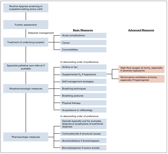
Summary : This flowchart outlines a stepwise approach to the outpatient management of dyspnea, including routine screening, assessment, treatment of underlying causes, referral to palliative care, and both nonpharmacologic and pharmacologic interventions, with escalation to advanced measures as needed. flowchart: # Nodes : • Routine dyspnea screening in outpatient setting (every visit) (rectangle) • Further assessment (rectangle) • Stepwise management (rectangle) • Treatment of underlying cause(s) (rectangle) • Basic Measures (bold, underlined heading) – Acute complications (rectangle) – Cancer (rectangle) – Comorbidities (rectangle) • Specialist palliative care referral if available (rectangle) • Nonpharmacologic measures (rectangle) – In descending order of preference: (italicized) • Airflow or fan (rectangle) • Supplemental O₂ if hypoxemic (rectangle) • Self-management strategies (rectangle) • Breathing techniques (rectangle) • Breathing postures (rectangle) • Physical therapy (rectangle) • Acupressure or reflexology (rectangle) • Pharmacologic measures (rectangle) – In descending order of preference: (italicized) • Opioids (typically oral for everyday dyspnea or prophylaxis of exertional dyspnea) (rectangle) • Corticosteroids if structural causes (rectangle) • Bronchodilators if bronchospasm (rectangle) • Benzodiazepines if severe anxiety (rectangle) • Advanced Measures (bold, underlined heading) – High-flow oxygen at home, especially if severely hypoxemic (rectangle, highlighted) – Noninvasive ventilation at home, especially if hypercapneic (rectangle, highlighted) # Connectors : • Downward arrows connect each step in the main vertical sequence: screening → assessment → stepwise management → treatment of underlying causes → specialist palliative care referral → nonpharmacologic measures → pharmacologic measures. • From "Stepwise management," branches lead to "Basic Measures" (with sub-branches for acute complications, cancer, comorbidities). • From "Nonpharmacologic measures," a branch from "Supplemental O₂ if hypoxemic" leads to "High-flow oxygen at home, especially if severely hypoxemic" under Advanced Measures. • From "Nonpharmacologic measures," a branch from the same level leads to "Noninvasive ventilation at home, especially if hypercapneic" under Advanced Measures. # Layout : • The flowchart is organized vertically, with main steps in a single column. • "Basic Measures" and "Advanced Measures" are presented as parallel columns, with "Advanced Measures" highlighted in a different color. • Sub-steps under "Basic Measures," "Nonpharmacologic measures," and "Pharmacologic measures" are listed in descending order of preference. # Analysis : • The flowchart emphasizes a structured, stepwise approach to dyspnea management, starting with routine screening and progressing through assessment, treatment, and both nonpharmacologic and pharmacologic interventions. • Referral to specialist palliative care is recommended if available. • Nonpharmacologic measures are prioritized before pharmacologic ones, with specific interventions listed in order of preference. • Advanced measures such as high-flow oxygen and noninvasive ventilation are reserved for severe cases, particularly with hypoxemia or hypercapnia. • The chart visually distinguishes between basic and advanced interventions, highlighting escalation pathways for more severe symptoms.
"dyspnea"[MeSH Terms] AND "diagnosis"[MeSH Terms]
Dyspnea (Breathlessness)
Definition
- "Tightness" or "choking"
- "Can't take a deep breath"
- "Suffocating" or "can't get enough air"
- "Feeling tired" with breathing effort
Pathophysiology
- Mismatch between respiratory motor drive and actual ventilatory output ("neuromechanical dissociation")
- Stimulation of mechanoreceptors in the lungs, chest wall, and airways
- Chemoreceptor activation by hypoxia (low O2) or hypercapnia (high CO2)
- Neural pathways that overlap with pain processing
Causes by Onset
Acute Dyspnea
| Cause | Key Features |
|---|---|
| Pulmonary embolism | Sudden onset, pleuritic chest pain, risk factors (DVT, immobility, cancer) |
| Pneumothorax | Sudden onset, unilateral chest pain, absent breath sounds |
| Acute heart failure / pulmonary edema | Orthopnea, PND, elevated BNP |
| Myocardial infarction | Chest pain, ECG changes, troponin rise |
| Pneumonia | Fever, productive cough, infiltrate on CXR |
| Severe asthma / COPD exacerbation | Wheezing, prolonged expiration |
| Pericardial tamponade | Beck's triad (hypotension, JVD, muffled heart sounds) |
| Drug overdose (e.g., salicylates) | History, toxicology |
| Upper airway obstruction | Stridor, inspiratory difficulty |
Subacute / Chronic Dyspnea
- COPD / chronic asthma - airways obstruction, barrel chest, prolonged expiration
- Heart failure (systolic or diastolic) - exertional dyspnea, PND, orthopnea, edema
- Interstitial lung disease - progressive exertional dyspnea, fine basal crackles
- Pulmonary hypertension - exertional dyspnea, right heart failure
- Severe anemia - dyspnea with fatigue; no lung/cardiac findings
- Deconditioning - sensation of heavy/rapid breathing; normal spirometry
- Neuromuscular disease - weak respiratory muscles, inability to lie supine
Special Types of Dyspnea
| Type | Description | Cause |
|---|---|---|
| Orthopnea | Dyspnea worsening when supine | Left heart failure, bilateral diaphragm paralysis |
| Paroxysmal nocturnal dyspnea (PND) | Wakes patient from sleep; relieved by sitting up | Left ventricular failure (classic), COPD secretion pooling |
| Platypnea | Dyspnea in upright position | Pulmonary vascular shunting (hepatopulmonary syndrome) |
| Trepopnea | Dyspnea in one lateral decubitus position | Pleural effusion, unilateral lung disease |
| Instant orthopnea | Inability to assume supine at all | Bilateral diaphragm paralysis |
Assessment
- Onset - sudden vs. gradual
- Severity - how many stairs/floors? On exertion or at rest?
- Position - orthopnea, platypnea?
- Triggers - dust, smoke, allergens, exertion
- Associated symptoms - wheeze, cough, chest pain, edema, fever
- Progression - importantly, patients may unknowingly limit activity to avoid dyspnea
Diagnostic Workup
- Chest X-ray - pneumonia, pulmonary edema, pneumothorax
- ECG - arrhythmia, MI, right heart strain (PE)
- BNP / NT-proBNP - heart failure
- D-dimer + CTPA - pulmonary embolism
- ABG - hypoxemia, hypercapnia, acid-base
- Troponin - ACS
- CBC - anemia
- Spirometry (chronic) - obstructive vs. restrictive pattern
- Echocardiography - cardiac function, wall motion, effusion
Management
Treat the Underlying Cause
Stepwise Outpatient Dyspnea Management
- Airflow / fan directed at face
- Supplemental O2 if hypoxemic (SpO2 < 90%)
- Self-management strategies, breathing techniques (pursed-lip breathing)
- Positioning, physical therapy
- Opioids (morphine, oral) - most evidence-based for refractory/palliative dyspnea; studies of 13 trials show benefit in advanced lung disease and terminal cancer
- Bronchodilators (SABA, LABA) - if bronchospasm present; long-acting beta-agonists have good-quality evidence in COPD
- Corticosteroids - structural/inflammatory causes
- Benzodiazepines - if severe anxiety component
- Nebulized opioids show no additional benefit over oral route
- High-flow nasal oxygen at home (severe hypoxemia)
- Non-invasive ventilation / BiPAP (hypercapnic respiratory failure)
Key Clinical Pearls
- Patients often unconsciously reduce activity to avoid dyspnea - always probe for functional limitation
- "Hunger for air" = heart failure; "increased effort" = airway obstruction or neuromuscular; "tightness" = bronchoconstriction; "heavy/rapid breathing" = deconditioning
- Dyspnea + severe anemia can mimic cardiac/pulmonary causes
- Acute diuresis relieves heart failure dyspnea but relief is not always proportional to diuresis volume
- Murray & Nadel's Textbook of Respiratory Medicine, block 4 & 5
- Goldman-Cecil Medicine, block 4
- Textbook of Family Medicine, 9e, block 1
- Recent systematic review on breathing techniques for respiratory illness (PMID 39477355, Eur Respir Rev 2024)